Neural Crest - Melanocyte Development
| Embryology - 13 May 2026 |
|---|
| Google Translate - select your language from the list shown below (this will open a new external page) |
|
العربية | català | 中文 | 中國傳統的 | français | Deutsche | עִברִית | हिंदी | bahasa Indonesia | italiano | 日本語 | 한국어 | မြန်မာ | Pilipino | Polskie | português | ਪੰਜਾਬੀ ਦੇ | Română | русский | Español | Swahili | Svensk | ไทย | Türkçe | اردو | ייִדיש | Tiếng Việt These external translations are automated and may not be accurate. (More? About Translations) |
Introduction

Melanocytes provide the pigment melanin to keratinocytes in the skin epithelium. These cells are neural crest in origin and recent research suggests that skin melaocytes are derived from the same population that Schwann cells are derived. Schwann cells wrap around nerve axon processes outside of the central nervous system. These cells in development originate from neural crest cells migrating out along the developing nerve fibers and these cells differentiate to form myelin sheaths that surround the mature nerve. The cells are named after their original discoverer a German physiologist Theodor Schwann (1810 - 1882).
In mammals, these pigmented cells can also be found in other tissues such as the: eyes, ears, heart, and central nervous system meninges. Melanocyte cells are a key topic in medical research as they are the cell transformed in melanoma.
See Integumentary System Development
| Neural Crest Links: neural crest | Lecture - Early Neural | Lecture - Neural Crest Development | Lecture Movie | Schwann cell | adrenal | melanocyte | peripheral nervous system | enteric nervous system | cornea | cranial nerve neural crest | head | skull | cardiac neural crest | Nicole Le Douarin | Neural Crest Movies | neural crest abnormalities | Category:Neural Crest | |||
|
Some Recent Findings
|
| More recent papers |
|---|
 This table allows an automated computer search of the external PubMed database using the listed "Search term" text link.
More? References | Discussion Page | Journal Searches | 2019 References | 2020 References Search term: Melanocyte Development <pubmed limit=5>Melanocyte Development</pubmed> |

Overview of Melanocyte Development in Mammals and Zebrafish[5]
Skin Pigmentation
Ephilis (freckle)
(pl., ephilides; freckle) Clinical term describing a "freckle", that is a small brown or tan mark on the skin. These inherited features result from a copy of variant Melanocortin 1 Receptor (MC1R) gene and are common on fair skinned Celtic children. Melanocytes produce locally more melanin, this can also increase following exposure to ultraviolet radiation in sunlight.
- Links: OMIM MC1R
Cafe-au-lait spots
(French, cafe-au-lait = coffee with milk; café-au-lait macule; birthmark) describes the characteristic colour of the hyperpigmented skin patch. The common name (birthmark) reflects the presence at birth (congenital) or appearing in early infancy. The pigment is produced locally by melanocytes, that produce all skin pigmentation.
Appear commonly as a solitary feature, multiple café-au-lait macules are associated with various genetic syndromes including Neurofibromatosis type 1 and 2.
- Links: OMIM - NF1
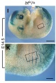
Mouse Melanocytes
Developing mouse ((bt9J/bt9J) melanoblast distribution in whole mount and trunk detail (green - β-galactosidase stained).[1]
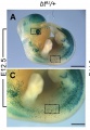
E12.5
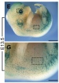
E13.5
E14.5
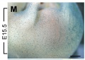
E15.5
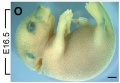
E16.5


Mouse Melanoblast Migration

|
|
See also Modeling melanoblast development [6]
- Links: Neural Crest Development
Zebrafish Melanocytes

Erbb3b gene is required to establish melanocyte stem cells in the embryo that are responsible for regenerating melanocytes after melanocytes are ablated in the larval zebrafish. Because this adult stem cell is not required for the development of embryonic melanocytes, we conclude that adult melanocyte stem cells develop in parallel to the embryonic tissues that they regulate.[7]
References
- ↑ 1.0 1.1 <pubmed>18454205</pubmed>| PLoS Genet.
- ↑ <pubmed>23333945</pubmed>
- ↑ <pubmed>21249204</pubmed>| PMC3020956 | PLoS One
- ↑ <pubmed>19837037</pubmed>
- ↑ <pubmed>25670789</pubmed>| Development
- ↑ <pubmed>22915137</pubmed>
- ↑ <pubmed>19578401</pubmed>| PMC2699538 | PLoS Genet.
Reviews
<pubmed>21310010</pubmed> <pubmed>20444197</pubmed> <pubmed>20211169</pubmed> <pubmed>18935965</pubmed>
Articles
<pubmed>20848220</pubmed> <pubmed>18703590</pubmed> <pubmed>16899407</pubmed>
Search PubMed
Search Dec 2010 "Melanocyte Development" All (2294) Review (443) Free Full Text (704)
Search Pubmed: Melanocyte Development
External Links
External Links Notice - The dynamic nature of the internet may mean that some of these listed links may no longer function. If the link no longer works search the web with the link text or name. Links to any external commercial sites are provided for information purposes only and should never be considered an endorsement. UNSW Embryology is provided as an educational resource with no clinical information or commercial affiliation.
- Bennett-Sviderskaya Laboratory
- The Wistar Institute - Melanoma Research
- Hoerter Research Lab - Zebrafish Model for Melanoma
Glossary Links
- Glossary: A | B | C | D | E | F | G | H | I | J | K | L | M | N | O | P | Q | R | S | T | U | V | W | X | Y | Z | Numbers | Symbols | Term Link
Cite this page: Hill, M.A. (2026, Mayıs 13) Embryology Neural Crest - Melanocyte Development. Retrieved from https://embryology.med.unsw.edu.au/embryology/index.php/Neural_Crest_-_Melanocyte_Development
- © Dr Mark Hill 2026, UNSW Embryology ISBN: 978 0 7334 2609 4 - UNSW CRICOS Provider Code No. 00098G