Endocrine - Pituitary Development
Introduction

Historically, this endocrine gland was called the "pituitary" as it was originally thought to produce mucous that discharged through the nose. We now know that this is not the function of the pituitary, or hypophysis which is an endocrine gland links the brain to peripheral endocrine organs and systems of the body through several specific hormones. The developmental origin of the hypophysis is also unique, epithelial origins from neural ectoderm (posterior) and from surface ectoderm (anterior).
During development, the boundary epitheilal ectoderm in the roof of the pharynx forms a pocket (Rathke's pouch) that comes into contact with the ectoderm of developing brain. Rathke's pouch is named after German embryologist and anatomist Martin Heinrich Rathke (1793 -1860).
Anatomically, the pituitary has 2 main parts posterior, or neurohypophysis and anterior, or adenohypophysis (the pars distalis, pars intermedia, and pars tuberalis). Between the two a specialized vascular (portal) system allows communication from the brain to peripheral endocrine organs and other systems. File:17thC-turkish-saddle3.jpg
The pituitary is located within the pituitary fossa of the sphenoid bone, anterior to the lamina terminalis and superior to the pharynx. The shape of the bone surrounding the pituitary led to the naming sella turcica (Latin sella = saddle, turcica = Turkish), as it resembled a saddle shape.
Pit1 (pituitary-specific transcription factor) is a transcription factor important for pituitary development and muations in this gene can lead to abnormalities in pituitary development and hormone production. (More? [../MolDev/factor/pit.htm Molecular Development Factors - Pit])
Anterior pituitary hormones - Thyroid-stimulating hormone (TSH), Adrenocorticotrophic hormone (ACTH), Luteinizing hormone (LH), Follicle-stimulating hormone (FSH), Somatotrophin/growth hormone (GH), Prolactin (PRL), Melanocyte-stimulating hormone (MSH)
Posterior pituitary hormones - Oxytocin, Arginine vasopressin
| Lecture - Head Development | original page
Development Overview

- Dual ectoderm origins
- Ectoderm - ectoderm roof of stomodeum, Rathke's pouch, adenohypophysis
- Neuroectoderm - prosenecephalon then diencephalon, neurohypophysis
Adenohypophysis
- Anterior wall proliferates - pars distalis
- Posterior wall little growth – pars intermedia
- Rostral growth around infundibular stem – pars tuberalis
Neurohypophysis
- Infundibulum – median eminence, infundibulum, pars nervosa
Pituitary Timeline
- Week 4 - hypophysial pouch, Rathke’s pouch, diverticulum from roof
- Week 5 - elongation, contacts infundibulum, diverticulum of diencephalon
- Week 6 - connecting stalk between pouch and oral cavity degenerates
- Week 10 - growth hormone and ACTH detectable
- Week 16 - adenohypophysis fully differentiated
- Week 20 to 24 - growth hormone levels peak, then decline
Pituitary Blood Vessel Development
pars distalis - vascularized by hypophysial portal vessels
A study in rats has identified the role of a known regulator of blood vessel development (Vascular Endothelial Growth Factor, VEGF) in the development of the pituitary portal vascular system. Nakakura T, Yoshida M, Dohra H, Suzuki M, Tanaka S. Gene expression of vascular endothelial growth factor-A in the pituitary during formation of the vascular system in the hypothalamic-pituitary axis of the rat. Cell Tissue Res. 2006 Apr;324(1):87-95
"The primary capillaries extended along the developing pars tuberalis, whereas the portal vessels penetrated into the pars distalis at E15.5 (rat) and subsequently expanded into the lobe to connect with the secondary capillary plexus, emerging in the pars distalis. ....study suggests that VEGF-A (Vascular Endothelial Growth Factor A) is involved in the development of the primary capillaries and in the vascularization of the pars distalis, but not in the portal vessels since the formation of portal vessels begins at E13.5 (rat), before the appearance of VEGF-A in the rostral region of the pars distalis."
The pars distalis is vascularized by hypophysial portal vessels that arise from the capillary beds in the median eminence of the hypothalamus (Murakami et al. 1987), and this hypophyseal portal system provides an important link for carrying hormonal information from the central nervous system to the pituitary. The capillaries of the pituitary gland are characterized by richly fenestrated endothelia.
Abnormalities
Anatomical abnormalities asssociated with the Rathke's pouch include a craniopharyngeal canal, from the anterior part of the fossa hypophyseos of the sphenoid bone to the under surface of the skull. The stomodeal end may also be present at the junction of the septum of the nose with the palate.
Abnormal functional development of the pituitary can lead to a wide range of other organ diseases due to the effect of hormones released from the pituitary on many other endocrine and non-endocrine organs (For example: dwarfism, hypothyroidism). (More? NIH Genes and Disease Chapter 41 - Endocrine)
There are several abnormalities associated with abnormal levels of the hormonal output of the pituitary due to the development of pituitary tumours (adenomas).
Growth hormone (GH) adenomas, which are benign pituitary tumors lead to chronic high GH output levels, that may lead to acromegaly.
Cushing's disease caused either by a pituitary adenoma produces excess adrenocorticotropic hormone (ACTH, corticotropin) or due to ectopic tumors secreting ACTH or corticotropin-releasing hormone (CRH).
Pituitary Adenoma Classification
Classification can be applied using specific criteria (clinical presentation, biochemical data, histology of growth pattern, tinctorial characteristics, proliferative activity, immunohistology marker expression, ultrastructure and molecular biology). The current classification used is the World Health Organization classification of 2000 recently updated in 2004.
Molecular
Scully KM, Rosenfeld MG. Pituitary development: regulatory codes in mammalian organogenesis. Science. 2002 Mar 22;295(5563):2231-5.
"During mammalian pituitary gland development, distinct cell types emerge from a common primordium. Appearance of specific cell types occurs in response to opposing signaling gradients that emanate from distinct organizing centers. These signals induce expression of interacting transcriptional regulators, including DNA binding-dependent activators and DNA binding-independent transrepressors, in temporally and spatially overlapping patterns. Together they synergistically regulate precursor proliferation and induction of distinct cell types. Terminal cell type differentiation requires selective gene activation strategies and long-term active repression, mediated by cell type-specific and promoter-specific recruitment of coregulatory complexes. These mechanisms imply the potential for flexibility in the ultimate identity of differentiated cell types."
Kioussi C, O'Connell S, St-Onge L, Treier M, Gleiberman AS, Gruss P, Rosenfeld MG. Pax6 is essential for establishing ventral-dorsal cell boundaries in pituitary gland development. Proc Natl Acad Sci U S A. 1999 Dec 7;96(25):14378-82.
"The transcription factor Pax6 (paired homeodomain) has been shown to be expressed transiently in the dorsal portion of the developing pituitary before the ventral/dorsal appearance of specific cell types. Transient dorsal expression of Pax6 could establish the boundary between dorsal and ventral cell types, based on the inhibition of Shh ventral signals."
Genes
PIT1 Pituitary-Specific Transcription Factor 1 - transcription factor responsible for pituitary development and hormone expression in mammals. OMIM 173110 | Gene Map Locus: 3p11 | is a pituitary-specific transcription factor responsible for pituitary development and hormone expression in mammals and is a member of the POU family of transcription factors that regulate mammalian development.
PitX1 Paired-Like Homeodomain Transcription Factor 1 - transcription factor expressed in pituitary primordium. Member of bicoid-related vertebrate homeobox genes. OMIM 602149 | Gene Map Locus: 5q31 |
PitX2 Paired-Like Homeodomain Transcription Factor 2 - transcription factor expressed in pituitary primordium and other anterior structures, including the eye Member of bicoid-related vertebrate homeobox genes. OMIM 601542 | Gene Map Locus: 4q25-q26 |
TPIT T-box transcription factor Pituitary OMIM 604614 | Gene Map Locus: 1q23-q24 |
VEGF Vascular Endothelial Growth Factor - mitogen growth factor for vascular endothelial cells. Role in pituitary vascular development. OMIM 192240 | Gene Map Locus: 1q23-q24 |
References
Online Textbooks
Endocrinology: An Integrated Approach Nussey, S.S. and Whitehead, S.A. Oxford, UK: BIOS Scientific Publishers, Ltd; 2001. table of Contents
Embryology of the pituitary gland
Blood supply of the hypothalamo-pituitary axis
NIH Genes & Disease Chapter 41 - Endocrine
Developmental Biology (6th ed) Gilbert, Scott F. Sunderland (MA): Sinauer Associates, Inc.; c2000.
Stages along the hypothalamus-pituitary-thyroid axis of salamanders
Molecular Biology of the Cell (4th Edn) Alberts, Bruce; Johnson, Alexander; Lewis, Julian; Raff, Martin; Roberts, Keith; Walter, Peter. New York: Garland Publishing; 2002.
table 15-1. Some Hormone-induced Cell Responses Mediated by Cyclic AMP
Alternative processing pathways for the prohormone proopiomelanocortin
Clinical Methods (3rd Edn) Walker, H.K.; Hall, W.D.; Hurst, J.W.; editors Stoneham (MA).
Tests of Pituitary or Target Gland Dysfunction
Health Services/Technology Assessment Text (HSTAT) Bethesda (MD): National Library of Medicine (US), 2003 Oct.
Pituitary Gland search Results
Search NLM Online Textbooks- "pituitary development" : Endocrinology | Molecular Biology of the Cell | The Cell- A molecular Approach
Reviews
- Scully KM, Rosenfeld MG. Pituitary development: regulatory codes in mammalian organogenesis. Science. 2002 Mar 22;295(5563):2231-5.
- Rosenfeld MG, Briata P, Dasen J, Gleiberman AS, Kioussi C, Lin C, O'Connell SM, Ryan A, Szeto DP, Treier M. Multistep signaling and transcriptional requirements for pituitary organogenesis in vivo. Recent Prog Horm Res. 2000;55:1-13; discussion 13-4
- Kioussi C, Carriere C, Rosenfeld MG. A model for the development of the hypothalamic-pituitary axis: transcribing the hypophysis. Mech Dev. 1999 Mar;81(1-2):23-35.
Articles
- Nakakura T, Yoshida M, Dohra H, Suzuki M, Tanaka S. Gene expression of vascular endothelial growth factor-A in the pituitary during formation of the vascular system in the hypothalamic-pituitary axis of the rat. Cell Tissue Res. 2006 Apr;324(1):87-95
- Kioussi C, O'Connell S, St-Onge L, Treier M, Gleiberman AS, Gruss P, Rosenfeld MG. Pax6 is essential for establishing ventral-dorsal cell boundaries in pituitary gland development. Proc Natl Acad Sci U S A. 1999 Dec 7;96(25):14378-82.
- Treier M, O'Connell S, Gleiberman A, Price J, Szeto DP, Burgess R, Chuang PT, McMahon AP, Rosenfeld MG. Hedgehog signaling is required for pituitary gland development. Development. 2001 Feb;128(3):377-86.
Search PubMed
Search April 2010
- Endocrine Development - All (14277) Review (4620) Free Full Text (3140)
Search Pubmed: pituitary development
External Links
- Gray's Anatomy 4. The Ductless Glands
- Garvan Institute Pituitary Research
Additional Images
Adult Histology
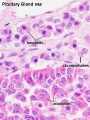
Pituitary - adenohypophysis
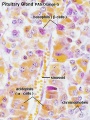
Pituitary - adenohypophysis
Pituitary - neurohypophysis


Terms
Glossary Links
- Glossary: A | B | C | D | E | F | G | H | I | J | K | L | M | N | O | P | Q | R | S | T | U | V | W | X | Y | Z | Numbers | Symbols | Term Link
Cite this page: Hill, M.A. (2024, June 16) Embryology Endocrine - Pituitary Development. Retrieved from https://embryology.med.unsw.edu.au/embryology/index.php/Endocrine_-_Pituitary_Development
- © Dr Mark Hill 2024, UNSW Embryology ISBN: 978 0 7334 2609 4 - UNSW CRICOS Provider Code No. 00098G