Category:Heart
UNSW Embryology
The links in the top section links to the original 2008 online notes pages for Cardiovascular System Development.
Cardiovascular Notes
Introduction | Abnormalities | Stage 13/14 | Stage 22 | Stage 22 Selected Highpower | Heart | Heart Rate | BloodBlood Vessels | Molecular | Lymphatic | Text only page | WWW Links | Postnatal | History - Harvey
Cardiovascular Movies
Heart Movies | Heart Looping | Atrial Septation | Realignment | Ventricular Septation | Heart Septation Models | Historic Heart Movie |
Other Cardiac and Vascular Movies Fetal Circulation (Before Birth) | Circulation (After Birth) | Aortic Branches to Glands (Kidneys only) | Aortic Branches to Glands (Gonads only)
Notes
- Pages section on this current page include lectures, laboratories, notes, quizzes and educational module sections that relate to cardiovascular development.
Subcategories
This category has the following 4 subcategories, out of 4 total.
Pages in category 'Heart'
The following 57 pages are in this category, out of 257 total.
(previous page) (next page)R
- Template:Ref-Mandarim-de-Lacerda1991
- Template:Ref-McBride1981
- Template:Ref-Morrill1916
- Template:Ref-Murray1919
- Template:Ref-NobaokRehman1941
- Template:Ref-Odgers1938
- Template:Ref-Patten1922
- Template:Ref-Patten1929
- Template:Ref-Patten1930
- Template:Ref-Patten1931
- Template:Ref-Patten1938
- Template:Ref-Patten1949
- Template:Ref-PMID1018009
- Template:Ref-PMID5165416
- Template:Ref-Pohlman1907
- Template:Ref-Pohlman1909
- Template:Ref-Retzer1920
- Template:Ref-Robinson1902
- Template:Ref-ScammonNorris1918
- Template:Ref-Schulte1916
- Template:Ref-Shaner1929
- Template:Ref-Shaner1930
- Template:Ref-Takahashi1923
- Template:Ref-Tandler1912
- Template:Ref-Vernall1962
- Template:Ref-Walmsley1931
- Template:Ref-Wang1917
- Template:Ref-Wang1918
- Template:Ref-Waterston1917
- Template:Ref-Watson1924
- Template:Ref-West1915
- Template:Ref-Witte1919
- Template:Ref-Yater1929
- Template:Ref-Yoshinaga1921
- Template:Ref-Zimmerman1927
- RPAH Cardiac Embryology 2014
T
U
V
Media in category 'Heart'
The following 200 files are in this category, out of 434 total.
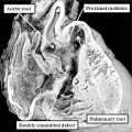
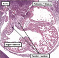
(previous page) (next page)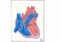 'Overriding' Aorta.PNG 840 × 575; 427 KB
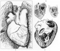
 Abbott 16-18.jpg 771 × 1,000; 164 KB
 Abbott 19.jpg 880 × 791; 171 KB
 Abbott 191.jpg 1,034 × 1,000; 234 KB
 Abbott 1915.jpg 554 × 776; 38 KB
 Abbott 20.jpg 854 × 800; 129 KB
 Abbott 201.jpg 1,150 × 1,000; 203 KB
 Abbott 21.jpg 619 × 1,000; 151 KB
 Abbott 211.jpg 600 × 600; 51 KB
 Abbott 212.jpg 600 × 600; 55 KB
 Abbott 213.jpg 600 × 600; 51 KB
 Abbott 214.jpg 600 × 600; 54 KB
 Abbott 215.jpg 600 × 600; 50 KB
 Abbott 216.jpg 600 × 600; 78 KB
 Abbott 22.jpg 947 × 800; 220 KB
 Abbott 23.jpg 884 × 800; 167 KB
 Abbott 231.jpg 914 × 800; 163 KB
 Abbott 24.jpg 588 × 800; 129 KB
 Abbott 241.jpg 827 × 800; 148 KB
 Abbott 25.jpg 585 × 810; 126 KB
 Abbott 251.jpg 908 × 1,000; 167 KB
 Abbott 26.jpg 933 × 728; 140 KB
 Abbott 261.jpg 953 × 823; 162 KB
 Abbott 27.jpg 628 × 796; 132 KB
 Abbott 271.jpg 979 × 901; 161 KB
 Abbott 28.jpg 607 × 754; 118 KB
 Abbott 281.jpg 681 × 1,000; 151 KB
 Abbott 29.jpg 606 × 597; 111 KB
 Abbott 291.jpg 1,030 × 800; 188 KB
 Abbott 30.jpg 500 × 674; 85 KB
 Abbott 301.jpg 779 × 800; 118 KB
 Abbott 31.jpg 684 × 535; 81 KB
 Abbott 311.jpg 1,134 × 800; 162 KB
 Abbott 32-34.jpg 846 × 800; 84 KB
 Abbott 32.jpg 504 × 374; 21 KB
 Abbott 33.jpg 504 × 374; 19 KB
 Abbott 34.jpg 504 × 374; 22 KB
 Abbott plate 05.jpg 671 × 1,000; 132 KB
 Abbott plate 51.jpg 618 × 800; 108 KB
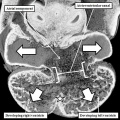
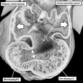
 Adult heart CT01.jpg 957 × 951; 212 KB
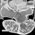
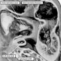
 Adult heart outflow tract CT01.jpg 747 × 747; 58 KB
 Adult heart outflow tract CT02.jpg 747 × 747; 65 KB
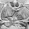
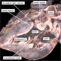
 Adult Heart Valves.jpg 1,475 × 1,070; 113 KB
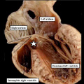
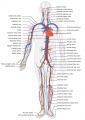 Adult human cardiovascular system.jpg 707 × 1,000; 151 KB
 Advanced Heart Development Timeline.jpg 1,772 × 769; 158 KB
 Anderson2016-fig01.jpg 800 × 800; 93 KB
 Anderson2016-fig02.jpg 800 × 788; 75 KB
 Anderson2016-fig03.jpg 800 × 800; 130 KB
 Anderson2016-fig04.jpg 800 × 800; 99 KB
 Anderson2016-fig05.jpg 796 × 573; 66 KB
 Anderson2016-fig06.jpg 800 × 800; 123 KB
 Anderson2016-fig07a.jpg 800 × 800; 193 KB
 Anderson2016-fig07b.jpg 800 × 800; 304 KB
 Anderson2016-fig08a.jpg 800 × 800; 112 KB
 Anderson2016-fig08b.jpg 800 × 800; 107 KB
 Anderson2016-fig09a.jpg 800 × 800; 106 KB
 Anderson2016-fig09b.jpg 800 × 800; 90 KB
 Anderson2016-fig10.jpg 800 × 800; 109 KB
 Anderson2016-fig11a.jpg 800 × 800; 108 KB
 Anderson2016-fig11b.jpg 800 × 800; 98 KB
 Anderson2016-fig12a.jpg 800 × 800; 120 KB
 Anderson2016-fig12b.jpg 800 × 800; 138 KB
 Anderson2016-fig13a.jpg 800 × 800; 115 KB
 Anderson2016-fig13b.jpg 800 × 800; 191 KB
 Anderson2016-fig14.jpg 800 × 800; 168 KB
 Anderson2016-fig15a.jpg 800 × 800; 142 KB
 Anderson2016-fig15b.jpg 800 × 800; 148 KB
 Anderson2016-fig16a.jpg 800 × 800; 193 KB
 Anderson2016-fig16b.jpg 800 × 800; 167 KB
 Anderson2016-fig17a.jpg 800 × 800; 98 KB
 Anderson2016-fig17b.jpg 800 × 800; 95 KB
 Anderson2016-fig18.jpg 800 × 800; 105 KB
 Anderson2016-fig19.jpg 800 × 800; 99 KB
 Anderson2016-fig20.jpg 800 × 800; 101 KB
 Anderson2016-fig21.jpg 762 × 800; 74 KB
 Anderson2016-fig22.jpg 800 × 779; 90 KB
 Anderson2016-fig23.jpg 783 × 800; 97 KB
 Anderson2016-fig24a.jpg 800 × 800; 107 KB
 Anderson2016-fig24b.jpg 800 × 800; 109 KB
 Anderson2016-fig25a.jpg 800 × 800; 122 KB
 Anderson2016-fig25b.jpg 800 × 800; 83 KB
 Anderson2016-fig26a.jpg 800 × 800; 126 KB
 Anderson2016-fig26b.jpg 800 × 800; 103 KB
 Anderson2016-fig27a.jpg 800 × 800; 117 KB
 Anderson2016-fig27b.jpg 800 × 800; 101 KB
 Anderson2016-fig28a.jpg 800 × 800; 117 KB
 Anderson2016-fig28b.jpg 800 × 800; 110 KB
 Anderson2016-fig29a.jpg 800 × 800; 111 KB
 Anderson2016-fig29b.jpg 800 × 800; 83 KB
 Anderson2016-fig30.jpg 647 × 800; 82 KB
 Anderson2016-fig31.jpg 800 × 800; 99 KB
 Anderson2016-fig32a.jpg 800 × 800; 108 KB
 Anderson2016-fig32b.jpg 800 × 800; 89 KB
 Anderson2016-fig33a.jpg 800 × 800; 110 KB
 Anderson2016-fig33b.jpg 800 × 800; 127 KB
 Anderson2016-fig34a.jpg 800 × 800; 105 KB
 Anderson2016-fig34b.jpg 800 × 800; 92 KB
 Anderson2016-fig35a.jpg 800 × 800; 118 KB
 Anderson2016-fig35b.jpg 800 × 800; 84 KB
 Anderson2016-fig36.jpg 800 × 800; 112 KB
 Anderson2016-fig37.jpg 800 × 800; 69 KB
 Anderson2016-fig38.jpg 800 × 800; 149 KB
 Anderson2016-fig39a.jpg 800 × 800; 86 KB
 Anderson2016-fig39b.jpg 800 × 800; 108 KB
 Anderson2016-fig40a.jpg 800 × 800; 102 KB
 Anderson2016-fig40b.jpg 800 × 800; 126 KB
 Anderson2016-fig41a.jpg 800 × 755; 92 KB
 Anderson2016-fig41b.jpg 800 × 800; 113 KB
 Anderson2016-fig42a.jpg 800 × 800; 90 KB
 Anderson2016-fig42b.jpg 800 × 800; 111 KB
 Anderson2016-fig43a.jpg 800 × 800; 60 KB
 Anderson2016-fig43b.jpg 800 × 800; 48 KB
 Anderson2016-fig44a.jpg 800 × 800; 83 KB
 Anderson2016-fig44b.jpg 800 × 800; 76 KB
 Anderson2016-fig45a.jpg 800 × 800; 62 KB
 Anderson2016-fig45b.jpg 800 × 800; 57 KB
 Anderson2016-fig46a.jpg 800 × 800; 100 KB
 Anderson2016-fig46b.jpg 800 × 800; 120 KB
 Anderson2016-fig47.jpg 800 × 800; 83 KB
 Anderson2016-fig48.jpg 800 × 800; 203 KB
 Anderson2016-fig49.jpg 800 × 800; 90 KB
 Anderson2016-fig50a.jpg 668 × 783; 139 KB
 Anderson2016-fig50b.jpg 798 × 786; 170 KB
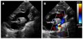 Aorta coarctation echocardiogram.jpg 601 × 283; 28 KB
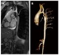 Aorta coarctation MRI.jpg 455 × 423; 25 KB
 Aortic arch and ductus arteriosus.jpg 600 × 720; 68 KB
 Aortic Stenosis.jpg 290 × 350; 16 KB
 Arey1924 fig371.jpg 1,000 × 626; 130 KB
 Atrial & Ventricular Septation 1.jpg 1,482 × 960; 97 KB
 Atrial & Ventricular Septation 2.jpg 1,482 × 1,075; 113 KB
 Atrial Septal Defect.jpg 287 × 350; 16 KB
 Atrial Septation.jpg 1,482 × 960; 90 KB
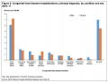 Australia Congenital heart disease 2016–17.png 1,296 × 989; 94 KB
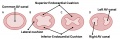 AV Canal Division (Superior View).jpg 1,487 × 489; 69 KB
 AV Canal Division.jpg 1,482 × 970; 93 KB
 AV Valves.jpg 1,183 × 1,085; 129 KB
 Bailey170.jpg 709 × 457; 67 KB
 Bailey171.jpg 954 × 507; 81 KB
 Bailey172.jpg 894 × 444; 82 KB
 Bailey173.jpg 888 × 620; 113 KB
 Bailey174.jpg 955 × 542; 88 KB
 Bailey175.jpg 885 × 306; 51 KB
 Bailey176.jpg 918 × 352; 62 KB
 Bailey177.jpg 943 × 873; 199 KB
 Bailey178.jpg 913 × 653; 125 KB
 Bailey179.jpg 892 × 794; 114 KB
 Bailey181.jpg 839 × 658; 67 KB
 Baroreceptor reflex cartoon.jpg 1,200 × 1,012; 182 KB
 Basic Heart Development Timeline.jpg 1,658 × 556; 73 KB
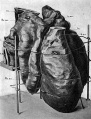
 Bedford01.jpg 734 × 1,000; 82 KB
 Bedford02.jpg 1,069 × 911; 266 KB
 Bremer1906 fig09.jpg 915 × 1,200; 208 KB
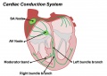 Cardiac Conduction System.jpg 1,201 × 862; 81 KB
 Cardiac connexins cartoon.jpg 990 × 621; 112 KB
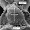
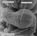
 Cardiac muscle EM01.jpg 1,072 × 735; 231 KB
 Cardiac muscle EM02.jpg 1,072 × 735; 224 KB
 Cardiac muscle EM03.jpg 849 × 615; 135 KB
 Cardiac muscle EM04.jpg 1,000 × 680; 191 KB
 Cardiac Muscle EM05.jpg 992 × 733; 158 KB
 Cardiac muscle histology.jpg 300 × 400; 42 KB
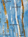
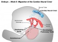 Cardiac Neural Crest Migration.jpg 1,517 × 1,116; 122 KB
 Cephalic plexus.png 600 × 557; 559 KB
 Cervical intersomitic vessels.png 600 × 462; 308 KB
 Chick Heart 002-icon.jpg 320 × 240; 5 KB
 Chicken heart 3D reconstruction from sections.jpg 1,000 × 571; 104 KB
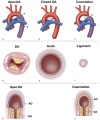
 Coarctation of the Aorta.jpg 289 × 350; 16 KB
 Cockle01.jpg 881 × 1,000; 158 KB
 Cockle02.jpg 1,459 × 929; 266 KB
 Cockle03.jpg 810 × 1,062; 158 KB
 Cockle04.jpg 740 × 1,065; 135 KB
 Complete atrioventricular canal.jpg 320 × 240; 22 KB
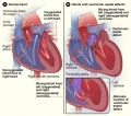 Congenital Heart Disease.jpg 475 × 421; 106 KB
 Corner1929 fig10-11.jpg 1,200 × 1,438; 730 KB
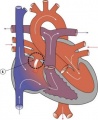
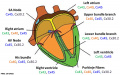
 Coronary arteries.png 800 × 472; 183 KB
 Davis1927 plate01.jpg 1,357 × 1,500; 501 KB
 Davis1927 plate02.jpg 1,357 × 1,500; 795 KB
 Dextrocardia heart position.jpg 400 × 533; 49 KB
 Dextrocardia.jpg 400 × 533; 30 KB
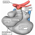
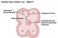
 Divisions of Early Heart Tube.jpg 1,105 × 978; 85 KB
 Double Outlet Right Ventricle.jpg 289 × 350; 16 KB
 Early Development of Heart Tube.jpg 1,475 × 1,099; 132 KB
 Early Heart Tube (Dorsal).jpg 1,282 × 1,124; 111 KB
 Early Heart Tube (Lateral).jpg 1,504 × 972; 110 KB
 Ectopia cordis.jpg 800 × 603; 37 KB
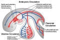 Embryonic Circulations.jpg 1,552 × 1,028; 171 KB
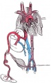
 Fetal blood flow 04.jpg 506 × 599; 50 KB
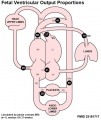
 Fetal circulation1.jpg 558 × 900; 76 KB
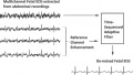 Fetal Electrocardiogram Enhancement 01.jpg 657 × 370; 40 KB
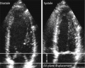 Fetal heart atrioventricular plane displacement 01.jpg 915 × 733; 55 KB
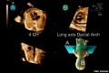 Fetal ultrasound ductal arch 01.jpg 800 × 533; 27 KB
 Folding animation 001.mov ; 1.32 MB
- Folding animation 002.mov ; 503 KB
 Frazer1916 fig01.jpg 878 × 879; 211 KB
 Frazer1916 fig02.jpg 904 × 892; 233 KB
 Frazer1916 fig03.jpg 1,469 × 828; 317 KB
 Frazer1916 fig04.jpg 1,221 × 1,020; 208 KB
 Functional Hypoplastic Left Heart.jpg 302 × 350; 18 KB
 Gray0458.gif 442 × 600; 34 KB
 Gray0458.jpg 693 × 911; 83 KB
 Gray0459.jpg 1,195 × 961; 258 KB


.jpg)
.jpg)


.jpg)