ANAT2341 Lab 6 - Postnatal
From Embryology
| Lab 6: Introduction | Trilaminar Embryo | Early Embryo | Late Embryo | Fetal | Postnatal | Abnormalities | Online Assessment |
Introduction

The Skull is a unique skeletal structure in several ways: embryonic cellular origin (neural crest), form of ossification (intramembranous and endochondrial) and flexibility (fibrous sutures).
- The cranial vault (which encloses the brain) bones are formed by intramembranous ossification.
- While the bones that form the base of the skull are formed by endochondrial ossification.
- The bones enclosing the brain have large flexible fibrous joints (sutures) which allow:
- the head to pass through the birth canal
- postnatal brain growth
- ossification continues postnatally, through puberty until mid 20s.
- in old age the sutures separating the vault plates are often completely ossified.
- Flexible fibrous sutures allow growth of the brain to be accomodated by calvarial plate growth.
- Recent molecular studies have show that noggin (a BMP antagonist) is involved in closure of these sutures.
Postnatal Skull

Skull CT Vertex, later and basal views.[1] |
Sutures and Fontanels
|
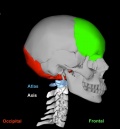
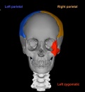
Additional Information
Head Growth
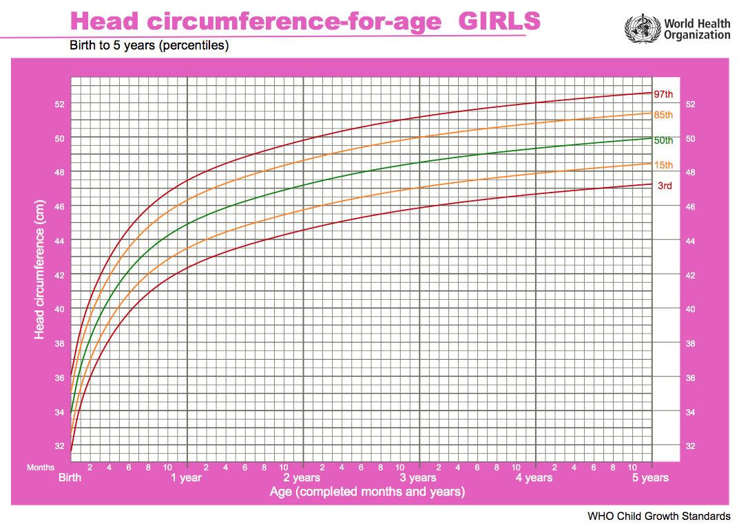
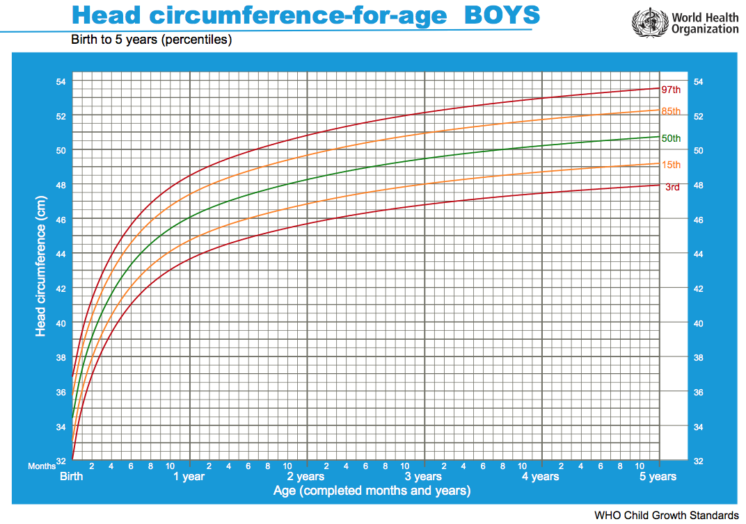
Head growth and corresponding charts differ slightly for girls and boys. Given as head circumference-for-age Birth to: 13 weeks, 2 years, 5 years.
| Girls | Boys |
|---|---|

|

|
| Chart PDF | WHO - Girls | Chart PDF | WHO - Boys |
- Links: Growth Charts | Neural Exam Movies | - Standard Head circumference-for-age | WHO Growth Standards
- Links: Growth Charts | Neural Exam Movies | - Standard Head circumference-for-age | WHO Growth Standards
Bone Histology
A histological image of a skull bone formation by Intramembranous ossification.

References
- ↑ <pubmed>21431034</pubmed>| PMC3056371 | Indian J Radiol Imaging.
| Lab 6: Introduction | Trilaminar Embryo | Early Embryo | Late Embryo | Fetal | Postnatal | Abnormalities | Online Assessment |
Glossary Links
- Glossary: A | B | C | D | E | F | G | H | I | J | K | L | M | N | O | P | Q | R | S | T | U | V | W | X | Y | Z | Numbers | Symbols | Term Link
Cite this page: Hill, M.A. (2026, September 25) Embryology ANAT2341 Lab 6 - Postnatal. Retrieved from https://embryology.med.unsw.edu.au/embryology/index.php/ANAT2341_Lab_6_-_Postnatal
- © Dr Mark Hill 2026, UNSW Embryology ISBN: 978 0 7334 2609 4 - UNSW CRICOS Provider Code No. 00098G