|
- International Classification of Diseases code 749.0
- Australian national rate (1982-1992) 4.8 - 6 /10,000 births.
- Of 1,530 infants 5.5% were stillborn and 11.5% liveborn died during neonatal period.
- slightly more common in twin births than singleton.
(Data: Congenital Malformations Australia 1981-1992 P. Lancaster and E. Pedisich ISSN 1321-8352)
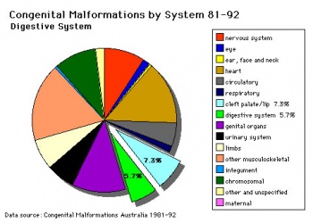 Cleft Palate - Australia (1981-1992) [9]
| Australian Palate Abnormalities (2002-2003)
|
| Cleft lip with or without cleft palate (9.2 per 10,000 births) ICD-10 Q36.0, Q36.1, Q36.9, Q37.0–Q37.5, Q37.8, Q37.9
|
A congenital anomaly characterised by a partial or complete clefting of the upper lip, with or without clefting of the alveolar ridge or the hard palate. Excludes a midline cleft of the upper or lower lip and an oblique facial fissure (going towards the eye).
- 17% of the affected pregnancies were terminated in early pregnancy or resulted in fetal deaths. Most of the fetal deaths or terminations of pregnancy (95%) had multiple abnormalities.
- more commonly seen in males than in females.
- babies born before 25 weeks of gestation, 150 per 10,000 births had this anomaly. Most babies (80.0%) were born at term with a birthweight of 2,500 grams or more.
- Maternal age group was not associated with the anomaly.
- Rates significantly higher among Indigenous women than non Indigenous women.
|
| Cleft palate without cleft lip (8.1 per 10,000 births) ICD-10 Q35.0–Q35.9
|
A congenital anomaly characterised by a closure defect of the hard and/or soft palate behind the foramen incisivum without a cleft lip. This anomaly includes sub-mucous cleft palate, but excludes cleft palate with a cleft lip, a functional short palate and high narrow palate.
- overall rate has increased to 9.1 when the rate was estimated using data from the four states that include TOP data. The reported number of fetal deaths or early terminations of pregnancy with this anomaly was small and these deaths or terminations could be due to other associated anomalies.
- proportion of females with this anomaly was higher (56.9%) than males.
- 52.7 per 10,000 babies born before 25 weeks of gestation.
- 83.0% were born at term and most of the babies (82.7%) had a birthweight of 2,500 grams or more.
- Women aged 40 years or older and women born in South Central America or the Caribbean region had the highest rates of affected births.
- Multiple births had a significantly higher rate of affected babies than singleton births.
- Rates did not differ significantly by Indigenous status or areas of residence.
|
- Links: Palate Development | Head Development | Gastrointestinal Tract - Abnormalities | ICD-10 GIT | Australian Statistics
|
- Reference: Abeywardana S & Sullivan EA 2008. Congenital Anomalies in Australia 2002-2003. Birth anomalies series no. 3 Cat. no. PER 41. Sydney: AIHW National Perinatal Statistics Unit.
|
Ten most frequently reported Birth Anomalies
- Hypospadias (More? Male movie | Genital Abnormalities - Hypospadia)
- Obstructive Defects of the Renal Pelvis (More? Renal System - Abnormalities)
- Ventricular Septal Defect (More? Cardiovascular Abnormalities - Ventricular Septal Defect)
- Congenital Dislocated Hip (More? Musculoskelal Abnormalities - Congenital Dislocation of the Hip (CDH))
- Trisomy 21 or Down syndrome - (More? Trisomy 21)
- Hydrocephalus (More? Hydrocephalus)
- Cleft Palate (More? Palate_Development)
- Trisomy 18 or Edward Syndrome - multiple abnormalities of the heart, diaphragm, lungs, kidneys, ureters and palate 86% discontinued (More? (More? Trisomy 18)
- Renal Agenesis/Dysgenesis - reduction in neonatal death and stillbirth since 1993 may be due to the more severe cases being identified in utero and being represented amongst the increased proportion of terminations (approximately 31%). (More? Renal System - Abnormalities)
- Cleft Lip and Palate - occur with another defect in 33.7% of cases.(More? Palate Development | Head Development)
(From the Victorian Perinatal Data Collection Unit in the Australian state of Victoria between 2003-2004)
Cleft Risk Variants - Two genes were identified from a recent genome-wide study.[4]
- MAFB is expressed in the mouse palatal shelf.
- ABCA4 is a member of a superfamily of transmembrane proteins, and mutations in ABCA4 play a major role in the etiology of Stargardt disease and related retinopathies. Gene produces an ATP-binding cassette (ABC) superfamily trans-membrane protein
- Links: OMIM - MAFB | OMIM - ABCA4
Folate - A recent study of periconceptional folate supplementation using the Cochrane Pregnancy and Childbirth Group's Trials Register (July 2010) identified no statistically significant evidence of any effects on prevention of cleft palate and cleft lip at birth.[10]
Development Overview
- week 4 - pharyngeal arch formation, first pharngeal arch contributes mandible and maxilla.
- week 6 - 7 - primary palate formation maxillary processes and frontonasal prominence.
- week 9 - secondary palate shelves fuse, separating oral and nasal cavities.
Embryonic Period
- (week 4) - pharyngeal arch formation in rostrocaudal sequence (1, 2, 3, 4 and 6)
- First pharyngeal arch - upper maxillary (pair) and lower mandibular prominences
- Late embryonic period - maxillary prominences fuse with frontonasal prominence forming upper jaw (maxilla and upper lip)
Fetal Period
- palatal shelves elevation
- palatal shelves midline fusion
|
|
Face Development
Begins week 4 centered around stomodeum, external depression at oral membrane
5 initial primordia from neural crest mesenchyme
- single frontonasal prominence (FNP) - forms forehead, nose dorsum and apex
- nasal placodes develop later bilateral, pushed medially
- paired maxillary prominences - form upper cheek and upper lip
- paired mandibular prominences - lower cheek, chin and lower lip
Neural Crest
- Mesenchyme invaded by neural crest generating connective tissue components
- cartilage, bone, ligaments
- arises from midbrain and hindbrain region
Frontonasal Process
The frontonasal process (FNP) forms the majority of the superior part of the early face primordia. It later fuses with the maxillary component of the first pharyngeal arch to form the upper jaw. Failure of this fusion event during the embryonic period leads to cleft lip. Under the surface ectoderm the process mesenchyme consists of two cell populations; neural crest cells, forming the connective tissues; and the mesoderm forming the endothelium of the vascular network.
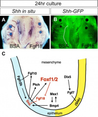
A chicken developmental model study has identified a specific surface region, the Frontonasal Ectodermal Zone (FEZ), initially induced by bone morphogenetic proteins that appears to regulate the future growth and patterning of the frontonasal process. The specific frontonasal ectodermal zone was located in the frontonasal process ectoderm flanking a boundary between Sonic hedgehog (Shh) and Fibroblast growth factor 8 (Fgf8) expression domains.[11]
Embryonic Palate
|
Human primary palate
- develops between embryonic stages 15 and 18.[12]
- fusion in the human embryo between stage 17 and 18, from an epithelial seam to the mesenchymal bridge.
|
|
- EM Links: Image - stage 16 | Image - stage 17 | Image - stage 18 | Image - stage 19 | Palate Development
Fetal Palate
Secondary palate, fusion in the human embryo in week 9. This requires the early palatal shelves growth, elevation, and fusion. There are many fusion events occurring during this period between each palatal shelf, to the primary palate, and also to the nasal septum.
palatal shelf elevation | secondary palate
Detail - hard and soft palate junction
Detail - hard palate seam
Head Growth
- continues postnatally - fontanelle allow head distortion on birth and early growth
- bone plates remain unfused to allow growth, puberty growth of face
Animal Models
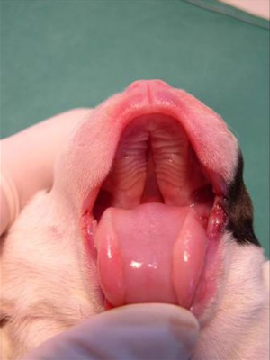 Newborn dog with cleft palate. Mouse Palate
- E11 - protrude from bilateral maxillary processes
- E12.5 - secondary palatal development begins
- E12.5-E14 - grow vertically along the developing tongue
- E14.5 - they elevate, meet, and fuse at the midline, to form an intact palate shelf, reflex opening and closing movements of the mouth
- E15.5 - palatal fusion is complete, mesenchymal condensation followed by osteogenic differentiation occurs.
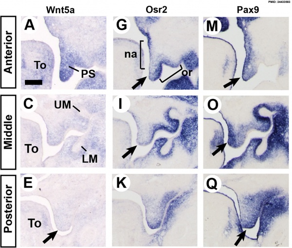
Mouse (E13.5) Palatal Shelf Wnt5a, Osr2 and Pax9 Expression.[13]
Image - Mouse E13.5 Bmp7 palate
|
|
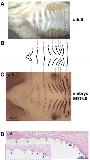
| Mouse ruga pattern (E16)
|
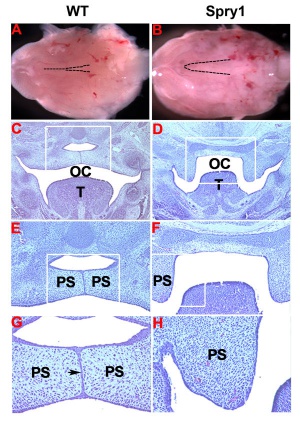
Mouse - Spry1 cleft palate
|
- Links: Mouse Development | Bone Morphogenetic Protein | Wnt | Pax
Molecular
Image - Mouse E13.5 Bmp7 palate PMID 23516636
Image - palate Bmp7 palate PMID 23516636
Image - palate detail Bmp7 palate PMID 23516636
- Links: Bone Morphogenetic Protein
References
- ↑ <pubmed>23613893</pubmed>| PLoS One.
- ↑ <pubmed>22022457</pubmed>| PLoS One.
- ↑ <pubmed>21246652</pubmed>
- ↑ 4.0 4.1 <pubmed>20436469</pubmed>
- ↑ <pubmed>17693063</pubmed>
- ↑ 6.0 6.1 6.2 <pubmed>22437671</pubmed>| PMC3928765 | J Appl Oral Sci.
- ↑ <pubmed>20537182</pubmed>| Orphanet J Rare Dis.
- ↑ <pubmed>21331089</pubmed>
- ↑ P. Lancaster and E. Pedisich, Congenital Malformations Australia 1981-1992, ISSN 1321-835.
- ↑ <pubmed>20927767</pubmed>
- ↑ <pubmed>18028903</pubmed>
- ↑ <pubmed>8227288</pubmed>
- ↑ <pubmed>24433583</pubmed>| BMC Dev Biol.
Journals
Reviews
Indian J Plast Surg. 2009 October; 42(Suppl):Cleft Lip and Palate Issue
<pubmed>22186724</pubmed>
<pubmed>19131313</pubmed>
<pubmed>16962647</pubmed>
<pubmed>3074914</pubmed>
<pubmed>8714286</pubmed>
Articles
<pubmed></pubmed>
<pubmed></pubmed>
<pubmed></pubmed>
<pubmed>20149609</pubmed>
<pubmed>19341725</pubmed>
Search PubMed
Search Pubmed: palate development | cleft palate development |
Additional Images
Nasal cavities and palate
Palate, tongue and Meckel's cartilage
Unilateral cleft lip and palate
Historic
Fig. 6. Showing the structures formed in the Lateral Nasal Processes.
Fig. 7. Coronal section of the skull of a 7th month human foetus to show the cartilages of the Lateral and Mesial Nasal Processes and the bones formed round them.
Fig. 8. Showing the ingrowth of the palatal plates of the two maxillary processes early in the 2nd month. (After Kollmann.) .
Fig. 9. Showing the Hard Palate at birth. The premaxillary part is formed from the Mesial Nasal Processes ; the remainder by the Palatal Plates of the Maxillary Processes.
Fig. 10, a, b, c. Showing what become of the skeletons of the Mandibular Arch (Meckel's Cartilage) and Maxillary Process (Palato-quadrate Cartilage).
Fig. 11. Showing the manner in which the development of the Maxillary Antrum affects the size of the palate and position of the molar teeth.
Terms
- cleft - An anatomical gap or space occuring in abnormal development in or between structures. Most commonly associated with cleft lip and cleft palate. Term is also used to describe the external groove that forms between each pharyngeal arch during their formation.
- cleft lip - An abnormality of face development leading to an opening in the upper lip. Clefting of the lip and or palate occurs with 300+ different abnormalities. Depending on many factors, this cleft may extend further into the oral cavity leading to a cleft palate. In most cases clefting of the lip and palate can be repaired by surgery.
- cleft palate - An abnormality of face development leading to an opening in the palate, the roof of the oral cavity between the mouth and the nose. Clefting of the lip and or palate occurs with 300+ different abnormalities. In most cases clefting of the lip and palate can be repaired by surgery. Palate formation in the embryo occurs at two distinct times and developmental processes called primary and secondary palate formation. This leads to different forms (classifications) and degrees of clefting.
- epithelial mesenchymal transition - (EMT, epitheliomesenchymal transformation) conversion of an epithelium into a mesenchymal (connective tissue) cellular organization.
- epitheliomesenchymal transformation - (epithelial mesenchymal transition) conversion of an epithelium into a mesenchymal (connective tissue) cellular organization.
- medial edge epithelial - (MEE) opposing palatal shelves adhere to each other to form this epithelial seam.
- palate - The roof of the mouth (oral cavity) a structure which separates the oral from the nasal cavity. Develops as two lateral palatal shelves which grow and fuse in the midline. Initally a primary palate forms with fusion of the maxillary processes with the nasal processes in early face formation. Later the secondary palate forms the anterior hard palate which will ossify and separate the oral and nasal cavities. The posterior part of the palate is called the soft palate (velum, muscular palate) and contains no bone. Abnormalities of palatal shelf fusion can lead to cleft palate.
- palatogenesis - The process of palate formation, divided into primary and secondary palate development.
- pharyngeal arch - (branchial arch, Greek, branchial = gill) These are a series of externally visible anterior tissue bands lying under the early brain that give rise to the structures of the head and neck. In humans, five arches form (1,2,3,4 and 6) but only four are externally visible on the embryo. Each arch has initially identical structures: an internal endodermal pouch, a mesenchymal (mesoderm and neural crest) core, a membrane (endoderm and ectoderm) and external cleft (ectoderm). Each arch mesenchymal core also contains similar components: blood vessel, nerve, muscular, cartilage. Each arch though initially formed from similar components will differentiate to form different head and neck structures.
- philtrum - (infranasal depression, Greek, philtron = "to love" or "to kiss") Anatomically the surface midline vertical groove in the upper lip. Embryonically formed by the fusion of the frontonasal prominence (FNP) with the two maxillary processes of the first pharyngeal arch. Cleft palate (primary palate) occurs if these three regions fail to fuse during development. Fetal alcohol syndrome is also indicated by flatness and extension of this upper lip region.
- T-box 22 - (TBX22) a transcription factor that cause X-linked cleft palate and ankyloglossia in humans. Tbx22 is induced by fibroblast growth factor 8 (FGF8) in the early face while bone morphogenic protein 4 (BMP4) represses and therefore restricts its expression. (More? OMIM - TBX22)
- Transforming Growth Factor-beta - (TGFβ) factors induces both epithelial mesenchymal transition and/or apoptosis during palatal medial edge seam disintegration.
External Links
External Links Notice - The dynamic nature of the internet may mean that some of these listed links may no longer function. If the link no longer works search the web with the link text or name. Links to any external commercial sites are provided for information purposes only and should never be considered an endorsement. UNSW Embryology is provided as an educational resource with no clinical information or commercial affiliation.
Glossary Links
- Glossary: A | B | C | D | E | F | G | H | I | J | K | L | M | N | O | P | Q | R | S | T | U | V | W | X | Y | Z | Numbers | Symbols | Term Link
Cite this page: Hill, M.A. (2026, March 13) Embryology Abnormal Development - Cleft Palate. Retrieved from https://embryology.med.unsw.edu.au/embryology/index.php/Abnormal_Development_-_Cleft_Palate
- What Links Here?
- © Dr Mark Hill 2026, UNSW Embryology ISBN: 978 0 7334 2609 4 - UNSW CRICOS Provider Code No. 00098G
|


