File:Bone-femur.jpg
From Embryology
Size of this preview: 478 × 599 pixels. Other resolution: 798 × 1,000 pixels.
Original file (798 × 1,000 pixels, file size: 150 KB, MIME type: image/jpeg)
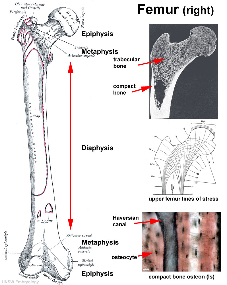
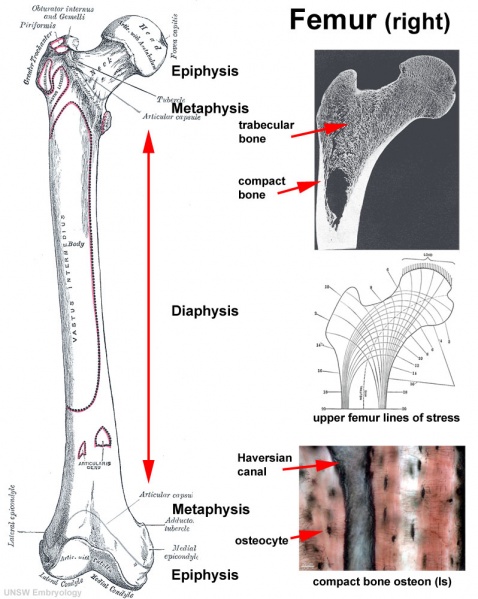
Human Femur Bone
- Human Femur (right, ventral view)
- Prepared for AE histology practical on bone formation based on images from Gray's Anatomy and UNSW histology slide set.
- Links: Bone Development | Image - Human Femur Bone
Cite this page: Hill, M.A. (2024, June 16) Embryology Bone-femur.jpg. Retrieved from https://embryology.med.unsw.edu.au/embryology/index.php/File:Bone-femur.jpg
- © Dr Mark Hill 2024, UNSW Embryology ISBN: 978 0 7334 2609 4 - UNSW CRICOS Provider Code No. 00098G
File history
Click on a date/time to view the file as it appeared at that time.
| Date/Time | Thumbnail | Dimensions | User | Comment | |
|---|---|---|---|---|---|
| current | 10:35, 23 September 2009 | | 798 × 1,000 (150 KB) | S8600021 (talk | contribs) | Bone - Femur (right) Prepared for histology practical on bone formation from Gray's Anatomy and UNSW images. http://embryology.med.unsw.edu.au/embryology/index.php?title=Bone_Development Other image versions: |
You cannot overwrite this file.
File usage
The following 3 pages use this file: