File:Keibel Mall 008.jpg
From Embryology

Size of this preview: 800 × 343 pixels. Other resolution: 1,200 × 515 pixels.
Original file (1,200 × 515 pixels, file size: 161 KB, MIME type: image/jpeg)
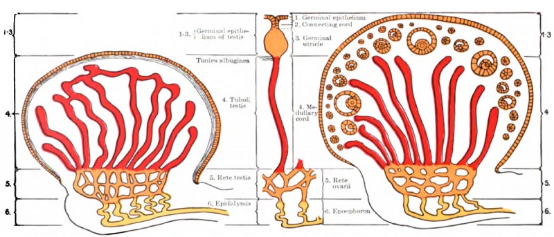
Fig. 8. Diagram showing a comparison of the testis and the ovary
Diagram showing a comparison of the testis and the ovary (based on the results of Winiwarter and Waldeyer). The germinal epithelium of the texts corresponds to 1-3 of the ovary.
- KM Figure Links: The Germ Cells | Segmentation | First Primitive Segment | Gastrulation | External Form | Placenta | Axial Skeleton | Limb Skeleton | Skull | Muscular System
| Historic Disclaimer - information about historic embryology pages |
|---|
 |
Glossary Links
- Glossary: A | B | C | D | E | F | G | H | I | J | K | L | M | N | O | P | Q | R | S | T | U | V | W | X | Y | Z | Numbers | Symbols | Term Link
Cite this page: Hill, M.A. (2024, May 23) Embryology Keibel Mall 008.jpg. Retrieved from https://embryology.med.unsw.edu.au/embryology/index.php/File:Keibel_Mall_008.jpg
- © Dr Mark Hill 2024, UNSW Embryology ISBN: 978 0 7334 2609 4 - UNSW CRICOS Provider Code No. 00098G
File history
Click on a date/time to view the file as it appeared at that time.
| Date/Time | Thumbnail | Dimensions | User | Comment | |
|---|---|---|---|---|---|
| current | 18:04, 12 February 2012 | | 1,200 × 515 (161 KB) | S8600021 (talk | contribs) | ==Fig. 8. Diagram showing a comparison of the testis and the ovary== Diagram showing a comparison of the testis and the ovary (based on the results of Winiwarter and Waldeyer). The germinal epithelium of the texts corresponds to 1-3 of the ovary. {{Keib |
You cannot overwrite this file.
File usage
The following 2 pages use this file: