Category:Student Image
From Embryology
Content in this category relates to images added to the site by students.
As shown by the text below:
Note - This image was originally uploaded as part of a student project and may contain inaccuracies in either description or acknowledgements.
--MarkHill 13:50, 4 February 2011 (EST) This category was added in February 2011 and may not appear on images uploaded before this time.
Pages in category 'Student Image'
The following 11 pages are in this category, out of 11 total.
Media in category 'Student Image'
The following 49 files are in this category, out of 650 total.
(previous page) (next page) Uveal Melanoma.gif 480 × 499; 169 KB
 V polarity.jpg 649 × 492; 93 KB
 Vaginal Ultrasonography in Sagittal Plane.jpg 975 × 554; 112 KB
 Varicocele induced cytoplasmic apoptosis.jpg 792 × 514; 90 KB
 Vegf-protects-vessels.JPG 600 × 403; 50 KB
 Ventricular Septal Defect (VSD).jpeg 358 × 402; 46 KB
 Vomeronasal Organ position.jpg 565 × 413; 49 KB
 Week 10 Midgut Herniation.png 865 × 603; 505 KB
 Week 11 fetal development of CNS.jpg 1,019 × 886; 213 KB
 Week 11 Midgut Herniation.png 865 × 744; 714 KB
 Week 6 embryonic development of CNS.jpg 1,209 × 794; 165 KB
 Week4.jpg 521 × 712; 75 KB
 Week5.jpg 717 × 1,459; 193 KB
 Week6.jpg 621 × 683; 89 KB
 Week7.jpg 1,027 × 1,121; 217 KB
 Wesley Whitten.jpg 300 × 458; 14 KB
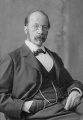 Wilhelm His History Wilhelm.jpeg 416 × 600; 67 KB
 Wnt Calcium ion pathway.JPG 1,252 × 1,134; 190 KB
 Wnt confocal microscopy.png 382 × 532; 200 KB
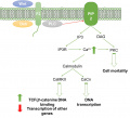
 Wnt signalling pathways.png 819 × 593; 260 KB
 WNT4 screening in the testis of the tammar wallaby.jpeg 3,543 × 2,657; 1.91 MB
 WomenwithSwyerSyndrome.png 360 × 382; 20 KB
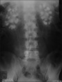 X-ray Abdomen bilateral nephrocalcinosis.jpg 490 × 649; 72 KB
 Z3289738 ANAT Lab 2.jpg 600 × 578; 52 KB
 Z3333865.gene expression.jpeg 598 × 640; 51 KB
 Z3333865.implantation.png 920 × 2,546; 2.87 MB
 Z3333865.pax2 and 8.jpg 1,200 × 1,501; 339 KB
 Z3333865.stages development inner ear.jpg 1,200 × 747; 182 KB
 Z3DCleft Lip Picture.jpg 640 × 480; 32 KB
 ZAnencephaly.jpg 372 × 550; 16 KB
 ZConvex Array Transducer.jpg 2,048 × 1,569; 441 KB
 ZDoppler Image of Fetal Aorta.jpg 640 × 480; 53 KB
 Zebrafish Melanoma.jpeg 1,280 × 1,213; 209 KB
 Zebrafish Melanoma.jpg 1,280 × 1,213; 209 KB
 Zebrafish.jpg 475 × 431; 51 KB
 ZFetus yawning.jpg 500 × 383; 35 KB
 ZHydrophone.jpg 800 × 503; 29 KB
 ZLinear Array Transducer.jpg 1,183 × 1,595; 221 KB
 Zona Pellucida and ZPC-ubiquitin.jpg 597 × 893; 348 KB
 ZPhased Array Transducerki.jpg 1,418 × 1,425; 214 KB
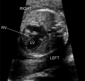 ZPulmonary Atresia.jpg 653 × 618; 85 KB
 ZScan Lines.jpg 985 × 974; 322 KB
 ZUltrasound Exam.JPG 1,600 × 1,200; 846 KB
 ZUltrasound Image of Fetal Aorta.jpg 640 × 480; 47 KB
 ZUltrasound machine.jpg 480 × 640; 93 KB
 ZUltrasound System.jpg 262 × 400; 49 KB
 Zygote - this image.png 372 × 166; 83 KB
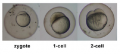
 Zygote replication z5039628.png 1,036 × 632; 510 KB
 Zygote.jpg 600 × 578; 52 KB


.jpeg)


