File:CNS secondary vesicles.jpg
Original file (987 × 562 pixels, file size: 81 KB, MIME type: image/jpeg)
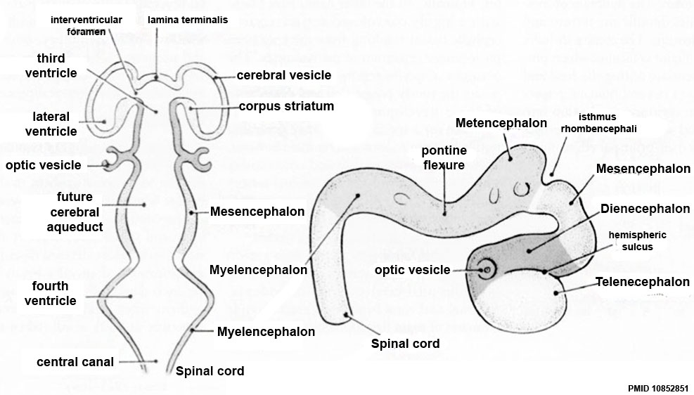
Central Nervous System - secondary brain vesicles
These 5 secondary vesicles developed from the initial 3 primary vesicles of the early neural tube, by division of the forebrain and hindbrain regions.
Secondary brain vesicles: telencephalon - diencephalon - mesencephalon - metencephalon - myelencephalon
Links: Neural System Development
Original file name: Figure-3CD-PMID10852851.jpg
Image: The more mature brain with five brain vesicles: the horizontal and lateral views correspond to GD 11.5 in rats and GD 33 ± 1 in humans.
Image Source: Critical periods of vulnerability for the developing nervous system: evidence from humans and animal models. Rice D, Barone S Jr. Environ Health Perspect. 2000 Jun;108 Suppl 3:511-33. Review. PMID: 10852851 | Environmental Health Perspectives - Critical Periods of Vulnerability for the Developing Nervous System: Evidence from Humans and Animal Models | PMC: 1637807 |
File history
Yi efo/eka'e gwa ebo wo le nyangagi wuncin ye kamina wunga tinya nan
| Gwalagizhi | Nyangagi | Dimensions | User | Comment | |
|---|---|---|---|---|---|
| current | 17:49, 18 Mayıs 2017 | | 987 × 562 (81 KB) | Z8600021 (talk | contribs) | |
| 02:02, 11 August 2009 | 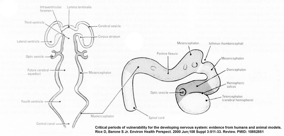 | 961 × 462 (58 KB) | MarkHill (talk | contribs) | CNS primary vesicles cartoon Figure-3CD-PMID10852851.jpg Image Source: Critical periods of vulnerability for the developing nervous system: evidence from humans and animal models. Rice D, Barone S Jr. Environ Health Perspect. 2000 Jun;108 Suppl 3:511-33 |
You cannot overwrite this file.
File usage
The following 27 pages use this file:
- 2009 Lecture 6
- 2010 BGD Lecture - Development of the Embryo/Fetus 2
- 2010 BGD Practical 6 - Week 5
- 2010 Lecture 6
- BGDA Lecture - Development of the Embryo/Fetus 2
- BGDA Lecture - Development of the Nervous System
- BGDA Practical 7 - Week 5
- Lecture - Ectoderm Development
- Lecture - Neural Development
- Neural - Amygdala Development
- Neural - Basal Ganglia Development
- Neural - Cerebellum Development
- Neural - Cerebrum Development
- Neural - Diencephalon Development
- Neural - Hippocampus Development
- Neural - Medulla Oblongata Development
- Neural - Metencephalon Development
- Neural - Myelencephalon Development
- Neural - Pons Development
- Neural - Rhinencephalon Development
- Neural - Spinal Cord Development
- Neural - Tectum Development
- Neural - Telencephalon Development
- Neural - Thalamus Development
- Neural System Development
- S
- Talk:BGDA Lecture - Development of the Nervous System


