File:Cerebral blood supply development 01.jpg: Difference between revisions
mNo edit summary |
|||
| Line 3: | Line 3: | ||
| | | | ||
Embryonic stage | Embryonic stage | ||
* 4-5 mm - the hindbrain (i.e., future posterior fossa) is supplied by two parallel neural arteries (or channels). These arteries obtain their blood supply from carotid-vertebrobasilar anastomoses given by the trigeminal artery (TA), the otic artery (OA), the hypoglossal artery (HA), and the proatlantal artery (ProA) | * [[Carnegie stage 13|4-5 mm]] - the hindbrain (i.e., future posterior fossa) is supplied by two parallel neural arteries (or channels). These arteries obtain their blood supply from carotid-vertebrobasilar anastomoses given by the trigeminal artery (TA), the otic artery (OA), the hypoglossal artery (HA), and the proatlantal artery (ProA) | ||
* 5-8 mm - the basilar artery (BA) forms from the consolidation of the neural arteries. | * [[Carnegie stage 14|5-8 mm]] - the basilar artery (BA) forms from the consolidation of the neural arteries. | ||
* 7-12 mm - the vertebral arteries (VA) forms from transverse anastomoses between cervical intersegmental arteries, beginning with the ProA and proceeding downward to the 6th intersegmental artery, | * [[Carnegie stage 15|7-12 mm]] - the vertebral arteries (VA) forms from transverse anastomoses between cervical intersegmental arteries, beginning with the ProA and proceeding downward to the 6th intersegmental artery, | ||
* 11-12 mm - (35 days) the development of the MCA is first identified as small buds originating proximal to the ACA on the anterior division of the primitive ICA. | * [[Carnegie stage 16|11-12 mm]] - (35 days) the development of the MCA is first identified as small buds originating proximal to the ACA on the anterior division of the primitive ICA. | ||
* 16-18 mm - the MCA becomes more prominent, the plexi fuse into a single artery and further branches pierce the cerebral hemisphere. | * [[Carnegie stage 19|16-18 mm]] - the MCA becomes more prominent, the plexi fuse into a single artery and further branches pierce the cerebral hemisphere. | ||
* 18 mm - the stem of the ACA gives rise to the olfactory artery. | * 18 mm - the stem of the ACA gives rise to the olfactory artery. | ||
* 21-24 mm - formation of the anterior communicating artery (ACOMM). | * [[Carnegie stage 21|21-24 mm]] - formation of the anterior communicating artery (ACOMM). | ||
| | | | ||
* '''A''' - In early phases of development the posterior circulation relies almost entirely from blood supply coming from the anterior circulation through carotid-vertebrobasilar anastomoses. | * '''A''' - In early phases of development the posterior circulation relies almost entirely from blood supply coming from the anterior circulation through carotid-vertebrobasilar anastomoses. | ||
Revision as of 16:53, 15 Haziran 2015
Cerebral Blood Supply Development
|
Embryonic stage
|
|
(above text modified from reference)
Reference
<pubmed>26060802</pubmed>| J Stroke.
Copyright
This is an Open Access article distributed under the terms of the Creative Commons Attribution Non-Commercial License (http://creativecommons.org/licenses/by-nc/3.0/) which permits unrestricted non-commercial use, distribution, and reproduction in any medium, provided the original work is properly cited.
Jos-17-144-g002-l.jpg
Cite this page: Hill, M.A. (2026, October 5) Embryology Cerebral blood supply development 01.jpg. Retrieved from https://embryology.med.unsw.edu.au/embryology/index.php/File:Cerebral_blood_supply_development_01.jpg
- © Dr Mark Hill 2026, UNSW Embryology ISBN: 978 0 7334 2609 4 - UNSW CRICOS Provider Code No. 00098G
File history
Yi efo/eka'e gwa ebo wo le nyangagi wuncin ye kamina wunga tinya nan
| Gwalagizhi | Nyangagi | Dimensions | User | Comment | |
|---|---|---|---|---|---|
| current | 13:24, 15 Haziran 2015 | 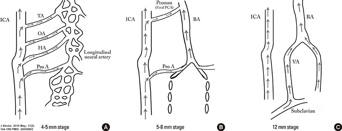 | 1,200 × 460 (67 KB) | Z8600021 (talk | contribs) | ==Cerebral Blood Supply Development== At the 4-5-mm embryonic stage, the hindbrain (i.e., future posterior fossa) is supplied by two parallel neural arteries (or channels). These arteries obtain their blood supply from carotid-vertebrobasilar anastomo... |
You cannot overwrite this file.
File usage
The following page uses this file:


