Thymus Development: Difference between revisions
mNo edit summary |
mNo edit summary |
||
| Line 22: | Line 22: | ||
|-bgcolor="F5FAFF" | |-bgcolor="F5FAFF" | ||
| | | | ||
* '''Multiple roles for HOXA3 in regulating thymus and parathyroid differentiation and morphogenesis in mouse'''<ref name="PMID25249461"><pubmed>25249461</pubmed></ref> "Previous studies have shown that the Hoxa3 null mutant lacks third pharyngeal pouch derivatives, the thymus and parathyroids by E18.5, and organ-specific markers are absent or downregulated during initial organogenesis. Our current analysis of the Hoxa3 null mutant shows that organ-specific domains did undergo initial patterning, but the location and timing of key regional markers within the pouch, including Tbx1, Bmp4 and Fgf8, were altered. Expression of the parathyroid marker Gcm2 was initiated but was quickly downregulated and differentiation failed; by contrast, thymus markers were delayed but achieved normal levels, concurrent with complete loss through apoptosis. To determine the cell type-specific roles of Hoxa3 in third pharyngeal pouch development, we analyzed tissue-specific mutants using endoderm and/or neural crest cell (NCC)-specific Cre drivers. Simultaneous deletion with both drivers resulted in athymia at E18.5, similar to the null. By contrast, the individual tissue-specific Hoxa3 deletions resulted in small, ectopic thymi, although each had a unique phenotype. Hoxa3 was primarily required in NCCs for morphogenesis. In endoderm, Hoxa3 temporally regulated initiation of the thymus program and was required in a cell-autonomous manner for parathyroid differentiation. Furthermore, Hoxa3 was required for survival of third pharyngeal pouch-derived organs, but expression in either tissue was sufficient for this function." [[Developmental Signals - Homeobox]] | |||
* '''Dynamics of thymus organogenesis and colonization in early human development'''<ref name=PMID23571219><pubmed>23571219</pubmed></ref> "The thymus is the central site of T-cell development and thus is of fundamental importance to the immune system, but little information exists regarding molecular regulation of thymus development in humans. ... In addition, we provide molecular evidence that the human thymic epithelium derives solely from the third pharyngeal pouch, as in the mouse, in contrast to previous suggestions." | * '''Dynamics of thymus organogenesis and colonization in early human development'''<ref name=PMID23571219><pubmed>23571219</pubmed></ref> "The thymus is the central site of T-cell development and thus is of fundamental importance to the immune system, but little information exists regarding molecular regulation of thymus development in humans. ... In addition, we provide molecular evidence that the human thymic epithelium derives solely from the third pharyngeal pouch, as in the mouse, in contrast to previous suggestions." | ||
* '''Thymus organogenesis'''<ref><pubmed>22266420</pubmed></ref>"A new study in the jawless fish, lampreys, indicates the existence of a primitive thymus in these surviving representatives of the most ancient vertebrates, providing strong evidence of co-evolution of T cells and thymus." | * '''Thymus organogenesis'''<ref><pubmed>22266420</pubmed></ref>"A new study in the jawless fish, lampreys, indicates the existence of a primitive thymus in these surviving representatives of the most ancient vertebrates, providing strong evidence of co-evolution of T cells and thymus." | ||
Revision as of 01:46, 25 March 2015
Embryology - 23 Sep 2026    Expand to Translate Expand to Translate
|
|---|
| Google Translate - select your language from the list shown below (this will open a new external page) |
|
العربية | català | 中文 | 中國傳統的 | français | Deutsche | עִברִית | हिंदी | bahasa Indonesia | italiano | 日本語 | 한국어 | မြန်မာ | Pilipino | Polskie | português | ਪੰਜਾਬੀ ਦੇ | Română | русский | Español | Swahili | Svensk | ไทย | Türkçe | اردو | ייִדיש | Tiếng Việt These external translations are automated and may not be accurate. (More? About Translations) |
Introduction


The thymus has a key role in the development of an effective immune system as well as an endocrine function.
The thymus has two origins for the lymphoid thymocytes and the thymic epithelial cells. The thymic epithelium begins as two flask-shape endodermal diverticula that form from the third pharyngeal pouch and extend lateralward and backward into the surrounding mesoderm and neural crest-derived mesenchyme in front of the ventral aorta. The immune system T cells are essential for responses against infections and much research concerns the postnatal development of T cells within the thymus.
The mature thymus epithelium has two main cell types: cortical thymic epithelial (cTECs) and medullary thymic epithelial cells (mTECs) or stromal cells. These thymic stromal cells provide signals for T cell differentiation.
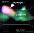
The thymic medulla phagocytes negatively selects auto reactive CD4+ and CD8+ thymocytes, eliminate T cells bearing autoreactive T cell antigen receptors (TCRs). This process to prevent autoimmunity is also known as "negative selection", see adult thymus T cell elimination movie.
| Immune Links: immune | blood | spleen | thymus | lymphatic | lymph node | Antibody | Med Lecture - Lymphatic Structure | Med Practical | Immune Movies | vaccination | bacterial infection | Abnormalities | Category:Immune | ||
|
Some Recent Findings

|
| More recent papers |
|---|
  This table allows an automated computer search of the external PubMed database using the listed "Search term" text link.
More? References | Discussion Page | Journal Searches | 2019 References | 2020 References Search term: Thymus Embryology <pubmed limit=5>Thymus Embryology</pubmed> |
Development Overview

The thymus and parathyroid are derived from 3rd pharyngeal pouches.
Development is a series of epithelial/mesenchymal inductive interactions between neural crest-derived arch mesenchyme and pouch endoderm. There is also the possibility that the surface ectoderm of 3rd pharyngeal clefts participates in thymus development.
Thymic epithelial cells (TECs) are derived from the endoderm of the third pharyngeal pouch.
Hassall's bodies form between 6 and 10 lunar months in humans. They appear after lymphopoiesis has been established and the cortex, medulla and the cortico-medullary junction are able to select of T lymphocytes undergoing progressive maturation. (Text modified from Bodey and Kaiser, 1997)
Experimental studies have shown that a neural crest contribution is also required during early thymic organogenesis.
Week 8
Late embryonic thymus development.

|

|
| Image shows general position of the developing thymus in entire embryo cross-section. The developing thymus is shown in the midline, located behind the sternum (right) and in front of the oesophagus and trachea. | Selected high power image of thymus from complete cross-section above. |
- Links: Carnegie stage 22
Development Changes


Human Fetal Thymus Weight Growth |
Overall Size Changes with age
|
Thymus Anatomy
- Superior mediastinum, anterior to heart
- Bilobed lymphoepithelial organ
- Contains reticular cells but no fibers
- Stem lymphocytes
- proliferate and differentiate
- forms long-lived T- lymphocytes
Thymus Cells
- Thymus Histology: Fetal Thymus overview | Fetal Thymus Medulla | Fetal Thymus Cortex | Adult Thymus | unlabeled fetal overview | unlabeled fetal medulla |unlabeled fetal thymic corpuscle |unlabeled fetal cortex | unlabeled adult overview | Category:Thymus | Immune System Development
- Reticular cells
- Abundant, eosinophilic, large, ovoid and light nucleus 1-2 nucleoli
- sheathe cortical capillaries
- form an epitheloid layer
- maintain microenvironment for development of T-lymphocytes in cortex (thymic epitheliocytes)
- Macrophages
- cortex and medulla
- difficult to distinguish from reticular cells in H&E
- Lymphocytes
- cortex and medulla - more numerous (denser) in cortex
- majority of them developing T-lymphocytes (= thymic lymphocytes or thymocytes)
T Lymphocyte
The following images are electron micrographs of T Lymphocyte (T cell) at different stages.
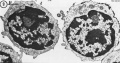
T and B Lymphocytes TEM
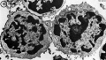
T and B Lymphocytes TEM
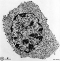
T2 Lymphocyte TEM
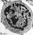
T2 Lymphocyte TEM
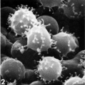
T lymphocyte rosettes
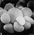
T lymphocyte rosettes
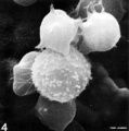
T lymphocyte rosettes
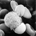
T lymphocyte rosettes
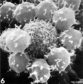
T lymphocyte rosettes


- Lymphocyte EM Images: T and B Lymphocytes 1 TEM | T and B Lymphocytes 2 TEM | T Lymphocyte SEM | B lymphocyte 1 TEM | B lymphocyte 2 TEM | B lymphocyte 3 TEM | Plasma Cell TEM | T2 Lymphocyte 1 TEM | T2 Lymphocyte 2 TEM | lymphocyte rosettes | T lymphocyte 1 | T lymphocyte 2 | T lymphocyte 3 | T lymphocyte 4 | T lymphocyte 5 | T lymphocyte 6 | B lymphocyte | B lymphocytes TEM | Immune System Development | Blood
Thymus Phagocytes
| <html5media height="530" width="490">File:Mouse adult thymus 01.mp4</html5media> | Movie shows how dying thymocytes are efficiently cleared by phagocytes.[6]
Examples of cell death in situ during negative selection. Purified CD4+CD8+ F5 thymocytes labeled with SNARF and Hoechst were introduced into LysM-GFP thymic slices then treated with 1 nM specific peptide for 30 min. The incubation was continued for the indicated times and the slices were imaged by time-lapse two-photon microscopy. The arrowheads point to dy|}ing thymocytes. Scale bars are 5 µm.
|

Fetal/Young Thymus

|

|
| Young medulla | Young cortex |
Thymic corpuscle
Hassall’s corpuscle - Mass of concentric epithelioreticular cells
Thymus Involution

Adult Thymus Histology |
A postnatal process defined as a decrease in the size, weight and activity of the gland with advancing age.
In a recent review[7], thymic involution was described as a result of high levels of circulating sex hormones, in particular during puberty, and a lower population of precursor cells from the bone marrow and finally changes in the thymic microenvironment. |
Hassall's Bodies
Hassall's bodies, also called Hassall's corpuscles
- form between 6 and 10 lunar months in humans.
- appear after lymphopoiesis has been established and the cortex, medulla and the cortico-medullary junction are able to select of T lymphocytes undergoing progressive maturation.
- within the thymus their number increases until puberty, then decreases.
- features are named after Arthur Hill Hassall (1817-1894) a British physician and chemist.
Function

Hassall's corpuscles express thymic stromal lymphopoietin (TSLP), suggesting that Hassall's corpuscles have a critical role in dendritic-cell-mediated secondary positive selection of medium-to-high affinity self-reactive T cells, leading to the generation of CD4(+)CD25(+) regulatory T cells within the thymus.[8]
Thymic stromal lymphopoietin is an epithelial cell-derived cytokine expressed in several tissues (skin, gut, lungs, and thymus) that signals through a TSLP receptor (TSLPR). This receptor is a heterodimer of the IL-7 receptor alpha chain and the TSLPR chain.
Disease Association
There has been one report showing changes in Hassall's bodies morphology associated with congenital heart defects.[9]
Histology
- Thymus Histology: Fetal Thymus overview | Fetal Thymus Medulla | Fetal Thymus Cortex | Adult Thymus | unlabeled fetal overview | unlabeled fetal medulla |unlabeled fetal thymic corpuscle |unlabeled fetal cortex | unlabeled adult overview | Category:Thymus | Immune System Development
The developing fetal thymus shown below is from a 20 week gestational age (GA), 18 week post-fertilization age, or second trimester stage of development.
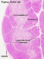
Fetal Thymus overview
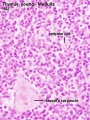
Fetal Thymus Medulla
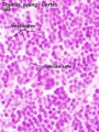
Fetal Thymus Cortex
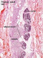
Adult Thymus
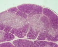
unlabeled fetal overview
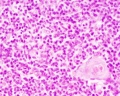
unlabeled fetal medulla
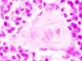
unlabeled fetal thymic corpuscle
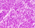
unlabeled fetal cortex
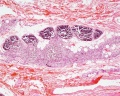
unlabeled adult overview


Molecular Development


Foxg1 and Isl1
Transcription factors that appear to have a role in early thymic epithelial cell (TEC) differentiation
Foxn1
Cited2
Cited2 deletion in the mouse is embryonic lethal with cardiovascular malformations, adrenal agenesis, cranial ganglia fusion, exencephaly, and left-right patterning defects.[10]
- "Examination of Lmo4-deficient embryos revealed partially penetrant cardiovascular malformations and hypoplastic thymus. Examination of Lmo4;Cited2 compound mutants indicated that there is a genetic interaction between Cited2 and Lmo4 in control of thymus development. Our data suggest that this may occur, in part, through control of expression of a common target gene, Tbx1, which is necessary for normal thymus development."
Eva and Six
Both Eva and Six have been implicated in thymus development.[11]
- Eya - human homolog of the Drosophila 'eyes absent' (Eya) gene.
- Six - vertebrate genes which are homologs of the Drosophila 'sine oculis' (so) gene.
References
- ↑ 1.0 1.1 1.2 1.3 <pubmed>22087235</pubmed>| PLoS One.
- ↑ <pubmed>25249461</pubmed>
- ↑ <pubmed>23571219</pubmed>
- ↑ <pubmed>22266420</pubmed>
- ↑ <pubmed>22057081</pubmed>
- ↑ <pubmed>23700386</pubmed>| PLoS Biol. | PLoS Biol. Immunobiology Collection
- ↑ <pubmed>20354268 </pubmed>
- ↑ <pubmed>16121185</pubmed>
- ↑ <pubmed>21125801</pubmed>
- ↑ <pubmed>20549734</pubmed>
- ↑ <pubmed>16530750</pubmed>
Reviews
<pubmed></pubmed> <pubmed></pubmed> <pubmed>24052146</pubmed> <pubmed>21862553</pubmed> <pubmed>18403191</pubmed> <pubmed>16846255</pubmed> <pubmed>16448532</pubmed> <pubmed>12969307</pubmed> <pubmed>11292256</pubmed>
Articles
<pubmed>21952680</pubmed> <pubmed>19306477</pubmed> <pubmed>11857615</pubmed>
Search PubMed: Thymus Development | Thymus Embryology
Terms
- Cytotoxic T lymphocytes - (CTLs, killer T cells) directly attack other cells carrying certain foreign or abnormal molecules on their surfaces including attacking viruses, that hide from other parts of the immune system growing inside infected cells.
- Helper T cells - (Th cells) coordinate immune responses by communicating with other cells. Some stimulate nearby B cells to produce antibodies, others call in phagocytes, and still others activate other T cells.
External Links
External Links Notice - The dynamic nature of the internet may mean that some of these listed links may no longer function. If the link no longer works search the web with the link text or name. Links to any external commercial sites are provided for information purposes only and should never be considered an endorsement. UNSW Embryology is provided as an educational resource with no clinical information or commercial affiliation.
- NIAID - Immune System
Glossary Links
- Glossary: A | B | C | D | E | F | G | H | I | J | K | L | M | N | O | P | Q | R | S | T | U | V | W | X | Y | Z | Numbers | Symbols | Term Link
Cite this page: Hill, M.A. (2026, September 23) Embryology Thymus Development. Retrieved from https://embryology.med.unsw.edu.au/embryology/index.php/Thymus_Development
- © Dr Mark Hill 2026, UNSW Embryology ISBN: 978 0 7334 2609 4 - UNSW CRICOS Provider Code No. 00098G


