Maternal-Fetal Medicine Trainees - Renal: Difference between revisions
| Line 82: | Line 82: | ||
===Kidney Nephron=== | ===Kidney Nephron=== | ||
'''Nephrogenesis''' is the process of generating new nephrons. | |||
{| border='0px' | {| border='0px' | ||
Revision as of 13:52, 22 July 2011
Introduction

| <Flowplayer width="320" height="230" autoplay="true">Renal_001.flv</Flowplayer> | Early Renal Development
This animation shows the process of early renal (kidney) development. Legend
|
Textbooks

|
Moore, K.L. & Persuad, T.V.N. (2008). The Developing Human: clinically oriented embryology (8th ed.). Philadelphia: Saunders.
The following chapter links only work with a UNSW connection and can also be accessed through this UNSW Library connection. |

|
Schoenwolf, G.C., Bleyl, S.B., Brauer, P.R. and Francis-West, P.H. (2009). Larsen’s Human Embryology (4th ed.). New York; Edinburgh: Churchill Livingstone.
The following chapter links only work with a UNSW connection and can also be accessed through this UNSW Library connection. |

|
Hill, M.A. (2011) UNSW Embryology (11th ed.). Sydney:UNSW.
|
- Links: More Embryology Textbooks
Renal Movies
|
|
|
| ||||||||||||
|
|
|
Urinary System Development


- The adult kidneys (the metanephroi) form from day 35, from a portion of the intermediate mesoderm called the metanephric blastema (or metanephric mesenchyme).
- They are induced to form by the ureteric buds, outgrowths from the end of the mesonephric ducts, which come into contact with the metanephric blastema.
- Upon contact, they begin to lengthen and bifurcate rapidly in the metanephric blastema – these branches differentiate into the collecting ducts.
- Both the ureteric buds and the metanephric blastema begin to differentiate; interestingly each induces differentiation in the other structure.
- The ureteric bud is induced by the metanephric blastema to form the collecting tubules, renal pelvis and ureters.
- The metanephric blastema is induced to form the nephrons.
Kidney and Mesonephric Duct
|
<wikiflv width="356" height="500" autoplay="true" position="left">Urogenital_sinus_001.flv|File:Urogenital_sinus_001 icon.jpg</wikiflv> |
First observe the development of the intermediate mesoderm.
|

Embryo Stage 13 (week 4-5)
Kidney Nephron
Nephrogenesis is the process of generating new nephrons.
| File:Renal_001 icon.jpg</wikiflv> | Early Renal Development
This animation shows the process of early renal (kidney) development. Legend
Sequence
|
Adult Histology
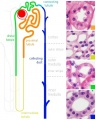
Adult nephron overview
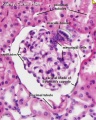
Glomerulus structure
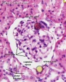
Vascular and renal poles


Development of the Kidney
| File:Renal blood 01 icon.jpg</wikiflv> |
|
Development of the Urinary Bladder
| File:Urogenital_septum_001 icon.jpg</wikiflv> | Division of the Cloaca
|
Development of the Urethra
- Further development of the urinary system varies depending on the sex of the embryo.
- Males - the pelvic urethra forms the membranous urethra, the prostatic urethra and penile urethra. (The sex of the above animation and sections is male)
- Females - the pelvic urethra forms the membranous urethra and the vestibule of the vagina.
Ten Most Frequently Reported Birth Anomalies
Based upon statistics from the Victorian Perinatal Data Collection Unit in Victoria (Australia) between 2003-2004.

|
Hypospadias (More? Development Animation - Genital Male External | Genital Abnormalities - Hypospadia) |

|
Obstructive Defects of the Renal Pelvis (obstructive defects of the renal pelvis, uteropelvic junction obstruction, pelvo-uterero junction obstruction) Term describing a developmental renal abnormality due to partial or complete blockage of the drainage of the kidney pelvis requiring surgical correction. The blockage can also have several causes including: unusual ureter twisting or bending, ureter compression by a blood vessel, malformations of the muscular wall. The blockage leads to an accumulation of urine in the affected region, with several potential effects: nephron damage from compression (hydronephrosis); decreased urine output leading to lack of amniotic fluid (oligohydramnios); respiratory development effects due to the lack of amniotic fluid.
(More? Renal System - Abnormalities | Renal System Development) |

|
Ventricular Septal Defect (More? Cardiovascular Abnormalities - Ventricular Septal Defect)
Heart Development Timeline (see Basic Cardiac Embryology) |

|
Congenital Dislocated Hip (More? Musculoskelal Abnormalities - Congenital Dislocation of the Hip (CDH))
(DHH, congenital dislocated hip, congenital hip dislocation, congenital hip dysplasia) Term describes a spectrum of musculoskeletal disorders of hip instability due either to the femoral head being able to move outside the acetabulum (luxation or dislocation), or abnormally within the acetabulum (subluxation or partial dislocation). This includes presentation following a normal examination of the hips in the newborn period (Ortolani and Barlow tests). When detected can be managed with splinting (Denis-Browne splint) allows the hip joint to develop normally and does not require surgery. If undetected and left untreated, the hip joint develops abnormally and surgical reduction is required. (More? Musculoskeletal System Development) |

|
Trisomy 21 or Down syndrome - (More? Trisomy 21) |

|
Hydrocephalus (More? Neural Abnormalities - Hydrocephalus | NINDS - Hydrocephalus Fact Sheet | Hydrocephalus Support Association | USA National Hydrocephalus Foundation) |

|
Cleft Palate (More? Development Animation - Palate 1 | Development Animation - Palate 2 | Cleft Palate) |

|
Trisomy 18 or Edward Syndrome - multiple abnormalities of the heart, diaphragm, lungs, kidneys, ureters and palate 86% discontinued (More? Trisomy 18) |
| Renal Agenesis/Dysgenesis - reduction in neonatal death and stillbirth since 1993 may be due to the more severe cases being identified in utero and being represented amongst the increased proportion of terminations (approximately 31%). (More? Renal Abnormalities - Renal Agenesis) | |

|
Cleft Lip and Palate - occur with another defect in 33.7% of cases. (More? Cleft Lip) |

Glossary Links
- Glossary: A | B | C | D | E | F | G | H | I | J | K | L | M | N | O | P | Q | R | S | T | U | V | W | X | Y | Z | Numbers | Symbols | Term Link
Cite this page: Hill, M.A. (2024, June 26) Embryology Maternal-Fetal Medicine Trainees - Renal. Retrieved from https://embryology.med.unsw.edu.au/embryology/index.php/Maternal-Fetal_Medicine_Trainees_-_Renal
- © Dr Mark Hill 2024, UNSW Embryology ISBN: 978 0 7334 2609 4 - UNSW CRICOS Provider Code No. 00098G