File:Keibel Mall 059-060.jpg: Difference between revisions
No edit summary |
mNo edit summary |
||
| (One intermediate revision by the same user not shown) | |||
| Line 1: | Line 1: | ||
==Fig. 59-60. Human Embryos== | |||
The Marburg fetus No.21. | |||
A fetus measuring 25 mm. in its greatest length js shown from the left side and from the ventral aspect in Figs. 59 and 60, reproduced from the Normentafel of Keibel and Elze. It may be regarded as standing between Figs. 24 and 25 (Fig. 34, y and z) of His's Normentafel. Again I would call attention to the position of the limbs. In Fig. 59 we see touch pads on the sole of the right foot. At the summit of the coccygeal tubercle there is a small knob, as in all well-preserved embryos of this stage, and it is also seen in the ventral view (Fig. 60), in which the physiological umbilical hernia is indicated by the coils of the intestine showing through the wall of the cord. | |||
{{Keibel_Mall Images}} | |||
[[Category:Human]] [[Category:Human Embryo]] | |||
Latest revision as of 14:33, 24 March 2014
Fig. 59-60. Human Embryos
The Marburg fetus No.21.
A fetus measuring 25 mm. in its greatest length js shown from the left side and from the ventral aspect in Figs. 59 and 60, reproduced from the Normentafel of Keibel and Elze. It may be regarded as standing between Figs. 24 and 25 (Fig. 34, y and z) of His's Normentafel. Again I would call attention to the position of the limbs. In Fig. 59 we see touch pads on the sole of the right foot. At the summit of the coccygeal tubercle there is a small knob, as in all well-preserved embryos of this stage, and it is also seen in the ventral view (Fig. 60), in which the physiological umbilical hernia is indicated by the coils of the intestine showing through the wall of the cord.
- KM Figure Links: The Germ Cells | Segmentation | First Primitive Segment | Gastrulation | External Form | Placenta | Axial Skeleton | Limb Skeleton | Skull | Muscular System
| Historic Disclaimer - information about historic embryology pages |
|---|
 |
Glossary Links
- Glossary: A | B | C | D | E | F | G | H | I | J | K | L | M | N | O | P | Q | R | S | T | U | V | W | X | Y | Z | Numbers | Symbols | Term Link
Cite this page: Hill, M.A. (2024, June 15) Embryology Keibel Mall 059-060.jpg. Retrieved from https://embryology.med.unsw.edu.au/embryology/index.php/File:Keibel_Mall_059-060.jpg
- © Dr Mark Hill 2024, UNSW Embryology ISBN: 978 0 7334 2609 4 - UNSW CRICOS Provider Code No. 00098G
File history
Click on a date/time to view the file as it appeared at that time.
| Date/Time | Thumbnail | Dimensions | User | Comment | |
|---|---|---|---|---|---|
| current | 23:29, 15 February 2012 | 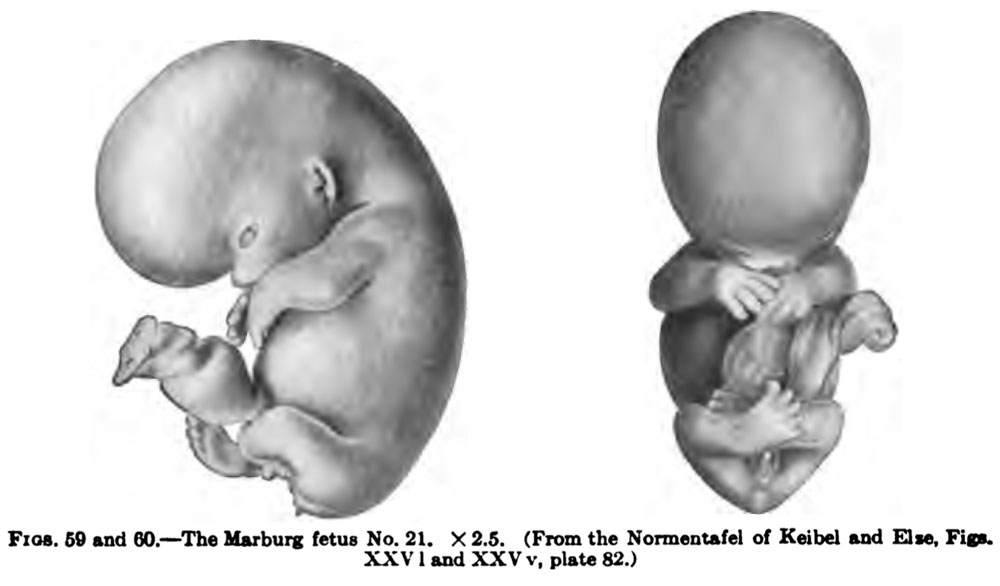 | 1,000 × 586 (54 KB) | S8600021 (talk | contribs) |
You cannot overwrite this file.
File usage
The following 2 pages use this file: