2010 Lecture 5: Difference between revisions
| (52 intermediate revisions by the same user not shown) | |||
| Line 1: | Line 1: | ||
=Mesoderm Development= | =Mesoderm Development= | ||
[[Image:Stage9sm.jpg|thumb|Carnegie stage 9 showing somite formation]] | [[Image:Stage9sm.jpg|thumb|Carnegie stage 9 showing somite formation]] | ||
[[Image:Stage 9 SEM1.jpg|thumb|Carnegie stage 9 scanning electron microscope image showing somite formation]] | [[Image:Stage 9 SEM1.jpg|thumb|Carnegie stage 9 scanning electron microscope image showing somite formation]] | ||
[[File:Stage11_sem100c.jpg|thumb|Carnegie stage 11 mesoderm]] | |||
== Introduction == | == Introduction == | ||
<wikiflv width="195" height="255" autostart="true" repeat="true">Mesoderm_001.flv|File:Mesoderm_001_icon.jpg</wikiflv> | {| | ||
| <wikiflv width="195" height="255" autostart="true" repeat="true">Mesoderm_001.flv|File:Mesoderm_001_icon.jpg</wikiflv> | |||
| This animation shows the migration of mesoderm throughout the embryonic disc during gastrulation. | |||
This animation shows the migration of mesoderm throughout the embryonic disc during gastrulation. | |} | ||
We have seen the following processes during early human development so far: fertilization and blastocyst development in the first week, implantation in the second week, early placentation and bilaminar to trilaminar in the third week. In the third to fourth week we will now follow the development of the trilaminar embryo as each layer begins to differentiate into the primordia of different tissues within the embryo. From this point onward the lectures will not be in a strict timeline format as we will have to follow each layer ('''ectoderm, mesoderm, endoderm''') forward through its early development, and then jump back to discuss the next layer. | We have seen the following processes during early human development so far: fertilization and blastocyst development in the first week, implantation in the second week, early placentation and bilaminar to trilaminar in the third week. In the third to fourth week we will now follow the development of the trilaminar embryo as each layer begins to differentiate into the primordia of different tissues within the embryo. From this point onward the lectures will not be in a strict timeline format as we will have to follow each layer ('''ectoderm, mesoderm, endoderm''') forward through its early development, and then jump back to discuss the next layer. | ||
| Line 18: | Line 17: | ||
'''Coelom''', meaning "cavity", and major fluid-filled cavities can be seen to form both within the embryo (intraembryonic coelom) and outside the embryo (extraembryonic coelom). The '''intraembryonic coelom''' is the single primitive cavity that lies within the mesoderm layer that will eventually form the 3 major anatomical body cavities ('''pericardial, pleural, peritoneal'''). | '''Coelom''', meaning "cavity", and major fluid-filled cavities can be seen to form both within the embryo (intraembryonic coelom) and outside the embryo (extraembryonic coelom). The '''intraembryonic coelom''' is the single primitive cavity that lies within the mesoderm layer that will eventually form the 3 major anatomical body cavities ('''pericardial, pleural, peritoneal'''). | ||
== Objectives == | |||
* Understanding of events during the third week of development | |||
* Understanding the process of notochord formation | |||
* Understanding the process of early somite development | |||
* Understanding the process of body cavity formation | |||
* Understanding the future fate of mesoderm components | |||
* Brief understanding of early heart formation | |||
== Lecture Summary == | |||
[[File:Trilaminar_embryo.jpg|thumb|The trilaminar embryo]] | |||
The four images below beginning in week 3 show cross-sections of the trilaminar embryo and the sequence of mesoderm development. | |||
[[Image:Mesoderm cartoon1.gif]] [[Image:Mesoderm cartoon2.gif]] | |||
[[Image:Mesoderm cartoon3.gif]] [[Image:Mesoderm cartoon4.gif]] | |||
==Notochord (Axial mesoderm)== | |||
<gallery> | |||
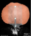
Image:Stage7_800x700px.jpg|Stage 7 embryonic disc | |||
Image:Stage7_primitive-streak-node.jpg|Stage 7 primitive-streak-node | |||
Image:Stage7_cloacal-oral-membranes.jpg|Stage 7 cloacal-oral-membranes | |||
Image:Stage7 notochord.jpg|Stage 7 notochord | |||
</gallery> | |||
==Mesoderm== | |||
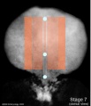
[[Image:Stage7_mesoderm.jpg|thumb|Stage 7 mesoderm]] | |||
** generated from epiblast cells migrating through the primitive streak | |||
** epiblast cells expressing fibroblast growth factor (FGF2) | |||
** forms a layer between ectoderm and endoderm with notochord down midline | |||
** present before neural tube formation | |||
* divides initially into 3 components | |||
<gallery> | |||
Image:Stage7_paraxial-mesoderm.jpg|Stage 7 paraxial mesoderm | |||
Image:Stage7_intermediate-mesoderm.jpg|Stage 7 intermediate mesoderm | |||
Image:Stage7_lateral-plate.jpg|Stage 7 lateral plate | |||
</gallery> | |||
* Paraxial mesoderm - somites - musculoskeletal structures | |||
* Intermediate mesoderm - kidney | |||
* Lateral plate mesoderm - body wall structures | |||
[[Image:Mesoderm cartoon1.gif]] | |||
==Mesenchyme== | |||
* Embryonic connective tissue, describes the cell morphology (Histology is not epithelial organization) | |||
** epithelial to mesenchymal transitions | |||
** mesenchymal to epithelial transitions | |||
==Paraxial Mesoderm== | |||
[[Image:Mesoderm cartoon2.gif]] | |||
** lies adjacent to notochord | |||
** Forms 2 components | |||
*** Head - unsegmented paraxial mesoderm | |||
*** Body - segmented paraxial mesoderm | |||
** Generates trunk muscles, skeleton, dermis of skin, blood vessels, connective tissue | |||
* Segmented Paraxial Mesoderm | |||
** segments called somites | |||
** first pair of somites (day 20) | |||
** segmentation imposes a pattern on | |||
** nerves, vasculature, vertebra.... | |||
** somites appear in ordered sequence cranial to caudal | |||
** appearance so regular used to stage the embryo | |||
** Hamburger & Hamilton 1951- chicken | |||
* thought to be generated by a "clock" (1 pair every 90 minutes) | |||
* neural tube begins to close at 4th somite level | |||
* 44 pairs of somites | |||
== Intermediate Mesoderm== | |||
[[Image:Mesoderm cartoon2.gif]] | |||
* lies between paraxial and lateral mesoderm | |||
* generates urogenital system | |||
** Wolffian duct, kidney | |||
** '''MH''' - covered in Kidney Development Lecture/Laboratory | |||
==Lateral Plate Development== | |||
[[Image:Mesoderm cartoon2.gif]] [[File:Stage7_lateral-plate.jpg|300px|Stage 7 lateral plate]] | |||
* lying at the surrounding edge of he embryonic disc | |||
* a cavity begins in this week to form within the mesoderm itself | |||
==Intraembryonic Coelom== | |||
[[Image:Mesoderm cartoon3.gif]] | |||
* small spaces (vacuoles) begin appearing within the lateral plate mesoderm | |||
* small spaces enlarge forming a single cavity within the lateral plate mesoderm | |||
** divides lateral plate mesoderm into 2 parts at about day 18-19 | |||
* this cavity is called the '''Intraembryonic Coelom''' | |||
** coelom is a general term for a "cavity" and can lie within the embryo (intraembryonic) and outside the embryo (extraembryonic) | |||
** later anatomical spaces within the embryo and fetus can also be described as coeloms | |||
* when the embryonic disc folds the intraembryonic coelom will form all 3 major body cavities: | |||
# Pericardial | |||
# Pleural | |||
# Peritoneal | |||
===Somatic mesoderm=== | |||
[[Image:Mesoderm cartoon3.gif]] | |||
The intraembryonic coelom divides the lateral plate into 2 portions | |||
* closest to ectoderm | |||
* body wall osteogenic, chrondrogenic and fibrogenic | |||
* except ribs and scapula | |||
===Splanchnic mesoderm=== | |||
* closest to endoderm | |||
* heart, smooth muscle of GIT and blood vessels | |||
===Heart=== | |||
* prechordal splanchnic mesderm forms cardiac mesoderm (week 4) | |||
** splanchnic mesoderm lying above the notochord | |||
* initially as a simple tube | |||
* first organ to form in the embryo | |||
[[Image:Mesoderm cartoon4.gif]] | |||
==Somite Formation== | |||
{| | |||
| [[File:Somitogenesis_01 icon.jpg|90px|link=Movie_-_Somitogenesis_01]] | |||
|- | |||
| [[Movie_-_Somitogenesis_01|Early somite induction signals in the mouse]] | |||
|} | |||
[[Image:Somite cartoon1.png]] [[Image:Somite cartoon2.png]] [[Image:Somite cartoon3.png]] [[Image:Somite cartoon4.png]] [[Image:Somite cartoon5.png]] | |||
* ball forms through epithelialization and interactions (cell-cell, cell-extracellular matrix, ECM) fibronectin, laminin | |||
* has 2 populations of cells - peripheral columnar and central mesenchymal | |||
* early somite has cavity- somitocoel, cavity is lost during growth | |||
* somite enclosed by ECM connected to nearby tissues | |||
===Somite Specification === | |||
* Different segmental level somites have to generate different segmental body structures? | |||
* somite has to form different tissues? | |||
* Somite Differentiation | |||
* Compartmentalization accompanied by altered patterns of expression of Pax genes within the somite | |||
Somite initially forms 2 main components | |||
** ventromedial- '''sclerotome''' forms vertebral body and intervertebral disc | |||
** dorsolateral - '''dermomyotome''' forms dermis and skeletal muscle | |||
===Somite Axial Specification=== | |||
* rostro-caudal axis appears regulated by Pax/Hox expression, family of DNA binding transcription factors | |||
* Movie: Somite Development | |||
===Sclerotome === | |||
* sclerotome later becomes subdivided | |||
* rostral and caudal halves separated laterally by von Ebner's fissure | |||
** half somites contribute to a single vertebral level body | |||
** other half intervertebral disc | |||
* therefore final vertebral segmentation ‚"shifts" | |||
===Dermomyotome=== | |||
* later divides into dorsal '''dermatome''' and ventral '''myotome''' | |||
** ('''MH''' - This topic of muscle and skeleton development will be covered in 2 later lectures [[2010_Lecture_13|Musculoskeletal Development]] and [[2010_Lecture_14|Limb Development]]) | |||
* lateral myotome edge migrates at level of limbs | |||
* upper limb first then lower | |||
* mixes with somatic mesoderm | |||
* dermotome continues to contribute cells to myotome | |||
===Myotome=== | |||
* Myotome component of Somite | |||
** epaxial myotome (dorsomedial quarter) forms the dorsal epimere (erector spinae) | |||
** hypaxial myotome (dorsolateral quarter) forms the ventral hypomere, 3 primary muscle layers which are different at neck, thorax and abdomen | |||
[[Image:Stage14 somites limbbuds.png|thumb|Stage 14 Embryo showing somites and limb buds (Week 5)]] | |||
Muscle | |||
* Myoblast determining transcription factor MyoD is first expressed in the dorsomedial quadrant of the still epithelial somite whose cells are not yet definitely committed | |||
** basic Helix Loop Helix | |||
** from myotome | |||
===Muscle Development Abnormalities=== | |||
* Duchenne Muscular Dystrophy | |||
** Embryonic muscle development normal and changes occur postnatally | |||
** X-linked dystrophy, large gene encoding cytoskeletal protein - Dystrophin | |||
** progressive wasting of muscle, die late teens | |||
* Becker Muscular Dystrophy, milder form, adult onset | |||
Axial Segmentation - Somite Specification Signals | |||
==Chicken Model - Somite formation== | |||
[[File:Chick33h.jpg|thumb|Hamburger & Hamilton Stage 10 (33 hours)]] | |||
* Chicken Stages -regular appearance of somites (somitomere to somite) allowed early experimenters to accurately stage the embryo [[Chicken Development]] | |||
* Advantages - accessible, easy to manipulate, limb grafts/removal, chimeras, developmental processes | |||
* taxon-Gallus gallus, develops and hatches in 20-21 days, fertilized eggs easily maintained in humidified incubators | |||
* Embryo Staged growth Series of Embryonic Chicken Growth Hamburger & Hamilton J. Morphology, 88 49 - 92, 1951 [[Hamburger Hamilton Stages]] | |||
* Other vertebrate animal models - Fish (Zebrafish), Frog (Xenopus) | |||
===Chick Embryo Mesoderm=== | |||
* Body Musculature - Myotome derivatives-mouse embryo | |||
* Lateral Plate Mesoderm | |||
* Limb Musculature | |||
* Dermomyotome- Muscle (MyoD) | |||
* MyoD Pax 3 | |||
===Somite Differentiation=== | |||
* migrating neural crest cells enter cranial half, will form dorsal root ganglia (DRG) | |||
* sclerotome bulges ventro-medially towards notochord, then surround and engulf notochord | |||
** mainly growth of surrounding tissues not movement of sclerotome | |||
* notochord forms nucleus pulposus of intervertebral disc (IVD) | |||
==Mesoderm Overview== | |||
{| | |||
| [[File:Trilaminar_embryo.jpg]] | |||
| [[File:Stage11 sem100c.jpg|stage 11 Embryo]] | |||
|- valign="top" | |||
|''' Week 3''' | |||
Trilaminar embryo | |||
Compare this week 3 trilaminar embryo with the week 4 embryo. | |||
(Note - 2 these images are not to scale) | |||
| '''Week 4''' | |||
Scanning electron micrograph of a cross-section of a human embryo at week 4 ([[Carnegie_stage_11|stage 11]]). | |||
Note the mesoderm structures now present and their relative position and size within the embryo. | |||
Compare the mesoderm structures to those formed by ectoderm (neural tube and epidermis) and endoderm (epithelia of developing gastrointestinal tract). | |||
|} | |||
==Take the Quiz== | |||
<quiz display=simple> | |||
{Mesenchyme refers to the middle layer of the trilaminar embryo | |||
|type="()"} | |||
- true | |||
+ false | |||
|| The the middle layer of the trilaminar embryo is the '''mesoderm''' (meaning middle layer), while most of these cells are mesemchymal in appearance, this term is used to describe the cell histological appearance/organization. | |||
{The intraembryonic coelom forms within : | |||
|type="()"} | |||
- somites | |||
+ lateral plate | |||
- neural tube | |||
- intermediate mesoderm | |||
||The intraembryonic coelom forms initially small spaces in the mesoderm layer and coalesce to form a single large "horseshoe-shaped" space within the '''lateral plate mesoderm''' around the embryonic disc. Both young somites (somitocoels) and the neural tube (neural tube lumen) do have cavities, but neither is called the intraembryonic coelom. | |||
{All paraxial mesoderm segments into somites. | |||
|type="()"} | |||
- true | |||
+ false | |||
|| While somites do form within paraxial mesoderm, this region remains unsegmented at the level of the head and therefore does not incorporate into somites. | |||
{Somites are developmental structures that contribute the following adult structures : | |||
|type="()"} | |||
- vertebra, notochord, dermis, skeletal muscle | |||
+ vertebra, intervertebral discs, dermis, skeletal muscle | |||
- kidney, body wall connective tissue, sensory ganglia | |||
- kidney, gastrointestinal tract smooth muscle, mesentry | |||
||Each somite has specific regions that contribute different components of the embryo. Sclerotome contributes the vertebral column ('''vertebra, intervertebral discs'''). Dermotome contributes the connective tissue layers of the skin ('''dermis''', hypodermis). Myotome ontributes the '''skeletal muscle''' of the body and limbs. | |||
</quiz> | |||
==Lecture Links== | |||
* '''Mesoderm Slides''' [[2009_Lecture_5|2009 Lecture]] | [http://embryology.med.unsw.edu.au/pdf/ANAT2341L6Mesoderms1.pdf Mesoderm Lecture 2008 - 1 slide/page ] | [http://embryology.med.unsw.edu.au/pdf/ANAT2341L6Mesoderms4.pdf Lecture 3 2008 Slides - 4 slides/page] | [http://embryology.med.unsw.edu.au/pdf/ANAT2341L6Mesoderms6.pdf Mesoderm Lecture 2008 Slides - 6 slides/page] | |||
* '''Mesoderm''' [http://embryology.med.unsw.edu.au/Movies/week4.htm Week 4 Movies] | [http://embryology.med.unsw.edu.au/Movies/mesoderm.htm Mesoderm Movies] | | |||
* '''Mesoderm Notes''' [http://embryology.med.unsw.edu.au/Notes/week4.htm Week 4] | [http://embryology.med.unsw.edu.au/Notes/week4_4.htm Week 4 - Somites] | [http://embryology.med.unsw.edu.au/Notes/coelom.htm Coelomic Cavity Development] | [http://embryology.med.unsw.edu.au/Notes/skmus.htm Musculoskeletal Development] | | |||
{{Template:2010ANAT2341}} | {{Template:2010ANAT2341}} | ||
Latest revision as of 01:46, 28 August 2010
Mesoderm Development


Introduction
| File:Mesoderm_001_icon.jpg</wikiflv> | This animation shows the migration of mesoderm throughout the embryonic disc during gastrulation. |
We have seen the following processes during early human development so far: fertilization and blastocyst development in the first week, implantation in the second week, early placentation and bilaminar to trilaminar in the third week. In the third to fourth week we will now follow the development of the trilaminar embryo as each layer begins to differentiate into the primordia of different tissues within the embryo. From this point onward the lectures will not be in a strict timeline format as we will have to follow each layer (ectoderm, mesoderm, endoderm) forward through its early development, and then jump back to discuss the next layer.
This lecture will look at mesoderm development and formation of the body cavities.
Mesoderm means the "middle layer" and it is from this layer that nearly all the bodies connective tissues are derived. In early mesoderm development a number of transient structures will form and then be lost as tissue structure is patterned and organised. Humans are vertebrates, with a "backbone", and the first mesoderm structure we will see form after the notochord will be somites.
Coelom, meaning "cavity", and major fluid-filled cavities can be seen to form both within the embryo (intraembryonic coelom) and outside the embryo (extraembryonic coelom). The intraembryonic coelom is the single primitive cavity that lies within the mesoderm layer that will eventually form the 3 major anatomical body cavities (pericardial, pleural, peritoneal).
Objectives
- Understanding of events during the third week of development
- Understanding the process of notochord formation
- Understanding the process of early somite development
- Understanding the process of body cavity formation
- Understanding the future fate of mesoderm components
- Brief understanding of early heart formation
Lecture Summary
The four images below beginning in week 3 show cross-sections of the trilaminar embryo and the sequence of mesoderm development.


Notochord (Axial mesoderm)
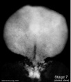
Stage 7 embryonic disc
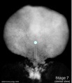
Stage 7 primitive-streak-node
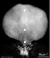
Stage 7 cloacal-oral-membranes
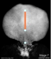
Stage 7 notochord


Mesoderm

- generated from epiblast cells migrating through the primitive streak
- epiblast cells expressing fibroblast growth factor (FGF2)
- forms a layer between ectoderm and endoderm with notochord down midline
- present before neural tube formation
- divides initially into 3 components

Stage 7 paraxial mesoderm
Stage 7 intermediate mesoderm
Stage 7 lateral plate


- Paraxial mesoderm - somites - musculoskeletal structures
- Intermediate mesoderm - kidney
- Lateral plate mesoderm - body wall structures
Mesenchyme
- Embryonic connective tissue, describes the cell morphology (Histology is not epithelial organization)
- epithelial to mesenchymal transitions
- mesenchymal to epithelial transitions
Paraxial Mesoderm
- lies adjacent to notochord
- Forms 2 components
- Head - unsegmented paraxial mesoderm
- Body - segmented paraxial mesoderm
- Generates trunk muscles, skeleton, dermis of skin, blood vessels, connective tissue
- Segmented Paraxial Mesoderm
- segments called somites
- first pair of somites (day 20)
- segmentation imposes a pattern on
- nerves, vasculature, vertebra....
- somites appear in ordered sequence cranial to caudal
- appearance so regular used to stage the embryo
- Hamburger & Hamilton 1951- chicken
- thought to be generated by a "clock" (1 pair every 90 minutes)
- neural tube begins to close at 4th somite level
- 44 pairs of somites
Intermediate Mesoderm
- lies between paraxial and lateral mesoderm
- generates urogenital system
- Wolffian duct, kidney
- MH - covered in Kidney Development Lecture/Laboratory
Lateral Plate Development

- lying at the surrounding edge of he embryonic disc
- a cavity begins in this week to form within the mesoderm itself
Intraembryonic Coelom
- small spaces (vacuoles) begin appearing within the lateral plate mesoderm
- small spaces enlarge forming a single cavity within the lateral plate mesoderm
- divides lateral plate mesoderm into 2 parts at about day 18-19
- this cavity is called the Intraembryonic Coelom
- coelom is a general term for a "cavity" and can lie within the embryo (intraembryonic) and outside the embryo (extraembryonic)
- later anatomical spaces within the embryo and fetus can also be described as coeloms
- when the embryonic disc folds the intraembryonic coelom will form all 3 major body cavities:
- Pericardial
- Pleural
- Peritoneal
Somatic mesoderm
The intraembryonic coelom divides the lateral plate into 2 portions
- closest to ectoderm
- body wall osteogenic, chrondrogenic and fibrogenic
- except ribs and scapula
Splanchnic mesoderm
- closest to endoderm
- heart, smooth muscle of GIT and blood vessels
Heart
- prechordal splanchnic mesderm forms cardiac mesoderm (week 4)
- splanchnic mesoderm lying above the notochord
- initially as a simple tube
- first organ to form in the embryo
Somite Formation
| Early somite induction signals in the mouse |


- ball forms through epithelialization and interactions (cell-cell, cell-extracellular matrix, ECM) fibronectin, laminin
- has 2 populations of cells - peripheral columnar and central mesenchymal
- early somite has cavity- somitocoel, cavity is lost during growth
- somite enclosed by ECM connected to nearby tissues
Somite Specification
- Different segmental level somites have to generate different segmental body structures?
- somite has to form different tissues?
- Somite Differentiation
- Compartmentalization accompanied by altered patterns of expression of Pax genes within the somite
Somite initially forms 2 main components
- ventromedial- sclerotome forms vertebral body and intervertebral disc
- dorsolateral - dermomyotome forms dermis and skeletal muscle
Somite Axial Specification
- rostro-caudal axis appears regulated by Pax/Hox expression, family of DNA binding transcription factors
- Movie: Somite Development
Sclerotome
- sclerotome later becomes subdivided
- rostral and caudal halves separated laterally by von Ebner's fissure
- half somites contribute to a single vertebral level body
- other half intervertebral disc
- therefore final vertebral segmentation ‚"shifts"
Dermomyotome
- later divides into dorsal dermatome and ventral myotome
- (MH - This topic of muscle and skeleton development will be covered in 2 later lectures Musculoskeletal Development and Limb Development)
- lateral myotome edge migrates at level of limbs
- upper limb first then lower
- mixes with somatic mesoderm
- dermotome continues to contribute cells to myotome
Myotome
- Myotome component of Somite
- epaxial myotome (dorsomedial quarter) forms the dorsal epimere (erector spinae)
- hypaxial myotome (dorsolateral quarter) forms the ventral hypomere, 3 primary muscle layers which are different at neck, thorax and abdomen

Muscle
- Myoblast determining transcription factor MyoD is first expressed in the dorsomedial quadrant of the still epithelial somite whose cells are not yet definitely committed
- basic Helix Loop Helix
- from myotome
Muscle Development Abnormalities
- Duchenne Muscular Dystrophy
- Embryonic muscle development normal and changes occur postnatally
- X-linked dystrophy, large gene encoding cytoskeletal protein - Dystrophin
- progressive wasting of muscle, die late teens
- Becker Muscular Dystrophy, milder form, adult onset
Axial Segmentation - Somite Specification Signals
Chicken Model - Somite formation

- Chicken Stages -regular appearance of somites (somitomere to somite) allowed early experimenters to accurately stage the embryo Chicken Development
- Advantages - accessible, easy to manipulate, limb grafts/removal, chimeras, developmental processes
- taxon-Gallus gallus, develops and hatches in 20-21 days, fertilized eggs easily maintained in humidified incubators
- Embryo Staged growth Series of Embryonic Chicken Growth Hamburger & Hamilton J. Morphology, 88 49 - 92, 1951 Hamburger Hamilton Stages
- Other vertebrate animal models - Fish (Zebrafish), Frog (Xenopus)
Chick Embryo Mesoderm
- Body Musculature - Myotome derivatives-mouse embryo
- Lateral Plate Mesoderm
- Limb Musculature
- Dermomyotome- Muscle (MyoD)
- MyoD Pax 3
Somite Differentiation
- migrating neural crest cells enter cranial half, will form dorsal root ganglia (DRG)
- sclerotome bulges ventro-medially towards notochord, then surround and engulf notochord
- mainly growth of surrounding tissues not movement of sclerotome
- notochord forms nucleus pulposus of intervertebral disc (IVD)
Mesoderm Overview

|

|
| Week 3
Trilaminar embryo Compare this week 3 trilaminar embryo with the week 4 embryo. (Note - 2 these images are not to scale) |
Week 4
Scanning electron micrograph of a cross-section of a human embryo at week 4 (stage 11). Note the mesoderm structures now present and their relative position and size within the embryo. Compare the mesoderm structures to those formed by ectoderm (neural tube and epidermis) and endoderm (epithelia of developing gastrointestinal tract). |
Take the Quiz
Lecture Links
- Mesoderm Slides 2009 Lecture | Mesoderm Lecture 2008 - 1 slide/page | Lecture 3 2008 Slides - 4 slides/page | Mesoderm Lecture 2008 Slides - 6 slides/page
- Mesoderm Week 4 Movies | Mesoderm Movies |
- Mesoderm Notes Week 4 | Week 4 - Somites | Coelomic Cavity Development | Musculoskeletal Development |
Glossary Links
- Glossary: A | B | C | D | E | F | G | H | I | J | K | L | M | N | O | P | Q | R | S | T | U | V | W | X | Y | Z | Numbers | Symbols | Term Link
Course Content 2010
Embryology Introduction | Cell Division/Fertilization | Lab 1 | Week 1&2 Development | Week 3 Development | Lab 2 | Mesoderm Development | Ectoderm, Early Neural, Neural Crest | Lab 3 | Early Vascular Development | Placenta | Lab 4 | Endoderm, Early Gastrointestinal | Respiratory Development | Lab 5 | Head Development | Neural Crest Development | Lab 6 | Musculoskeletal Development | Limb Development | Lab 7 | Kidney | Genital | Lab 8 | Sensory | Stem Cells | Stem Cells | Endocrine | Lab 10 | Late Vascular Development | Integumentary | Lab 11 | Birth, Postnatal | Revision | Lab 12 | Lecture Audio | Course Timetable
Cite this page: Hill, M.A. (2024, June 26) Embryology 2010 Lecture 5. Retrieved from https://embryology.med.unsw.edu.au/embryology/index.php/2010_Lecture_5
- © Dr Mark Hill 2024, UNSW Embryology ISBN: 978 0 7334 2609 4 - UNSW CRICOS Provider Code No. 00098G