Musculoskeletal System - Joint Development: Difference between revisions
| Line 25: | Line 25: | ||
===Movement=== | ===Movement=== | ||
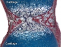
[[File:Cartilage_histology_005.jpg|thumb|Mouse neck joint articular cartilage. [[Cartilage Histology]]]]]] | |||
* Hinge - (elbow and knee) Flexion/Extension | * Hinge - (elbow and knee) Flexion/Extension | ||
Revision as of 07:41, 6 September 2011
Introduction

In the adult, the region where two skeletal bones meet and articulate is called a "joint", that are classified based upon their: anatomical structure, mobility and shape.
In the embryo, the majority of the vertebrate skeleton is initially formed as a cartilage template, that is later replaced by bone except at the interface between two adjacent bones, leaving in the adult a layer of cartilage in this region. The musculoskeletal system consists of skeletal muscle, bone, and cartilage and is mainly mesoderm in origin with some neural crest contribution.
Some Recent Findings
|
Joint Types

Classification
- Fibrous (synarthrodial) - immoveable joints found in cranial vault and teeth
- Cartilagenous (synchondroses and sympheses) - partially moveable joints
- Synovial (diarthrosis) - freely moveable joints are the most common found in the skeleton
Movement

]]
- Hinge - (elbow and knee) Flexion/Extension
- Pivot - (neck, atlas and axis bones) Rotation of one bone around another
- Ball and Socket - (shoulder and hip)
- Saddle - (thumb)
- Condyloid - (wrist joints)
- Gliding - (intercarpal joints) Gliding movements
Synovial Joint Development
Skeletal joint cavity development (cavitation) occurs along planes of the future articular surfaces of synovial joints. A number of different markers have been shown to be present in the interzone at the time of cavitation (hyaluronan and hyaluronan synthase, but not chondroitin sulphates).
Fibroblast-like cells (and/or adjacent chondrocytes) with uridine-diphospho glucose dehydrogenase (UDPGD) activity contribute to glycosaminoglycan levels (increases in hyaluronan). These cells are located on the intimal surface of the synovial lining and have been suggested as the possible cavitation mechanism, switching from cellular cohesion to dissociation.[3]
Joint Abnormalities
FGFR-Related Craniosynostosis Syndromes
Pfeiffer syndrome, Apert syndrome, Crouzon syndrome, Beare-Stevenson syndrome, FGFR2-related isolated coronal synostosis, Jackson-Weiss syndrome, Crouzon syndrome with acanthosis nigricans (AN), and Muenke syndrome
Multiple Epiphyseal Dysplasia
- Links: [http://www.ncbi.nlm.nih.gov/bookshelf/br.fcgi?book=gene&partid=1123#edm-ad GeneReviews - Multiple Epiphyseal Dysplasia)
Temporomandibular Disorders
Osteoarthritis
References
Online Textbooks
Developmental Biology Gilbert, Scott F. Sunderland (MA): Sinauer Associates, Inc. ; c2000 Forming the joints
Reviews
Articles
<pubmed>15492776</pubmed> <pubmed>10645964</pubmed> <pubmed>7544653</pubmed> <pubmed>7525525</pubmed>
Search PubMed
Search July 2010 "Joint Development" All (19900) Review (3137) Free Full Text (3325)
Search Pubmed: Joint Development
Additional Images

Adult axial skeleton

Adult appendicular skeleton

Bone structure
Developing vertebra

Endochondral bone

Fetal head lateral (12 weeks)

Fetal head medial (12 weeks)

Fetal head section (12 weeks)


Terms
Glossary Links
- Glossary: A | B | C | D | E | F | G | H | I | J | K | L | M | N | O | P | Q | R | S | T | U | V | W | X | Y | Z | Numbers | Symbols | Term Link
Cite this page: Hill, M.A. (2024, May 23) Embryology Musculoskeletal System - Joint Development. Retrieved from https://embryology.med.unsw.edu.au/embryology/index.php/Musculoskeletal_System_-_Joint_Development
- © Dr Mark Hill 2024, UNSW Embryology ISBN: 978 0 7334 2609 4 - UNSW CRICOS Provider Code No. 00098G