2010 BGD Practical 6 - Week 4: Difference between revisions
| Line 18: | Line 18: | ||
===Limb Buds=== | ===Limb Buds=== | ||
Limb buds form from ectoderm and mesoderm (somitic and somatic) components and are the "paddle-like" projections from the trunk which will form all the upper and lower limb components. (An overview of limb development will be covered in week 8). | Limb buds form from ectoderm and mesoderm (somitic and somatic) components and are the "paddle-like" projections from the trunk which will form all the upper and lower limb components. (An overview of limb development will be covered in [[2010_BGD_Practical_6_-_Week_8|week 8]]). | ||
===Cardiogenesis=== | ===Cardiogenesis=== | ||
Revision as of 12:25, 16 May 2010
Introduction
Practical 6: Week 3 | Week 4 | Week 5 | Week 6 | Week 7 | Week 8 | Quiz
Introduction
Key Events of Human Development during the fourth week (week 4) following fertilization or Clinical week 6 (LMP).
These notes cover the fourth week of embryonic development, which is the beginning of organogenesis, (specific tissues and systems are beginning to differentiate) from the trilaminar embryo. With many parallel processes, descriptions begin to get complicated! Many of the described processes begin and extend over a broader range of time. Some developmental processes will be discussed later in the practical to simplify matters.
Ectoderm on the embryo surface undergoes segmentation: The central portion of the embryonic disc forms the neural plate, the edge of this plate forms neural crest and the remainder outside this forms the epitheium of the skin and other structures.
Neurogenesis
- Central Nervous System (CNS) - the neural plate undergoes morphological changes to form the primitive central nervous system. An epithelial layer of cells which contributes all neural (brain, spinal cord, peripheral nervous system) and the external epithelium (surface layer of the skin) of the embryo. Neurogenesis begins towards the end of week 3, when the neural tissues separate from this germ cell layer.
- Peripheral Nervous System (PNS) - the neural crest cells in the body region migrate and spread to different regions of the embryo forming the PNS and many other embryonic tissues. Neural crest cells in the head region form skeletal and other structures.
Placodes
In the head region, ectoderm small patches form pairs of specialised placodes that eventually contribute to specific sensory components, cranial ganglia and the anterior pituitary (adenohypophysis).
Limb Buds
Limb buds form from ectoderm and mesoderm (somitic and somatic) components and are the "paddle-like" projections from the trunk which will form all the upper and lower limb components. (An overview of limb development will be covered in week 8).
Cardiogenesis
Within the embryo mesoderm, the heart tube and vascular development continues. Cardiogenesis will be covered in week 5, when septation begins.
Neurogenesis
- Central Nervous System (CNS) - the neural plate undergoes morphological changes to form the primitive central nervous system. An epithelial layer of cells which contributes all neural (brain, spinal cord, peripheral nervous system) and the external epithelium (surface layer of the skin) of the embryo. Neurogenesis begins towards the end of week 3, when the neural tissues separate from this germ cell layer.
- Peripheral Nervous System (PNS) - the neural crest cells in the body region migrate and spread to different regions of the embryo forming the PNS and many other embryonic tissues. Neural crest cells in the head region form skeletal and other structures.
Placodes
In the head region, ectoderm small patches form pairs of specialised placodes that eventually contribute to specific sensory components, cranial ganglia and the anterior pituitary (adenohypophysis).
Embryo Stage 13
Movies - Embryo Carnegie stage 13 - These are rotating embryo animations based upon reconstruction of serial slice images.
| Gastrointestinal | Cardiovascular | Central Nervous System |
Week 4 Movies
Note that many of the movies start in week 4 and continue on through later embryonic development.
Ectoderm
| Neural Plate | Neural Tube |
Mesoderm
| Mesoderm | Somite Structures | Vertebra |
Endoderm
![]()
Practical 6: Week 3 | Week 4 | Week 5 | Week 6 | Week 7 | Week 8 | Quiz
Additional Information
| Additional Information - Content shown under this heading is not part of the material covered in this class. It is provided for those students who would like to know about some concepts or current research in topics related to the current class page. |
Detailed Week by Week
The following information is a detailed timeline of embryonic development between week 3 to 8 and content does not form part of the current practical class.
Embryo Stages and Events
| Day | Stage | Event |
| Stage 10 |  
| |
| Heart begins to beat in Humans by day 22-23, first functioning embryonic organ formed. | ||
| Stage 11 | 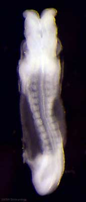
Thyroid thyroid median endodermal thickening in the floor of pharynx Neural rostral (or cephalic) neuropore closes within a few hours; closure is bidirectional, it takes place from the dorsal and terminal lips and may occur in two areas simultaneously. The two lips, however, behave differently. Optic ventricle appears | |
| Stage 12 | 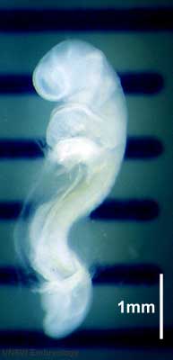
Pituitary Week 4 hypophysial pouch, Rathke’s pouch, diverticulum from roof GIT - Liver septum transversum forming liver stroma and hepatic diverticulum forming hepatic trabeculae PMID: 9407542 Neural caudal neuropore takes a day to close (closure is approximately at future somitic pair 31/sacral vertebra 2) Neural secondary neurulation begins Neural Crest cardiac crest, neural crest from rhombomeres 6 and 7 that migrates to pharyngeal arch 3 and from there the truncus arteriosus PMID: 17848161 Neural Crest vagal neural crest enter the foregut (20-25 somite stage) | |
| Stage 13 | 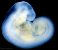 Neural the neural tube is normally completely closed, ventricular system now separated from amniotic fluid. Neural crest at spinal level is segregating, and spinal ganglia are in series with the somites. Spinal cord ventral roots beginning to develop. PMID: 3354839 Neural the neural tube is normally completely closed, ventricular system now separated from amniotic fluid. Neural crest at spinal level is segregating, and spinal ganglia are in series with the somites. Spinal cord ventral roots beginning to develop. PMID: 3354839
telencephalon cavity appears GIT - Liver epithelial cord proliferation enmeshing stromal capillaries PMID: 9407542 Sense - Smell Crest comes from the nasal platesPMID: 15604533 Skin 4 weeks - simple ectoderm epithelium over mesenchyme Skin 1-3 months ectoderm- germinative (basal) cell repeated division of generates stratified epithelium; mesoderm- differentiates into connective tissue and blood vessels |
Glossary Links
- Glossary: A | B | C | D | E | F | G | H | I | J | K | L | M | N | O | P | Q | R | S | T | U | V | W | X | Y | Z | Numbers | Symbols | Term Link
- 2010 BGD: Lecture 1 | Lecture 2 | Practical 3 | Practical 6 | Practical 12
Cite this page: Hill, M.A. (2024, June 14) Embryology 2010 BGD Practical 6 - Week 4. Retrieved from https://embryology.med.unsw.edu.au/embryology/index.php/2010_BGD_Practical_6_-_Week_4
- © Dr Mark Hill 2024, UNSW Embryology ISBN: 978 0 7334 2609 4 - UNSW CRICOS Provider Code No. 00098G