Endocrine - Pituitary Development: Difference between revisions
| Line 36: | Line 36: | ||
==Pituitary Timeline== | |||
* Week 4 - hypophysial pouch, Rathke’s pouch, diverticulum from roof | * Week 4 - hypophysial pouch, Rathke’s pouch, diverticulum from roof | ||
* Week 5 - elongation, contacts infundibulum, diverticulum of diencephalon | * Week 5 - elongation, contacts infundibulum, diverticulum of diencephalon | ||
| Line 43: | Line 43: | ||
* Week 16 - adenohypophysis fully differentiated | * Week 16 - adenohypophysis fully differentiated | ||
* Week 20 to 24 - growth hormone levels peak, then decline | * Week 20 to 24 - growth hormone levels peak, then decline | ||
== Pituitary Blood Vessel Development == | |||
pars distalis - vascularized by hypophysial portal vessels | |||
A study in rats has identified the role of a known regulator of blood vessel development (Vascular Endothelial Growth Factor, VEGF) in the development of the pituitary portal vascular system. [http://www.ncbi.nlm.nih.gov:80/entrez/query.fcgi?cmd=Retrieve&db=pubmed&dopt=Abstract&list_uids=16411082 Nakakura T, Yoshida M, Dohra H, Suzuki M, Tanaka S.] Gene expression of vascular endothelial growth factor-A in the pituitary during formation of the vascular system in the hypothalamic-pituitary axis of the rat. Cell Tissue Res. 2006 Apr;324(1):87-95 | |||
"The primary capillaries extended along the developing pars tuberalis, whereas the portal vessels penetrated into the pars distalis at E15.5 (rat) and subsequently expanded into the lobe to connect with the secondary capillary plexus, emerging in the pars distalis. ....study suggests that VEGF-A (Vascular Endothelial Growth Factor A) is involved in the development of the primary capillaries and in the vascularization of the pars distalis, but not in the portal vessels since the formation of portal vessels begins at E13.5 (rat), before the appearance of VEGF-A in the rostral region of the pars distalis." | |||
The pars distalis is vascularized by hypophysial portal vessels that arise from the capillary beds in the median eminence of the hypothalamus (Murakami et al. 1987), and this hypophyseal portal system provides an important link for carrying hormonal information from the central nervous system to the pituitary. The capillaries of the pituitary gland are characterized by richly fenestrated endothelia. | |||
== Abnormalities == | |||
Anatomical abnormalities asssociated with the Rathke's pouch include a '''craniopharyngeal canal''', from the anterior part of the fossa hypophyseos of the sphenoid bone to the under surface of the skull. The stomodeal end may also be present at the junction of the septum of the nose with the palate. | |||
Abnormal functional development of the pituitary can lead to a wide range of other organ diseases due to the effect of hormones released from the pituitary on many other endocrine and non-endocrine organs (For example: dwarfism, hypothyroidism). (More? NIH Genes and Disease [http://www.ncbi.nlm.nih.gov/books/bv.fcgi?rid=gnd.chapter.41 Chapter 41 - Endocrine]) | |||
There are several abnormalities associated with abnormal levels of the hormonal output of the pituitary due to the development of pituitary tumours (adenomas). | |||
Growth hormone (GH) adenomas, which are benign pituitary tumors lead to chronic high GH output levels, that may lead to acromegaly. | |||
Cushing's disease caused either by a pituitary adenoma produces excess adrenocorticotropic hormone (ACTH, corticotropin) or due to ectopic tumors secreting ACTH or corticotropin-releasing hormone (CRH). | |||
'''Pituitary Adenoma Classification''' | |||
Classification can be applied using specific criteria (clinical presentation, biochemical data, histology of growth pattern, tinctorial characteristics, proliferative activity, immunohistology marker expression, ultrastructure and molecular biology). The current classification used is the World Health Organization classification of 2000 recently updated in 2004. | |||
'''Molecular markers''' | |||
'''Pituitary-specific transcription factor 1''' - (Pit1) in GH-, prolactin- or TSH-secreting adenomas. (More? [../MolDev/factor/pit.htm Molecular Development Factors - Pit] | [http://www.ncbi.nlm.nih.gov/entrez/dispomim.cgi?id=173110 OMIM - Pit1]) | |||
'''T-box factor 19''' - (TBX19 or TPIT) in ACTH-producing adenomas. (More? [http://www.ncbi.nlm.nih.gov/entrez/dispomim.cgi?id=604614 OMIM - TBX19]) | |||
'''Splicing Factor 1''' - (SF1) in gonadotroph, null cell and oncocytic adenomas. (More? [http://www.ncbi.nlm.nih.gov/entrez/dispomim.cgi?id=601516 OMIM - SF1]) | |||
'''Links:''' [http://www.ncbi.nlm.nih.gov/entrez/dispomim.cgi?id=201400 OMIM - ACTH Deficiency] | [http://www.med.uc.edu/neurorad/webpage/bua.html Pituitary Adenoma scan] | |||
== References == | == References == | ||
Revision as of 19:11, 25 April 2010
Introduction

Historically, this endocrine gland was called the "pituitary" as it was originally thought to produce mucous that discharged through the nose. We now know that this is not the function of the pituitary, or hypophysis which is an endocrine gland links the brain to peripheral endocrine organs and systems of the body through several specific hormones. The developmental origin of the hypophysis is also unique, epithelial origins from neural ectoderm (posterior) and from surface ectoderm (anterior).
During development, the boundary epitheilal ectoderm in the roof of the pharynx forms a pocket (Rathke's pouch) that comes into contact with the ectoderm of developing brain. Rathke's pouch is named after German embryologist and anatomist Martin Heinrich Rathke (1793 — 1860).
Anatomically, the pituitary has 2 main parts posterior, or neurohypophysis and anterior, or adenohypophysis (the pars distalis, pars intermedia, and pars tuberalis). Between the two a specialized vascular (portal) system allows communication from the brain to peripheral endocrine organs and other systems. File:17thC-turkish-saddle3.jpg
The pituitary is located within the pituitary fossa of the sphenoid bone, anterior to the lamina terminalis and superior to the pharynx. The shape of the bone surrounding the pituitary led to the naming sella turcica (Latin sella = saddle, turcica = Turkish), as it resembled a saddle shape.
Pit1 (pituitary-specific transcription factor) is a transcription factor important for pituitary development and muations in this gene can lead to abnormalities in pituitary development and hormone production. (More? [../MolDev/factor/pit.htm Molecular Development Factors - Pit])
Anterior pituitary hormones - Thyroid-stimulating hormone (TSH), Adrenocorticotrophic hormone (ACTH), Luteinizing hormone (LH), Follicle-stimulating hormone (FSH), Somatotrophin/growth hormone (GH), Prolactin (PRL), Melanocyte-stimulating hormone (MSH)
Posterior pituitary hormones - Oxytocin, Arginine vasopressin
| Lecture - Head Development | original page
Development Overview

- Dual ectoderm origins
- Ectoderm - ectoderm roof of stomodeum, Rathke's pouch, adenohypophysis
- Neuroectoderm - prosenecephalon then diencephalon, neurohypophysis
Adenohypophysis
- Anterior wall proliferates - pars distalis
- Posterior wall little growth – pars intermedia
- Rostral growth around infundibular stem – pars tuberalis
Neurohypophysis
- Infundibulum – median eminence, infundibulum, pars nervosa
Pituitary Timeline
- Week 4 - hypophysial pouch, Rathke’s pouch, diverticulum from roof
- Week 5 - elongation, contacts infundibulum, diverticulum of diencephalon
- Week 6 - connecting stalk between pouch and oral cavity degenerates
- Week 10 - growth hormone and ACTH detectable
- Week 16 - adenohypophysis fully differentiated
- Week 20 to 24 - growth hormone levels peak, then decline
Pituitary Blood Vessel Development
pars distalis - vascularized by hypophysial portal vessels
A study in rats has identified the role of a known regulator of blood vessel development (Vascular Endothelial Growth Factor, VEGF) in the development of the pituitary portal vascular system. Nakakura T, Yoshida M, Dohra H, Suzuki M, Tanaka S. Gene expression of vascular endothelial growth factor-A in the pituitary during formation of the vascular system in the hypothalamic-pituitary axis of the rat. Cell Tissue Res. 2006 Apr;324(1):87-95
"The primary capillaries extended along the developing pars tuberalis, whereas the portal vessels penetrated into the pars distalis at E15.5 (rat) and subsequently expanded into the lobe to connect with the secondary capillary plexus, emerging in the pars distalis. ....study suggests that VEGF-A (Vascular Endothelial Growth Factor A) is involved in the development of the primary capillaries and in the vascularization of the pars distalis, but not in the portal vessels since the formation of portal vessels begins at E13.5 (rat), before the appearance of VEGF-A in the rostral region of the pars distalis."
The pars distalis is vascularized by hypophysial portal vessels that arise from the capillary beds in the median eminence of the hypothalamus (Murakami et al. 1987), and this hypophyseal portal system provides an important link for carrying hormonal information from the central nervous system to the pituitary. The capillaries of the pituitary gland are characterized by richly fenestrated endothelia.
Abnormalities
Anatomical abnormalities asssociated with the Rathke's pouch include a craniopharyngeal canal, from the anterior part of the fossa hypophyseos of the sphenoid bone to the under surface of the skull. The stomodeal end may also be present at the junction of the septum of the nose with the palate.
Abnormal functional development of the pituitary can lead to a wide range of other organ diseases due to the effect of hormones released from the pituitary on many other endocrine and non-endocrine organs (For example: dwarfism, hypothyroidism). (More? NIH Genes and Disease Chapter 41 - Endocrine)
There are several abnormalities associated with abnormal levels of the hormonal output of the pituitary due to the development of pituitary tumours (adenomas).
Growth hormone (GH) adenomas, which are benign pituitary tumors lead to chronic high GH output levels, that may lead to acromegaly.
Cushing's disease caused either by a pituitary adenoma produces excess adrenocorticotropic hormone (ACTH, corticotropin) or due to ectopic tumors secreting ACTH or corticotropin-releasing hormone (CRH).
Pituitary Adenoma Classification
Classification can be applied using specific criteria (clinical presentation, biochemical data, histology of growth pattern, tinctorial characteristics, proliferative activity, immunohistology marker expression, ultrastructure and molecular biology). The current classification used is the World Health Organization classification of 2000 recently updated in 2004.
Molecular markers
Pituitary-specific transcription factor 1 - (Pit1) in GH-, prolactin- or TSH-secreting adenomas. (More? [../MolDev/factor/pit.htm Molecular Development Factors - Pit] | OMIM - Pit1)
T-box factor 19 - (TBX19 or TPIT) in ACTH-producing adenomas. (More? OMIM - TBX19)
Splicing Factor 1 - (SF1) in gonadotroph, null cell and oncocytic adenomas. (More? OMIM - SF1)
Links: OMIM - ACTH Deficiency | Pituitary Adenoma scan
References
Reviews
Articles
Search PubMed
Search April 2010
- Endocrine Development - All (14277) Review (4620) Free Full Text (3140)
Search Pubmed: pituitary development
Additional Images
Adult Histology
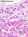
Pituitary - adenohypophysis
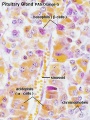
Pituitary - adenohypophysis
Pituitary - neurohypophysis


Terms
Glossary Links
- Glossary: A | B | C | D | E | F | G | H | I | J | K | L | M | N | O | P | Q | R | S | T | U | V | W | X | Y | Z | Numbers | Symbols | Term Link
Cite this page: Hill, M.A. (2024, June 1) Embryology Endocrine - Pituitary Development. Retrieved from https://embryology.med.unsw.edu.au/embryology/index.php/Endocrine_-_Pituitary_Development
- © Dr Mark Hill 2024, UNSW Embryology ISBN: 978 0 7334 2609 4 - UNSW CRICOS Provider Code No. 00098G