2010 Foundations Lecture - Introduction to Human Development: Difference between revisions
m (Redirected page to Foundations Lecture - Introduction to Human Development) |
|||
| (103 intermediate revisions by 2 users not shown) | |||
| Line 1: | Line 1: | ||
#REDIRECT [[Foundations_Lecture_-_Introduction_to_Human_Development]] | |||
==Introduction== | ==Introduction== | ||
[[File:Foundsmall.jpg|left]] [[Image:Mark_Hill.jpg|thumb|Dr Mark Hill]] | [[File:Foundsmall.jpg|left]] [[Image:Mark_Hill.jpg|thumb|Dr Mark Hill]] | ||
Human development is one of the most exciting topics to study not only as a medical student, but also for our fundamental understanding of the human body. This lecture is going to take you briefly through key concepts in human development, these will later be explored in more detail through the BGD course. The lecture will be followed by a practical class introducing online resources for independent study and working through similar embryology concepts. | Human development is one of the most exciting topics to study not only as a medical student, but also for our fundamental understanding of the human body. | ||
This lecture is going to take you briefly through key concepts in human development, these will later be explored in more detail through the BGD course. I will be using simplified terms in the lecture slides (with developmental term in brackets). | |||
The lecture will be followed by a practical class introducing online resources for independent study and working through similar embryology concepts. | |||
<center>Note this is an archive copy of 2010 lecture, many links and features may not work. Use the link below to see the current lecture version.</center> | |||
<center>[[Foundations_Lecture_-_Introduction_to_Human_Development|'''Current Foundations Lecture 2014''']]</center> | |||
==Aims== | ==Aims== | ||
| Line 18: | Line 29: | ||
{| border='0px' | {| border='0px' | ||
|- | |- | ||
| <Flowplayer height=" | | <Flowplayer height="225" width="180" autoplay="true">Human development 001.flv</Flowplayer> | ||
| valign="top" |This animation begins at the two cell (blastomere) stage following fertilization and takes you through an overview of the entire 9 months of human development in just over a minute! | | valign="top" |This animation begins at the two cell (blastomere) stage following fertilization and takes you through an overview of the entire 9 months of human development in just over a minute! | ||
| Line 33: | Line 44: | ||
==UNSW Embryology Online== | ==UNSW Embryology Online== | ||
[[Image:Front-page-image.jpg|200px]] | {| border='0px' | ||
|- | |||
| [[File:UNSW_Embryo_front_page.jpg|200px|link=http://embryology.med.unsw.edu.au/]] | |||
| [[Image:Front-page-image.jpg|200px|link=http://php.med.unsw.edu.au/embryology/]] | |||
|- | |||
| [http://embryology.med.unsw.edu.au/ Original Website] | |||
| [http://php.med.unsw.edu.au/embryology/ New Website] | |||
|- | |||
|} | |||
==Textbooks== | |||
* There are many different excellent embryology textbooks | |||
* I have included 2 that cover the clinical topics as well. | |||
* [[Embryology_Textbooks|More Textbooks?]] | |||
===The Developing Human: Clinically oriented embryology=== | |||
{| border='0px' | |||
|- | |||
| [[File:The Developing Human, 8th edn.jpg|120px]] | |||
| '''Citation:''' The developing human : clinically oriented embryology 8th ed. Moore, Keith L; Persaud, T V N; Torchia, Mark G Philadelphia, PA : Saunders/Elsevier, c2008. | |||
Links: [http://locatorplus.gov/cgi-bin/Pwebrecon.cgi?DB=local&v1=1&ti=1,1&Search_Arg=101293798&Search_Code=0359&CNT=20&SID=1 NLM ID: 101293798] | [http://www.ncbi.nlm.nih.gov/nlmcatalog/535855 NLM Holding] | [http://orkney.library.unsw.edu.au:1701/primo_library/libweb/action/display.do?fn=display&doc=UNSW_Aleph001191400&vid=LRD&afterPDS=true UNSW Library] | [http://www.us.elsevierhealth.com:80/product.jsp?isbn=9781416037064 publisher page] | |||
|- | |||
|} | |||
===Larsen's human embryology=== | |||
{| border='0px' | |||
|- | |||
| [[File:Larsen's human embryology 4th edn.jpg|120px]] | |||
| '''Citation:''' Larsen's human embryology 4th ed. Schoenwolf, Gary C; Larsen, William J, (William James). Philadelphia, PA : Elsevier/Churchill Livingstone, c2009. | |||
Links: NLM ID: 101309446 [http://www.ncbi.nlm.nih.gov/nlmcatalog/550102 NLM Holding] | [http://orkney.library.unsw.edu.au:1701/primo_library/libweb/action/display.do?fn=display&doc=UNSW_Aleph001215898&vid=LRD&afterPDS=true UNSW Library] | [http://www.us.elsevierhealth.com:80/product.jsp?isbn=9780443068119 publisher page] | |||
|- | |||
|} | |||
==Four Basic Tissue Types== | |||
# Epithelial | |||
# Connective | |||
# Muscular | |||
# Nervous | |||
* How do they develop? | |||
* Where do they come from? | |||
==Human Reproductive Cycle== | |||
* Meiosis in gonad produces haploid gametes (egg and sperm) | |||
===Female=== | |||
* [[Menstrual Cycle]] a regular cycle of reproduction (28 days) | |||
* begins at puberty | |||
* release of 1 egg (oocyte) every cycle | |||
* Endocrine controlled (HPG axis) | |||
**Hypothalamus | |||
**Pituitary | |||
**Gonad | |||
[[File:XXhpgaxis.jpg|300px]] [[File:Menstrual_cycle.png|500px]] | |||
===Male=== | |||
* continuous production of spermatozoa | |||
* begins at puberty | |||
* release millions of spermatazoa | |||
==Ovary== | |||
* Paired organs | |||
* lying in the peritoneal cavity | |||
[[File:Human_right_ovary_and_tube_1.jpg|400px]] | |||
[[File:Ovary10x.jpg|400px]] | |||
==Ovulation== | |||
[[File:Menstrual_cycle.png|thumb|Human Menstrual Cycle]] | |||
* [[O#ovulation|ovulation]] is the release of the egg (oocyte) | |||
* middle of the [[Menstrual Cycle|menstrual cycle]] | |||
[[File:Ovulation_icon.jpg|150px|link=Movie_-_Ovulation 01]] [[File:Follicle 001 icon.jpg|150px|link=Development_Animation_-_Ovulation]] | |||
==Fertilization== | |||
* the process of the 2 [[H#haploid|haploid]] [[G#gamete|gametes]] (egg and sperm) fusing and combining genetic material. | |||
* '''conceptus''' - the entire product of fertilization | |||
[[File:Fertilization 001 icon.jpg|150px|link=Movie_-_Fertilization 01]] | |||
[[File:Fertilization 002 icon.jpg|150px|link=Development_Animation_-_Fertilization]] | |||
[[File:Early_zygote.jpg]] | |||
==Early Development== | |||
* occurs during [[Week_1|week 1]] following fertilization | |||
* last menstrual period (LMP) week 3 | |||
* mitosis to form solid ball of cells (morula), then hollow ball (blastocyst) | |||
[[File:Early_zygote.jpg]] [[File:Stage2.jpg]] | |||
[[File:Human-oocyte_to_blastocyst.jpg]] | |||
* | ==Week 1 Development== | ||
* occurs freely floating in uterus | |||
[[File:Week1 001 icon.jpg|90px|link=Development_Animation_-_Week_1]] | |||
[[File:Week1_summary.jpg]] | |||
==Week 2 Development== | |||
* Implantation | |||
* initial attachment to uterine wall | |||
* invasion of uterine wall | |||
[[File:Week2_001 icon.jpg|120px|link=Development_Animation_-_Implantation]] | |||
[[File:Chorion 001 icon.jpg|120px|link=Development Animation - Chorionic Cavity]] | |||
==Notes== | ==Pregnancy== | ||
===Detect Pregnancy=== | |||
[[File:Pregnancy_test.gif|thumb|pregnancy test]] | |||
[[File:Ovary_corpus_luteum.jpg|thumb|Ovary - corpus luteum secretes hormone to support pregnancy.]] | |||
* Clinically can be detected following implantation (week 2) | |||
* Last Menstrual Period (LMP) - today ? ....... Birth Date - January 30, 2011 | |||
[http://embryology.med.unsw.edu.au/Medicine/FlabEmbryo_1.htm#DueDate Calculate a new Birth Date] (I need to update calculator for 2010) | |||
===Gestation Calculation=== | |||
* First pregnancy (primipara) 274 days, just over 39 weeks | |||
* Subsequent pregnancies (multipara) 269 days, 38.4 weeks | |||
Median duration of gestation assumed from ovulation to delivery | |||
* Historic - Franz Carl Naegele (1777-1851), first rule for estimating pregnancy length | |||
* Current - Ultrasound, the most accurate staging method | |||
===Trimesters=== | |||
* Divide the pregnancy into 3 "blocks" of about 3 months (trimesters) | |||
* First Trimester - embryonic period (organogenesis) | |||
* Second and Trimester - fetal period (growth) | |||
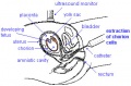
==Implantation Sites== | |||
===Abnormal Implantation=== | |||
[[File:Tubal_pregnancy.gif|right]] | |||
* Ectopic Sites | |||
** external surface of uterus, ovary, bowel, gastrointestinal tract, mesentery, peritoneal wall | |||
** If not spontaneous then, embryo has to be removed surgically | |||
* Uterine - tubal pregnancy (most common ectopic) | |||
===Normal Implantation=== | |||
* Uterine body | |||
** posterior, anterior, superior, lateral (most common posterior) | |||
** inferior implantation - placenta overlies internal os of uterus '''Placenta Previa''' | |||
===Early Placenta=== | |||
* interaction between implanting conceptus and uterine wall (endometrium) | |||
* The uterine lining following implantation (Decidua) | |||
** forms 3 distinct regions, at approx 3 weeks | |||
** Decidua Basalis - implantation site | |||
** Decidua Capsularis - enclosing the conceptus | |||
** Decidua Parietalis - remainder of uterus | |||
* uterine cavity is lost by 12 weeks | |||
==Placenta== | |||
* Materno/fetal organ | |||
* No exchange of blood | |||
* Many different roles | |||
** can be "sampled" as part of a prenatal diagnostic test | |||
==Embryonic Development== | |||
* Embryonic Period - Week 1 to 8 (first trimester) | |||
* Establish the basic structure of organs and tissues (Organogenesis) | |||
* development and growth of the placenta (Placentation) | |||
[[File:Human_Carnegie_stage_1-23.jpg]] | |||
[[File:Embryo stages 002 icon.jpg|120px|link=Embryonic Development]] | |||
==Week 3== | |||
[[File:Stage7-sem4.jpg|right|300px]] | |||
* 3 Key processes commence | |||
===Gastrulation=== | |||
[[File:Trilaminar embryo.jpg|thumb|Trilaminar embryo]] | |||
[[File:Inner cell mass cartoon.jpg|thumb|300px|Trilaminar embryo and tissue origins]] | |||
* the formation of the 3 layer embryo (trilaminar embryo) | |||
** All tissues of the body are formed from these 3 embryonic tissue layers (germ layers) | |||
# '''Ectoderm''' (epithelium) | |||
# '''Mesoderm''' (connective tissue) | |||
# '''Endoderm''' (epithelium) | |||
* simplified explanation of the 3 layer contributions | |||
===Ectoderm=== | |||
* forms the central and peripheral nervous system and epithelium of the skin | |||
===Endoderm=== | |||
* forms gastrointestinal tract organs and the epithelium of the gastrointestinal and respiratory tracts | |||
===Mesoderm=== | |||
* forms the body connective tissues: blood, bone, muscle, connective tissue skin, gastrointestinal and respiratory tracts | |||
===Somitogenesis=== | |||
* segmentation of the mesoderm | |||
* forms the axial body plan | |||
===Neuralation=== | |||
* segmentation of the ectoderm | |||
* separates the neural tissue from the skin (epidermis) | |||
[[File:Week3_folding icon.jpg|90px|link=Development Animation - Week 3]] | |||
==Week 4== | |||
* heart formation (cardiogenesis) | |||
* first functioning organ | |||
[[File:Amnion 001 icon.jpg|120px|link=Development Animation - Amniotic Cavity]] | |||
[[File:Chick_Heart_001-icon.jpg|120px|link=Movie_-_Normal Chick Heart]] | |||
[[File:Mouse_CT_E11.5_movie-icon.jpg|120px|link=Movie_-_CT_Mouse_E11.5]] | |||
==Week 4-8== | |||
* early development of the other organs, tissues and limbs | |||
==Week 9 - 38 == | |||
* Second and Third Trimester (Fetal Period) | |||
* Continuing growth and differentiation of organs formed in embryonic period | |||
** some organs have a later development - neural, genital, respiratory, bones | |||
** some continue to develop after birth - neural, genital, respiratory, bones | |||
* growth in size, length (Second Trimester) | |||
* growth in weight (Third Trimester) | |||
<gallery> | |||
File:Fetal_head_medial.jpg|Fetal Head 12 cartilage and bone formation (12 week) | |||
File:Fetal_head_section.jpg|Fetal Head head structures and the brain (12 week) | |||
File:Endochondral_bone.jpg|Fetal knee region | |||
</gallery> | |||
[[File:fetal growth icon.jpg|120px|link=Fetal_Development]] | |||
[[File:Ultrasound12wk_3D_image2.jpg|120px|link=Ultrasound_-_ Fetus 12 week]] | |||
[[File:19weeklabel1.jpg|120px|link=Ultrasound_-_ Fetus 19 week]] | |||
==Birth== | |||
[[File:Galletti1770_birth.jpg|thumb|Historic teaching model of birth]] | |||
* birth (parturition) is a complex physiological process between the fetus and mother | |||
* thought to be initiated by the fetus | |||
===Maternal Birth Stages=== | |||
# Dilatation | |||
# Expulsion | |||
# Placental | |||
# Recovery | |||
===Newborn=== | |||
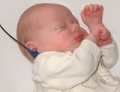
[[File:Newborn.jpg|thumb|Newborn infant (perinatal period)]] | |||
Newborn ([[P#perinatal|perinatal]]) needs to activate many systems and establish independent regulation ([[H#homeostasis|homeostasis]]) | |||
* '''Lung function''' - Fluid drainage, Gas exchange, muscular activity | |||
* '''Circulatory changes''' - Closure of 3 vascular shunts | |||
* '''Thermoregulation''' - metabolic rate, fat metabolism | |||
* '''Nutrition''' - gastrointestinal tract function, peristalsis | |||
* '''Waste''' - kidney function | |||
* '''Endocrine function''' - loss of placenta, maternal hormones | |||
==Critical Periods of Development== | |||
===Abnormal Development=== | |||
Three main causes: | |||
# Genetic | |||
# Environmental | |||
# Unknown | |||
* First trimester most critical | |||
* Different effect depending on time of insult (teratogen) | |||
[[File:Human-critical_periods.jpg]] | |||
===Diagnosis=== | |||
* Prenatal diagnosis - number of different techniques (non-invasive, invasive) for determining normal development | |||
* Neonatal diagnosis ([[A#APGAR|APGAR test]], [[G#Guthrie test|Guthrie test]]) | |||
* Maternal diagnosis - often pregnancy will expose maternal health problems | |||
<gallery> | |||
File:Amniocentesis.jpg|Amniocentesis | |||
File:Cvs.jpg|Chorionic Villus Sampling | |||
File:Ultrasound12wk_3D.jpg|Ultrasound | |||
File:Apgar.jpg|Apgar scoresheet | |||
File:Gutherie_card_icon.jpg|Gutherie card | |||
File:Congenital dislocation hip.jpg|Xray congenital dislocation hip | |||
File:Newborn hearing test.jpg|Newborn hearing test | |||
</gallery> | |||
[[:Media:Dancing_baby.mov|Finished!]] now here is a [http://www.youtube.com/watch?v=oHg5SJYRHA0 Link to Exam Question.....] | |||
==Revision Notes== | |||
* You don't need to know everything today, its an introduction. | |||
* Don't confuse "germ cell layers" (ectoderm, mesoderm, endoderm) with "germ cells" (egg, spermatazoa). | * Don't confuse "germ cell layers" (ectoderm, mesoderm, endoderm) with "germ cells" (egg, spermatazoa). | ||
* Remember the difference between "clinical weeks" (last menstral period) and "embryonic weeks" (from fertilization, 2 weeks later). | * Remember the difference between "clinical weeks" (last menstral period) and "embryonic weeks" (from fertilization, 2 weeks later). | ||
| Line 53: | Line 341: | ||
[[2010 Foundations Practical - Introduction to Human Development]] | [[2010 Foundations Practical - Introduction to Human Development]] | ||
{{ | <center>[[Foundations_Lecture_-_Introduction_to_Human_Development|'''Current Foundations Lecture 2014''']]</center> | ||
{{Glossary}} | |||
{{Footer}} | |||
[[Category:Medicine]] [[Category:Foundations]] | [[Category:Medicine]] [[Category:Foundations]] | ||
Latest revision as of 08:51, 18 September 2014
Introduction


Human development is one of the most exciting topics to study not only as a medical student, but also for our fundamental understanding of the human body.
This lecture is going to take you briefly through key concepts in human development, these will later be explored in more detail through the BGD course. I will be using simplified terms in the lecture slides (with developmental term in brackets).
The lecture will be followed by a practical class introducing online resources for independent study and working through similar embryology concepts.
Aims
- Purpose of learning embryology
- Basic facts about early human development
- Appreciate differences between the conceptus, embryo and fetus
- General understanding of the term “critical periods” of development
Concepts: Fertilization, Early conceptus, Germ layers, Embryo, Tissue origins, Timetable/stages of development, Fetus, Placenta
Background Lectures: Cell Structure (structure and function), Cell Division (mitosis, meiosis, lifespan, cell death), 4 Basic Tissues (Epithelial, Connective, Muscular, Nervous)
Links: Embryology Textbooks | 2009 Lecture | 2009 Lecture Slides
Animated overview
| <Flowplayer height="225" width="180" autoplay="true">Human development 001.flv</Flowplayer> | This animation begins at the two cell (blastomere) stage following fertilization and takes you through an overview of the entire 9 months of human development in just over a minute!
|
UNSW Embryology Online
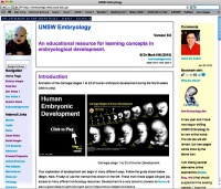
|
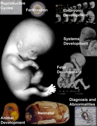
|
| Original Website | New Website |
Textbooks
- There are many different excellent embryology textbooks
- I have included 2 that cover the clinical topics as well.
- More Textbooks?
The Developing Human: Clinically oriented embryology

|
Citation: The developing human : clinically oriented embryology 8th ed. Moore, Keith L; Persaud, T V N; Torchia, Mark G Philadelphia, PA : Saunders/Elsevier, c2008.
|
Larsen's human embryology

|
Citation: Larsen's human embryology 4th ed. Schoenwolf, Gary C; Larsen, William J, (William James). Philadelphia, PA : Elsevier/Churchill Livingstone, c2009.
|
Four Basic Tissue Types
- Epithelial
- Connective
- Muscular
- Nervous
- How do they develop?
- Where do they come from?
Human Reproductive Cycle
- Meiosis in gonad produces haploid gametes (egg and sperm)
Female
- Menstrual Cycle a regular cycle of reproduction (28 days)
- begins at puberty
- release of 1 egg (oocyte) every cycle
- Endocrine controlled (HPG axis)
- Hypothalamus
- Pituitary
- Gonad


Male
- continuous production of spermatozoa
- begins at puberty
- release millions of spermatazoa
Ovary
- Paired organs
- lying in the peritoneal cavity


Ovulation
- ovulation is the release of the egg (oocyte)
- middle of the menstrual cycle
![]()
![]()
Fertilization
- the process of the 2 haploid gametes (egg and sperm) fusing and combining genetic material.
- conceptus - the entire product of fertilization
![]()
![]()

Early Development
- occurs during week 1 following fertilization
- last menstrual period (LMP) week 3
- mitosis to form solid ball of cells (morula), then hollow ball (blastocyst)


Week 1 Development
- occurs freely floating in uterus
![]()

Week 2 Development
- Implantation
- initial attachment to uterine wall
- invasion of uterine wall
![]()
![]()
Pregnancy
Detect Pregnancy


- Clinically can be detected following implantation (week 2)
- Last Menstrual Period (LMP) - today ? ....... Birth Date - January 30, 2011
Calculate a new Birth Date (I need to update calculator for 2010)
Gestation Calculation
- First pregnancy (primipara) 274 days, just over 39 weeks
- Subsequent pregnancies (multipara) 269 days, 38.4 weeks
Median duration of gestation assumed from ovulation to delivery
- Historic - Franz Carl Naegele (1777-1851), first rule for estimating pregnancy length
- Current - Ultrasound, the most accurate staging method
Trimesters
- Divide the pregnancy into 3 "blocks" of about 3 months (trimesters)
- First Trimester - embryonic period (organogenesis)
- Second and Trimester - fetal period (growth)
Implantation Sites
Abnormal Implantation

- Ectopic Sites
- external surface of uterus, ovary, bowel, gastrointestinal tract, mesentery, peritoneal wall
- If not spontaneous then, embryo has to be removed surgically
- Uterine - tubal pregnancy (most common ectopic)
Normal Implantation
- Uterine body
- posterior, anterior, superior, lateral (most common posterior)
- inferior implantation - placenta overlies internal os of uterus Placenta Previa
Early Placenta
- interaction between implanting conceptus and uterine wall (endometrium)
- The uterine lining following implantation (Decidua)
- forms 3 distinct regions, at approx 3 weeks
- Decidua Basalis - implantation site
- Decidua Capsularis - enclosing the conceptus
- Decidua Parietalis - remainder of uterus
- uterine cavity is lost by 12 weeks
Placenta
- Materno/fetal organ
- No exchange of blood
- Many different roles
- can be "sampled" as part of a prenatal diagnostic test
Embryonic Development
- Embryonic Period - Week 1 to 8 (first trimester)
- Establish the basic structure of organs and tissues (Organogenesis)
- development and growth of the placenta (Placentation)

![]()
Week 3

- 3 Key processes commence
Gastrulation


- the formation of the 3 layer embryo (trilaminar embryo)
- All tissues of the body are formed from these 3 embryonic tissue layers (germ layers)
- Ectoderm (epithelium)
- Mesoderm (connective tissue)
- Endoderm (epithelium)
- simplified explanation of the 3 layer contributions
Ectoderm
- forms the central and peripheral nervous system and epithelium of the skin
Endoderm
- forms gastrointestinal tract organs and the epithelium of the gastrointestinal and respiratory tracts
Mesoderm
- forms the body connective tissues: blood, bone, muscle, connective tissue skin, gastrointestinal and respiratory tracts
Somitogenesis
- segmentation of the mesoderm
- forms the axial body plan
Neuralation
- segmentation of the ectoderm
- separates the neural tissue from the skin (epidermis)
![]()
Week 4
- heart formation (cardiogenesis)
- first functioning organ
![]()
![]()
![]()
Week 4-8
- early development of the other organs, tissues and limbs
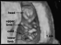
Week 9 - 38
- Second and Third Trimester (Fetal Period)
- Continuing growth and differentiation of organs formed in embryonic period
- some organs have a later development - neural, genital, respiratory, bones
- some continue to develop after birth - neural, genital, respiratory, bones
- growth in size, length (Second Trimester)
- growth in weight (Third Trimester)

Fetal Head 12 cartilage and bone formation (12 week)

Fetal Head head structures and the brain (12 week)

Fetal knee region


![]()

Birth

- birth (parturition) is a complex physiological process between the fetus and mother
- thought to be initiated by the fetus
Maternal Birth Stages
- Dilatation
- Expulsion
- Placental
- Recovery
Newborn

Newborn (perinatal) needs to activate many systems and establish independent regulation (homeostasis)
- Lung function - Fluid drainage, Gas exchange, muscular activity
- Circulatory changes - Closure of 3 vascular shunts
- Thermoregulation - metabolic rate, fat metabolism
- Nutrition - gastrointestinal tract function, peristalsis
- Waste - kidney function
- Endocrine function - loss of placenta, maternal hormones
Critical Periods of Development
Abnormal Development
Three main causes:
- Genetic
- Environmental
- Unknown
- First trimester most critical
- Different effect depending on time of insult (teratogen)

Diagnosis
- Prenatal diagnosis - number of different techniques (non-invasive, invasive) for determining normal development
- Neonatal diagnosis (APGAR test, Guthrie test)
- Maternal diagnosis - often pregnancy will expose maternal health problems
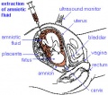
Amniocentesis
Chorionic Villus Sampling
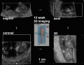
Ultrasound
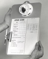
Apgar scoresheet
- Gutherie card icon.jpg
Gutherie card
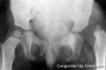
Xray congenital dislocation hip
Newborn hearing test


Finished! now here is a Link to Exam Question.....
Revision Notes
- You don't need to know everything today, its an introduction.
- Don't confuse "germ cell layers" (ectoderm, mesoderm, endoderm) with "germ cells" (egg, spermatazoa).
- Remember the difference between "clinical weeks" (last menstral period) and "embryonic weeks" (from fertilization, 2 weeks later).
- Revise meiosis and the difference between mechanism and timecourse for oogenesis and spermatogenesis in generating haploid gametes.
- With abnormalities, think about the types of prenatal dianostic techniques that are now available, the 2 major types (genetic and environmental) and the effect of maternal age/lifestyle.
2010 Foundations Practical - Introduction to Human Development
Glossary Links
- Glossary: A | B | C | D | E | F | G | H | I | J | K | L | M | N | O | P | Q | R | S | T | U | V | W | X | Y | Z | Numbers | Symbols | Term Link
Cite this page: Hill, M.A. (2024, June 17) Embryology 2010 Foundations Lecture - Introduction to Human Development. Retrieved from https://embryology.med.unsw.edu.au/embryology/index.php/2010_Foundations_Lecture_-_Introduction_to_Human_Development
- © Dr Mark Hill 2024, UNSW Embryology ISBN: 978 0 7334 2609 4 - UNSW CRICOS Provider Code No. 00098G