User:Z3332339
Welcome to the 2014 Embryology Course!
- Links: Timetable | How to work online | One page Wiki Reference Card | Moodle
- Each week the individual assessment questions will be displayed in the practical class pages and also added here.
- Copy the assessment items to your own page and provide your answer.
- Note - Some guest assessments may require completion of a worksheet that will be handed in in class with your student name and ID.
| Individual Lab Assessment |
|---|
|
| Lab 12 - Stem Cell Presentation Assessment | More Info | |
|---|---|---|
| Group | Comment | Mark (10) |
| 1/8 |
|
7 |
| 2 |
|
7.5 |
| 3 |
|
7.5 |
| 4 |
|
8.5 |
| 5 |
|
8.5 |
| 6 |
|
8.5 |
| 7 |
|
7.5 |
Lab Attendance
Lab1 --Z3332339 (talk) 12:45, 6 August 2014 (EST)
http://www.ncbi.nlm.nih.gov/pubmed
<pubmed>25084016</pubmed>
Lab2 --Z3332339 (talk) 11:13, 13 August 2014 (EST)
Lab 3 --Z3332339 (talk) 11:12, 20 August 2014 (EST)
Lab 4--Z3332339 (talk) 11:05, 27 August 2014 (EST)
Lab 5--Z3332339 (talk) 11:05, 3 September 2014 (EST)
Lab 6--Z3332339 (talk) 11:12, 10 September 2014 (EST)
Lab 7--Z3332339 (talk) 11:05, 17 September 2014 (EST)
Lab 8--Z3332339 (talk) 11:24, 24 September 2014 (EST)
Lab 9--Z3332339 (talk) 11:10, 8 October 2014 (EST)
Lab 10--Z3332339 (talk) 12:05, 15 October 2014 (EST)
Lab 11--Z3332339 (talk) 11:16, 22 October 2014 (EST)
Lab 12--Z3332339 (talk) 11:08, 29 October 2014 (EST)
Lab Assessment 1
A role for carbohydrate recognition in mammalian sperm-egg binding The primary focus of this article is on the first stage of fertilization, the binding of sperm to the specialised extracellular matrix of the egg, known as the zona pelluicda (ZP). The article suggests that the mammalian egg cell has a specialised carbohydrate site on the ZP for which the sperm recognises and binds to, enabling the fusion of genetic information between these two gametes.
The article explains how it was previously thought that data obtained from mouse sperm-egg interactions could explain human sperm-cell binding. However, recent research has suggested that the mouse model cannot be directly applied to the human model. Thus, this research paper investigates sperm-ZP interactions, using humans as the predominant model in finding the specific requirements for human sperm-egg binding which couldn’t previously be explained by the mouse model.
This article also uses a review that focused on the identification of the egg binding proteins associated with the binding of human sperm to the egg. Their findings concluded identifying the role for carbohydrate recognition on the ZP. These carbohydrates have specific sequences that cause restriction of ZP glycosylation in humans that could not otherwise be explained in mouse and pig models or are not the same for humans. This finding suggests that the regulation of glycosylation could be directly correlated with the degree of organismal complexity. Evidence favouring this concept would require the sequencing of ZP glycoproteins from other mammals at different levels of the evolutionary ladder, which could be are areas of future directions for this research.
Examining the temperature of embryo culture in in vitro fertilization: a randomized controlled trial comparing traditional core temperature (37°C) to a more physiologic, cooler temperature (36°C)
The study undertaken in this article was to determine if better clinical outcomes of IVF resulted from embryo cultures in cooler temperatures (36 degrees) as oppose to the traditional core temperature of (37 degrees).
The method of investigation: retrieving eight or more oocytes from a female of 42 years of age, with infertile couples (n=52). These mature oocytes were divided into two groups to be cultured at different temperatures; one group at 36 degrees, the other at 37 degrees. The rate of development and expansion of blastocysts (volume), fertilization, aneuploidy and sustained implantation were the factors measured to in order to determine which of these conditions clinically improved the environment best for embryonic development. This could potentially change the temperatures of which in vitro fertilization takes places in clinics in the future.
However, the results concluded that IVF culture at 36 degrees does not improve the conditions for blastulation and pregnancy rates in human in IVF. Thus, maintaining the existing temperature or changing it to 26 degrees does not alter the effects or success of IVF.
--Mark Hill These articles are good and your descriptions are appropriate. We will discuss in later tutorials how to format the referencing correctly. Help:Reference_Tutorial (5/5)
Lab Assessment 2
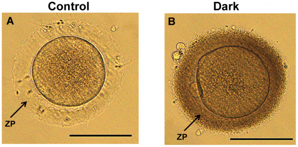
Oocytes with Dark Zona Pelluica affect fertility
Human mature oocytes with a normal (A) and dark (B) zona pelluicda. Oocytes with a DZP (dark zona pelluicda) have demonstrated a lower success of fertlization and implantation in clinical pregnancy rates in IVF/ICSI cycles. Patients with normal zona pellucida (NZP) were used as the control group.
Reference
<pubmed>24586757</pubmed>| PLoS One.
Shi W, Xu B, Wu L-M, Jin R-T, Luan H-B, et al. (2014) Oocytes with a Dark Zona Pellucida Demonstrate Lower Fertilization, Implantation and Clinical Pregnancy Rates in IVF/ICSI Cycles. PLoS ONE 9(2): e89409. doi:10.1371/journal.pone.0089409
Copyright
© 2014 Shi et al. This is an open-access article distributed under the terms of the Creative Commons Attribution License, which permits unrestricted use, distribution, and reproduction in any medium, provided the original author and source are credited.
--Mark Hill This is a good image for the assessment and I have made some minor changes to the information associated with the file. You do not need to include the copyright and student template on your page, just with the image. (5/5)
- Note - This image was originally uploaded as part of an undergraduate science student project and may contain inaccuracies in either description or acknowledgements. Students have been advised in writing concerning the reuse of content and may accidentally have misunderstood the original terms of use. If image reuse on this non-commercial educational site infringes your existing copyright, please contact the site editor for immediate removal.
Lab Assessment 3
2.Identify Current Research, Models and Findings
Physiological factors in fetal lung growth
<pubmed>3052746</pubmed>
This article looks at the current findings of different physiological factors that affect normal neonatal, functioning lungs upon during fetal development. The size of the paired organ to be able to exchange carbon dioxide with oxygen for the very first time at birth, is crucial to be able to withstand that pressure. As we know surfactant, is a lipid-protein composite. It is crucial to the function of the neonatal lung because:
A. Its high viscosity and low surface tension stabilize the diameter of the alveoli and prevent their collapse after each expiration.
B. Because the alveoli remain partially open, they are expanded on inspiration with much less expenditure of energy. [ANAT 2241 LEC 11-Respriation]
However, current research suggests that the production of surfactant which is reliant on hormonal factors, have little influence on fetal lung growth. In contrast, the following physiological lung growth factors were found to permit the lungs to express their inherent growth potential.
[this will be looked at further as the research project progresses]
Lung morphogenesis revisited: old facts, current ideas
<pubmed>11002333</pubmed>
Classical ideas -4 basic rules vs their review
Genetic control of lung development
<pubmed>12890942</pubmed>
Current concepts of lung development
Effects of hormones on fetal lung development
<pubmed>15550344</pubmed>
The fetal respiratory system as target for antenatal therapy
<pubmed>24753844</pubmed>
--[[User:Z8600021|Mark Hill] These references are more than appropriate (5/5).
Lab Assessment 4
1. An example of a use of Stem Cell Cord Therapy
<pubmed>25101638</pubmed>
Human mesenchymal stem cells MSCs (human embryonic tissue) have been used on animal models such as mice for their therapeutic qualities involved in regenerating liver tissue. This paper specifically looks at the possibility of using MSC to be used to treat degenerating organs after the discovering that MSC can be used as a substitute for liver acute failure on the mouse model. The human umbilical cord MSCs (hUCMSCs) have the capability to differentiate into hepatocyte-like cells due to their multipotence, meaning the that all the functions of the typical hepatocyte such as secretion of albumin and storage of glycogen can now be carried out from the hUCMSCs.
To imitate the environment for hUCMSCs to proliferate and differentiate into functional hepatocyte-like cells (iHeps), hUCMSCs were exposed to the growth factors, cytokinesis and chemicals. The induced i-heps demonstrated similar morphology to that of human hepatocytes, however the more significant part was evaluating their hepatic functions. Demonstration of hepatocyte function of i-Heps in vitro, is summarised in their findings as they compared i-Heps to hUCMS.
1) More glycogen was stored in i-Heps than in hUCMSC
2) 12 times more urea was produced by i-Heps than hUCMSC
3) lower levels of glycogen were stored in hUCMSC
This has had a significant clinical research relevance in treating acute liver failure and the possibility of treating other diseases as well.
2.Vascular shunts present in the embryo but closed postnatally
- The Foramen ovale -located between the right and left atrium.
- The Ductus arteriosus - located between the pulmonary artery and descending aorta.
- The Ductus venosus - located in the liver between the umbilical vein and IVC.
--[[User:Z8600021|Mark Hill] Very good (5/5)
Lab Assessment 5
Abnormality of Respiratory development: Asthma
Asthma is a disease that affects 10% of the population in Australia according to the Asthma organisation of Australia. [1].This prevalence in Australia is significantly high compared to other countries. However, the cause for our high ranking amongst other countries is unknown. In this research paper, a strong association between low birth weight, short gestational age and fetal growth restriction is shown to influence the development of asthma in children.
A primary part of their research involved a cohort study on infants born between 1979-2005, and following up during different stages of their development postnatally; 3 years old, first hospitalisation for asthma, 18th birthday etc. A majority of the subjects were hospitalized for asthma during their follow up that was consistent with their 3 findings that influenced infant hospitalisation because of the disease.
One conclusion from the study was that pre-term neonates may have under developed lungs that are smaller than the fetuses who completed the full gestation period (38weeks). Incompetent lungs could be due to restricted growth factors, inhibiting full lung capacity. Fetuses that were born small yet completed the gestational period, were infants unaffected by asthma and hence hospitalisation from it. As predicted, the risk of hospitalization for childhood asthma was proportional to lower birth weights, with only 1kg making a remarkable difference. This was a similar case for shorter gestational age.
References
<pubmed>24602245</pubmed>
--[[User:Z8600021|Mark Hill] Very good (5/5)
Lab Assessment 7
One of the most important developmental aspects of the male gonads is the descent of the testes. Recent research has discovered that the normal descent of the testes during male gonad development can be interrupted when exposed to paracetamol, aspirin, and Indomethacin (a nonsteroidal anti-inflammatory drug) causing cryptorchidism. Cryptorchidism is an abnormality of either unilateral or bilateral testicular descent, occurring in up to 30% premature and 3-4% term males. Descent may complete post-natally in the first year, failure to descend can result in sterility [1] . The aim of this research article was to determine whether common analgesic (pain relief drugs as mentioned above) disrupted the morphology and endocrine function of the human testis [2] .
Amongst the outcomes measured from comparing human fetal testes exposed to analgesic and those were not exposed to analgesic, were testosterone and the anti-Müllerian hormone. The number of testicular cells was then counted through histological and image analysis, as the testing of this occurred in vitro. The conclusion from this research identified that when fetuses were exposed to analgesic from pregnancy this cause disturbances in the fetal testis. These disturbances increase when small, critical age windows, such as when male gonad development takes place.
Data from a recent study of male human fetal (between 10 and 35 weeks) gonad position [3]
- 10 to 23 weeks - (9.45%) had migrated from the abdomen and were situated in the inguinal canal
- 24 to 26 weeks - (57.9%) had migrated from the abdomen
- 27 to 29 weeks - (16.7%) had not descended to the scrotum
Thus, what is advised by this article is a caution concerning consumption of analgesics such as aspirin, indomethacin, and paracetamol during pregnancy that may cause an inhibition of normal fetal testes morphology and endocrine function.
References
The embryonic layers and tissues that contribute to developing teeth.
The stages in tooth development include:
- Lamina
- Placode
- Bud
- Cap
- Bell
The tissues that contribute to developing teeth include:
- Odontoblasts
- Ameloblasts
- Periodontal ligament
--[[User:Z8600021|Mark Hill] This is more about testis anatomy rather than endocrine function. Your tooth summary does not identify embryonic origins of tooth components. (1/5)
Lab Assessment 8
Embryonic Development of the Ovary
Indifferent Stage
Much of the gonad development between males and females is analogous during embryonic development. Differentiation of the gonads (testis or ovary) occur late in embryonic development. Sexual differentiation is determined early on, where double X chromosomes in embryo will trigger the female gonad development whereas, an inherent XY chromosome will determine that the sex of this embryo will be a male. More particularly, the expression of the SRY gene on the Y chromosome determines the gender of the conceptus and signals pathways for male gonad development. Thus, when the SRY gene is not expressed, the human embryo will follow the gonad development of females.
Another contributing factor to gonad development after sex determining genes is hormone production. For example, at the urogenital sinus the presence dihyrdrotestosterone (DHT) determines males development and the absence of dihyrdrotestosterone (DHT) determines female development.
Differentiation Stage
• In the absence of the Y chromosome, female development occurs
• somatic support cells differentiate into follicle cells (instead of sertoli cells in males)
• From the intermediate mesoderm, the development of the Müllerian duct Müllerian duct persists and is stimulated to differentiate into the uterine tube, the uterus and the upper vagina. However, mesonephric ducts degenerate. The opposite occurs for the opposite sex.
• Presence of dihyrdrotestosterone (DHT)
• Absence of Anti- Müllerian hormone (AMH), since sertoli cells are not differentiated by SRY gene
• Some of the other essential genes involved in ovarian development include Wnt-4 and DAX-1
• Cortical cords extend from the surface of the developing ovary into the underlying mesenchyme during early fetal period
• As these cortical cords increase in size, primordial germ cells begin to arise> these then become primordial follicles> which contain an oogonium> proliferate and enter first meiotic division to for primary oocytes
• By the 10th week, ovaries are histologically identifiable
Human Ovary Timeline
• 24 days - intermediate mesoderm, pronephros primordium
• 28 days - mesonephros and mesonephric duct
• 35 days - uteric bud, metanephros, urogenital ridge
• 42 days - cloacal divison, gonadal primordium (indifferent)
• 49 days - paramesonephric duct, gonadal differentiation
• 56 days - paramesonephric duct fusion (female)
• 100 days - primary follicles (ovary)
Hill, M.A. (2014) Embryology Ovary Development. Retrieved October 7, 2014, from https://php.med.unsw.edu.au/embryology/index.php?title=Ovary_Development
--[[User:Z8600021|Mark Hill] You have used the existing online sources rather than research literature. At the beginning of the course I made it clear that these assessments were based upon the "research literature". (3/5)
Lab Assessment 9
Group 2
The introduction to this page was will written and the information was clear and to the point. Each component of the renal system was mentioned in your group’s introduction which gave an overall/holistic preview of the information that is evidently discussed underneath. There is a developmental timeline showing the key events of renal development at the embryonic, fetal and post-natal stages. Perhaps consider presenting this information in a table. The historic findings section, however, was lacking information. This section needs to be further researched and added to make this project complete. Your choice of content, clear structure, headings and images is evident that your group is working well and have a good understanding of this topic area. However, there is no hand drawn image yet. The image chosen form Langman’s Medical Embryology is a great image to show as it demonstrates the progressive stages of kidney ascent, perhaps you could consider re-drawring that image rather than just immediately upload it from the textbook. Your descriptions and information presented can be understood at the peer level. It is both engaging and informative, well done!! There is also a good balance between text and images that are appealing for the reader. The information presented in the first half of your project is ample however this is not coherent with the second half of your project page, where descriptions are not as developed.
There is a great selection of images that are used in your group project. Most of these images are correctly cited and have been uploaded in the correct manner. Some images are just missing the student template image:----
- Note - This image was originally uploaded as part of an undergraduate science student project and may contain inaccuracies in either description or acknowledgements. Students have been advised in writing concerning the reuse of content and may accidentally have misunderstood the original terms of use. If image reuse on this non-commercial educational site infringes your existing copyright, please contact the site editor for immediate removal.
You can view this in edit mode and add it to your images.
There are a great number of resources that are used in this project, and all your references are correctly cited. As your project is still underway, I am sure that you will add additional references and also make it one complete this at the end of your project. Overall, I enjoyed reading about the renal system on presented by your group and I am confident you will earn high marks for your project. Best of wishes group 2!
Group 3
Your introduction to the gastrointestinal system provided a clear overview of what your project is about. I think it would be a good idea to couple this introduction with an image that shows the pathway and divisions of the GIT. The timeline shown is fantastic, it is not only extensive, but it divides the GIT into regions of the foregut, midgut and hindgut as well as the weeks in which key development events take place. It is in simple, easy to read language, at an element of teaching at the peer level- great work! There is also a reference next to each of these events which reflects the amount of research that took place-well done guys!
Your page includes a table with statistics- the percentage of herniated foetuses which adds credibility to your work and gives the reader information on how frequent this abnormality occurs. Your section for current does not have a lot of information, there is only one reference available for your recent findings. This section of your project needs to be further researched before the submission date. There is more than one hand drawn image is which fantastic! The colours used for it are a bit too bright, however, this shouldn't be too difficult to change, perhaps just adjust the brightness of the picture on paint, or whichever program the picture opens up with on your computer (this is just a very minor critique. The fact that your group project has more than one student hand drawn image shows adherence to the requirement for the project guidelines.
It was great to see only one reference list, as opposed to different reference lists for each section in the project. Your reference list appears to be long, with 24 references however, 16 of these references part of the timeline. More research papers need to be included to make what is already an amazing project, better! A video of the GIT and the rotations that occur during development would be rotations would be great visual representation of this system due to the nature of its development course. Perhaps you could find one off YouTube or create one.
Overall, this is a good project page, well done group and best of wishes!
Group 4
Your group project is of excellent quality, there are just a few minor things to take in to consideration if you wish.
Firstly, well done in creating a timeline in a table format that seperates the key events that occur between male and female gonad development. This is exactly the type of information I wouldve expected to see if I was interested in looking up information about the differences between male and female internally and externally and when these events take place. There is, however, much information about the male in this table and not as much information as there is in the female. You might also like to consider selecting one type of font for your table, just so that it looks a little neater.
Your section of current research and findings looks fantastic, with a lot of text, but sadly not enough images! Good work though with the and drawn image! There are a few hand drawn images on this group project page so well done for that! There are a few parts in the project where an image still needs to be uploaded/formatted but it looks like you are aware of these things with mention of [draw image here] as an example. In this same section, there is a great number of dot points, perhaps try to part of it in paragraphs so that not all the information is simply presented in dot point form. You can tell you have done a lot of research here, so well done.
In the historic finding section, there is a lot of text and only one image (a hand draw one, which is really good!). However, the amount of text is not matched with a visual component such as more images, or a diagram or table. Perhaps increase the amount of visual things in this section so there is appropriate balance-awesome work really!
Great choice of a youtube video! It showed the different stages of gonad development, both at the indifferentiation stage and when the gonads differentiate, into male and female. However, the video is quite long, it is approximately 10 minutes long, would you perhaps consider a shorter video? or trimming the video down? With that being said, I do think it provides a great visual for the key developmental aspects, so great choice there!
The abnormalities section is well researched. The abnormalities are listed from the most common to those that are rare, that a great way of giving the reader a general idea of its frequency in society. There are any abnormalities described in this section, and information is presented equally for both sexes as well as abnormalities that affect both sexes. There is also an excellent hand drawn image from the textbook that is correctly cited and contains the appropriate copyright information as well permission for this image to be reused after 6 months.
One last thing, before submission place all your references in one reference list.
Well done project group 4! I enjoyed reading about your project. All the best!
Group 5
Group 5, you have a brilliant introduction, introducing the reader to what your page is about. Your introduction contains information for each of the parts involved in the integumentary system such as skin, glands, hair nails and teeth. There is a clear structure to your project with clear headings and sub-headings. This makes the reader find information about a particular part in your project more easily.
There is an extensive list of references, which demonstrates, a great effort towards researching your projects system. Some of the references however, need to be put into in the correct format. There are different reference lists under the different sections of your group project and as I understand why, I'm sure these are just small things that will be fixed before the final submission.
I have to commend you on your table, it is more than sufficient. It not only clear describes a clear transition from week to week changes in development of the integumentary system. The table however, needs to be reformatted to fit the window of the page and likewise, the pictures inside the table as there are too small to be seen without opening up the image. There are other images also on the page were too small such as "The stages of embryonic teeth development". These are just minor changes that need to be made before your groups final submission.
At the start of your project, all descriptions were matched with an image. This provided an appropriate balance between written text and visual representations. However, in the historic findings section, this balance was not seen as there are no images for this section.
Your page also contains information that is teaching at the peer level-well done guys.
Under your section of some recent findings, there are blocks of information in purple; I'm not sure as to the reasoning behind this, as the other parts in your project do not have the same background.
I particularly liked how in the introductory paragraph you mentioned what topics you will be covering; including abnormalities associated with the Integumentary system and delivered this information under the abnormalities section, where treatments and managements of these abnormalities were put forward!
Overall, good work guys!
Group 6
Great work, it looks like your group has a clear mindset and direction to where your group project is going, even if it is not there yet. One of the images next to the timeline section was too small to be view without actually opening up the actual image. There are a few images on your page, and also a few tables, perhaps uploading a few more images that correspond to the text would make your project page more visualling appealing.
There is table in your introduction- Table 1. Summarises the hormones released by the human pineal gland and their role in embryonic and foetal development. This is a great way to summarise information you have discovered after your research. However, as there is only one line of information, the information you have gathered needs to be added in order to make your group page complete.
Under the heading of Pineal gland, there is a sub-section labelled 'timeline', however there are only three points under this and no time course, or time frame included. The timeline needs to be further developed. You may also consider putting this information into a table and referencing articles from which you found this information.
There is not a lot of information for abnormalities. Only an uncompleted table and a references list exist. This information needs to be filled out the sooner the better. There seems to be a sub-section about abnormalities for each endocrine organ, but this does not contain much information-see Pineal gland and Hypothalamus sections. Perhaps your group would consider, just having one section in your project for all the abnormalities associated with the endocrine system.
There are several reference lists within your group project page that need to be put together to create just one reference list, I understand why this is at the moment, just remember to change before your final group submission.
Overall, well done group 6!
Group 7
Your group's project page has a good introduction with a description entailing what your page is about and the information your page covers.
There is a very good layout, with a combination of text, tables, dot points and images- well done!Some of the images however a slightly small (Images under Brain development) or too big (image under development during fetal period) and need to be reformatted.
Whilst your group mentioned that the neural tube differentiates into the proencephalon (forebrain), the mesencephalon (midbrain) and the rhombencephalon (hindbrain), there was no mention of the different brain flexures in your project. This an important aspect of brain development as it divides the three primary vesicles into 5 secondary primary vesicles (which you did mention) and how the cephalic flexure separates the brain from the spinal cord.
The images use in your project are excellent and provide a visual to the information that you describe. However, there are many images from your group project that are from the lecture notes, perhaps try and mix up your selection to incorporate images from other sources such as research articles on pubmed. There also appears to be an image deleted under the Abnormalites, and this formatting would need to be fixed up, but this is just a small thing.
Well done for your extensive reference list that you have. Although it is split in respective parts of your project, under different headings, don't forget to make it into one before the final submission.
Group 8
The introduction to your page is extremely funny, but this is completely irrelevant to the project and should be taken out before you submit the assignment. There are long blocks of texts on the page, with no tables or any pictures sadly. There should be a some images/digarams/videos for each heading. There is a number of good headings, with information within that needs to be further developed. There is great potential for this group project to develop further.
There is a Heading labelled, 'Muscle development general timeline' however, underneath this section, there is only a small paragraph with no timeline whatsoever. If you don't want to have a timeline in this section of your project, then remove the word 'timeline from this heading'. However, I think a timeline would be a great way to show an overview of the key events of the muscoskeletal system.
There is an broad, and long section of information under "background embryonic development". Just remember that our projects are about fetal development and not the embryonic stage of the system our project is about. The time spent on writing this section could have been spent on working on other parts of the assignment that require greater attention.
Towards the end of the references list, there are references that have not properly been citied. There also exists a format error in your reference list that would need to be fixed before the final group submission.
--[[User:Z8600021|Mark Hill] There are fair peer assessments. (9/10)
Lab Assessment 10
<pubmed>25324764</pubmed>
Amongst the five senses is vision. For one of the most important processes of development, visuomotor development of the eyes takes place at the embryonic and fetal stages but rapidly develops after birth. Although there may not be much visual stimuli inside the maternal environment, the foetus is still able to see as visually excitable cortical areas already exist before extrinsic stimuli are present. External stimuli for example, can be facial recognition or other visual cues. Fetal eye movements although observed in utero, there is no receptive brain activity detected from visual stimuli. Thus, the aim of this study was to make the link between the spontaneous eye movements that are observed and the signalling network back to the frontal cerebral areas of the brain.
Research Methods
In order for this research group to carry out their investigation, they selected seven foetuses between 30-36 weeks and mothers with an average age of 32.29 years. MRI imaging was conducted to ensure no pathological brain development existed and this consent was approved by from the maternal participants on behalf of the unborn fetus.
The movements by the fetus were then tracked by fMRI and data was computed. As a result of this mapping, eye centre locations and lens centre locations were determined. Correspondingly, the head axis was defined as the symmetry axis between the two eyes.
Research Findings
After obtaining fMRI data and ICA information, the positions of the eyes were determined. The relationship of single-subject component time courses with the eye movement regressor was calculated. Four fetal eye movement patterns were initially characterized based on early ultrasound observations. Some of the results included:
Type I eye movements were described as single, transient deviations consisting of a bulb deviation, and a slower return back to the resting position, single but prolonged eye movements
Type II, complex sequences of eye movements to different directions without periodicity
Sensory Notes page:
https://embryology.med.unsw.edu.au/embryology/index.php/Sensory_System_Development
--[[User:Z8600021|Mark Hill] I have now read 4 students using this same article. Your summary is quite good. (4/5)
Lab Assessment 11
<pubmed>19637940</pubmed>
Induced pluripotent stem cells (iPS) are stem cells that resemble embryonic stem cells (ES) because of their capacity to generate all cell types within the body. In this research paper, they examine whether a somatic cell, once returned to it pluripotent state can gain the ability to reprogram other somatic cells. By applying somatic cell nuclear transfer (SCNT) and reprogramming by cell fusion of two different cells and consequently two different genomes to create a hybrid. As expected and as in most cases the phenotype of the less differentiate fusion partner dominated the phenotypes of the more-differentiated partner. Thus embryonic cells belonging to the mouse have the ability to reprogram the somatic genome of human embryonic cells following both cell fusions of each species. Their findings concluded that indeed once the nucleus of somatic cell is reprogrammed, it possesses the capacity and pluirpotency to reprogram other somatic cells by cell fusion and it shares this trait with those of embryonic stem cells (ES).
--[[User:Z8600021|Mark Hill] Good summary. (5/5)

