Paper - The sexual differences of the fetal pelvis
| Embryology - 23 Apr 2024 |
|---|
| Google Translate - select your language from the list shown below (this will open a new external page) |
|
العربية | català | 中文 | 中國傳統的 | français | Deutsche | עִברִית | हिंदी | bahasa Indonesia | italiano | 日本語 | 한국어 | မြန်မာ | Pilipino | Polskie | português | ਪੰਜਾਬੀ ਦੇ | Română | русский | Español | Swahili | Svensk | ไทย | Türkçe | اردو | ייִדיש | Tiếng Việt These external translations are automated and may not be accurate. (More? About Translations) |
Thomson A. The sexual differences of the fetal pelvis. (1899) J Anat Physiol. 33(3): 359-380.
| Historic Disclaimer - information about historic embryology pages |
|---|
| Pages where the terms "Historic" (textbooks, papers, people, recommendations) appear on this site, and sections within pages where this disclaimer appears, indicate that the content and scientific understanding are specific to the time of publication. This means that while some scientific descriptions are still accurate, the terminology and interpretation of the developmental mechanisms reflect the understanding at the time of original publication and those of the preceding periods, these terms, interpretations and recommendations may not reflect our current scientific understanding. (More? Embryology History | Historic Embryology Papers) |
The Sexual Differences of the Fetal Pelvis
By Professor Arthur Thomson, Oxford.
(PLATES XIII.- XV.).
THE conflicting statements in many of the standard text-books of midwifery, together with an almost entire absence of any reference to the subject in most anatomical works, led me to make a series of observations on the pelvis, with the object of determining whether or no there were any sexual differences which coul be recognised during foetal life. At this time I was ignorant of Fehling’s work on the subject, and unfortunately I have not been able to obtain access to his original papers, having to content myself meanwhile with abstracts from different sources. This, perhaps, is not an unmixed evil, for my results, obtained independently, strikingly confirm what I assume is Fehling’s main contention, that the differences in form and appearances are such as to enable the observer to discriminate between the pelvis of the male and female as early as the third month of foetal life. To Fehling} therefore, belongs the credit of having first pointed out this remarkable fact, a fact which seems to have met with but scant recognition by both anatomists and gynaecologists. In a work published so recently as 1888,? it is stated that “There is, however, nothing which would enable us to distinguish with even an approach to certainty between the male and female pelvis ‘until the period of puberty ”; and in the more recent American text-book 3 (1896) the following passage occurs :—“ The distinctive characteristics of sex are acquired after puberty, although, according to Fehling, indications of these peculiarities are present even at birth,”—a very half-hearted recognition of the facts.
1 “ Die Form des Beckens beim Fetus u. Neugeborenen,” Zettschr. fur Geburt.
u. G’ync'iek., Bd. ix. and X. 2 System of Midwifery, Leisliman. 3 Textbook of Obstetrics, Norris, 1896.
voL. XXXIII. (N.s. voL. XIII.)
Schauta} whilst referring to Fehling and Litzmann’s 2 observations, scarcely credits. them with the importance which ‘they deserve. Nor do We get much more information from the anatomists. Quain passes it by without reference. Macalister, the only English anatomist, so far as I know, who mentions it, is satisfied with a brief note to the effect that “These sex characters are discernible even at birth ”; whilst Humphry states that “ It is not till after puberty that the distinctive peculiarities of the male and female pelvis, particularly the preponderance of the transverse diameter, are recognised.”
Testut and Poirier ignore the subject, the latter contenting himself with a reference to Fehling’s work on the obliquity of the foetal pelvis.
In view of this state of things, it is not surprising that gynaecologists and others interested in the mechanism of the female pelvis have been at great pains to account for its peculiarity of growth. The mechanical effects of pressure and the influence of posture and muscular action have been the favourite explanations; and whilst willing to admit that any or all of these may exercise an important influence on the form of the adult as compared with the foetal pelvis, it is difficult to see how the same forces are to lead to different results in the two sexes. Matthews Duncan} whose views on the development of the pelvis have met with wide acceptance, evidently felt this, for in accounting for the fact that the male pelvis does not respond to the same forces in a similar way to the female, he puts forward the somewhat unsatisfactory suggestion that “ The changes are less marked, for in it (the masculine pelvis) the bones are thicker and stronger and stouter, and earlier consolidated with each other. These conditions are at once the signs and causes of the peculiarities of a masculine pelvis.” Hermann Meyer‘
1 M1'ille.r’s Handbuch der Gebzcrtshiil/e.
'3 Die Formen dcs Beclcens, 1861.
3 Researches in Obstetrics.
‘ Lehrbuch der Pkysiologischen Anatomzle.
also seemed to regard some explanation of this difference necessary, for he assumed that during growth the female pelvis is more plastic than the male.
The purpose of this paper is, however, not to discuss the influences to which the pelvis may be subjected after birth, but rather to emphasise the fact already pointed out by Fehling, that at a comparatively early period in the development of the foetus the sexual differences are as pronounced and characteristic as they are in the adult. If this can be proved, the explanations, however ingenious, hitherto advanced, become needless and unnecessary, the factors which determine the subsequent growth of the pelvis exercising their influences on male and female alike.
For the sake of comparison, it may be well to tabulate the differences generally recognised between the male .and female pelvis under the heading -
1. The pelvis as a whole: its relative proportions in height, width, and the slope of its walls.
2. The false pelvis. The form and mode of expansion of the iliac fossae.
3. The true pelvis:
(at) Its form and diameters, particularly the inlet.
(6) The sacrum.
(c) The ischium ; projection of‘ischial spines.
(cl) The pubes ; body, and angle of pubic arch.
(e) The great sacro-sciatic notch. besides some minor points, which will be dealt with subsequently in the text.
Before proceeding to discuss these points in detail, it is advisable to say something regarding the specimens on which these observations are based, and at the same time to point out some of the obvious sources of error which have led to wrong conclusions in the past. The pelves for the present inquiry were taken from foetuses which had been previously hardened in spirit or formalin. After being carefully cleaned and prepared by my Assistant, Mr Chas. Robertson, it was found that they retained their form sufficiently well for all practical purposes, the coccyx alone excepted. This, however, was a matter of little moment, and does not invalidate any of the results obtained. The specimens have all been conserved as wet preparations, and the measurements made, and the photographs obtained, have been taken from the moist specimens. This is a matter of great importance, as the conclusions arrived at from the examination of dried specimens are absolutely fallacious, the shrinkage of the cartilage and the drying and contraction of the ligaments having produced distortion to such an extent as to render worthless any conclusions based upon them. It is necessary to emphasise this, as Galabin} in criticising Fehling’s conclusions, says, “ but almost all foetal pelves do show in some degree characters corresponding to the change enumerated above, and it is impossible that any changes in drying should always occur in the same direction.” As a matter of fact, this is precisely what does happen, so that it is well to guard against such a source of error.
The measurements taken were those selected by Sir William Turner in his “ Challenger ” Monograph, but it was felt that in dealing with structures of so small a size, the relative proportions and angles could be but roughly estimated, the difference of a millimetre in the foetal condition corresponding with a much larger difference in the adult. Besides the difficulty of estimatingmeasurements so small, it was felt that the results obtained hardly conveyed the differences in form and size which were apparent to the eye. For this reason it was decided to adopt a graphic method of comparison, which would enable the reader to. estimate for himself the characteristics of the pelves of either sex. The results obtained must speak for themselves. In order, however, ‘to render clearer some of the more important features,. diagrams have been carefully prepared by the enlargement of the original negatives, and the results are represented in schematicform. In taking the photographs, a lens of long focus and narrow angle was employed, so as to reduce as far as possible the distortion resulting from forced perspective. In this respect, therefore, the photographs approach, as near as ca11 be, orthographic representations.
Taking first the proportions of the pelvis as a whole (see table), it will be seen that the breadth-height index is high, viz., 856
for the females and 824 for the males; in other words, this
1 A Manual of Midwifery, London, 1891, p. 28.
About vii.
About vi. About v. About iv. L months. months. months. months. Average‘ H I ‘ M. F. ' M. F. M. F. M. F. M. F. ’ I ~ 1 Breadth of Pelvis, . . 66 58 53 51 49 48 36'5 35 51'1 48 Height of Pelvis, . . 53'5 49 45'5 44 39 41 '5 30 30 42 41 Breadth—Height Index, 81'1 84 '4 85'8 ‘ 86 '2 79'5 86 '4 83'3 85 '7 82 '4 85 '6 Breadth between Ant. - l Sup. Iliac Spines, . 61 55 50'5 47 '5 45'5 -‘ 45 35 32 48 44'8 Breadth between Post. Su . Iliac Spines, . 19 19 13 13 12 _ 15 9'5 105 13'3 14'3 Brea th between Ischial 1 Tuberosities, . . . 25 26 '5 22 ’ 22'5 18 19 13 15 19'5 20'7 Breadth between Ischial ' Spines, . . . . 19 1.9 11'5 17'5 9'5 11 8 8'5 12 14 I Greatest diameter of} 12'5 11 11 10 8 9'5 5'5 5'5 9'2 9 L Cotyloid Cavity, - 12-5 '11’ 16 8'5 8'5 8'5 5-5 .5 9-1 82 Vertical diameter of Obturator Foramen, . . 11 10 '5 9'5 10 6'5 7 5'5 4'5 8'1 8 Transverse diameter of I Obturator Foramen, 7'5 8 ’ 6'5 6 5 ‘ 5 4 3 8'7 5'5 Sub-pubic Angle, . . 56° 70° 50° - 65° 50° A 60° 46° 76° 50'5° 67 '7° 1. Brim, Transverse Diameter, . . 28 27 23 21 '5 19'5 20 14'5 15 21 '2 20'8 Brim, Conjugate, . 23 23 175 19 16'5 17 14 11 17'7 17'5 Pelvic or Brim Index, . 82'1 85 76 90 86'8 85 100 73 86'2 83 '2 Q Brim, Oblique diameters, right, . . . . 26 27 21 21 20 20 13'5 15 20'1 20 '7 1 Brim, Oblique diameters, ' left, . . . . 26 27 21 21 20 20 13 '5 15 20 '1 20 '7 Inferior Sagittal diameter, 17 22 18'5 20 17 18 '5 15 13 '5 16 '8 18 '5 Coccygeo-pubic diameter, 15 17 '5 15'5 15 '5 15'5 13 '5 15 11 15'2 14'3 Intertuberal diameter, . 1835 20 14'5 19 11 13 '5 8 11 13 15 '8 Depth of Pubic Sym- ' physis, . . . . 10'5 10 10 10 8 8 6 6'5 8'6 8'6 Depth of Pelvic Cavity, . 24 '5 23 '5 22 21 19 18 14 15’ 19'8 18 '8 Inter-obturator width, 10 10 6 '5 8 7 7 5 6 7 '1 7 '7 Inter-cotyloid width, . 34 33 '5 25 '5 27 22'5 22 17 15 24'7 24'3 Width between Ant. Inf. Iliac Spines, . 47'5 44'5 38 345 32'5 32'5 23 24 35'2 3633 IliumHeight Length, 32 80 27'5 27 23 25 '5 19 18 '5 25'3 25 '2 Breadth, . . . 37 32 '5 32 31 '5 27 27 21 '5 20 '5 29 '3 27 '8 Iliac Index, . . . 115 108 118 115 117 108 113 113 116 111 Breadth of Innominate ‘ 1 Bone, . . . 39 37 * 34'5 3.3 28 31 22 22 30'8 307 Length of O8 Pubis, 19-5 18 4 14-5 14 12-5 13 9 9 138 13-5 Pubo-innom. Index, 50 48 ' 42'6 42 44'6 42 40'9 40 44'5 43 Length of Ischium, . 17 16 14 14 ‘ 12 12 8 8 12'7 12'5 Innominate Index, . 73'5 75 76'6 75 73'6 , 75 73'3 73 74'2 74'5 Ischio-innom. Index, . 32 32 31'1 31 30'7 29’7 26'6 26 '6 301 298 Length of Sacrum, . 25'2 27 22'5 24'5 22'5 ‘ 21 '5 16'5 18 21 '6 22'7 Breadth of Sacrum, 29 '5 28 22'5 , 24'5 22'5 21 '5 16'5 16 '5 22'7 22'6 Sacral Index, 118 103 100 ' 100 100 100 100 96 104 99'7
In the above table the measurements are given in millimetres.
means that the height of the foetal pelvis is great in proportion to its Width,—the breadth-height index of Europeans averaging about 74 and 79, for females and males respectively. It follows from this that during the growth of the pelvis from the foetal to the adult form there is a greater proportionate growth in width than in height. It will be noted, however, that in thefoetal condition the breadth-height index is higher in the female(856) than in the male (824), the converse of what holds good in the adult, where the breadth-height index (79) exceeds that of the female (747 5), so that it would appear that during growth the female pelvis increases in width more rapidly than in height, a circumstance no doubt associated with the peculiarity of the form and size of the hinder portion of the ilium, to be hereafter referred to. At the same time, it may be pointed out that whilst the proportions between the height and width of the foetal pelvis differ from the corresponding proportion in the adult, it is the case that in the foetal condition the diameters of the male pelvis are absolutely greater than the female. The comparatively small number of specimens I have been able to examine, apart from those figured and measured in this paper, and the difficulty of being surethat the male and female specimens are precisely the same age, renders it advisable not to put too much reliance on these figures.
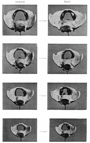
In the foetus the splay of the lateral walls of the pelvis is greater in the male than in the female. Thus the sides of the malepelvis, as indicated. by lines drawn through the points of extreme width of the iliac crests and the ischial tuberosities (see fig. 1), are set at an angle on an average of about 64°, while the sides of the female pelvis are inclined at an angle of about 53° to each other. It is noteworthy that this difference in the splay of the pelvic walls is characteristic of the different sexes in the adult, the female pelvis being frequently described as a short segment of a long cone, as contrasted with the male, which is a long seg-. ment of a short cone. iThisdifi’erence in the splay of the sides of the pelvis necessarily reacts on the proportions of the true and thefalse pelves, whereas in the female, owing to the nearer approach to parallelism in the disposition of the sides of the girdle, thelower segment (z'.e., the true pelvis) bears a greater proportion to the upper segment ('£.e., the false pelvis); in the male, owing tothe greater splay of the pelvic wall as a whole, the proportion of the lower segment to the upper is much less than in the female. I have been led by my observations to place considerable
an A‘ 1' IV.
Females. Months. Males. FIG. 1.—Foetal pelves.
importance on the proportion of the Width of the ilium (taken from the anterior superior to the posterior superior iliac spine) to the total pelvic height, assuming the latter equal 100. This may be determined in adults from Verneau’s 1 figures, as follows :_
Height of Breadth of
Verneau. Pelvis. Ilium.
Males, . . 220 mm. : 164 mm. : : 100 : 74°5 Females, . 197 ,, : 156 ,, : : 100 : 79:1 In the foetal pelves the proportions are Males, . . 42 mm. : 293 mm. : : 100 : 69‘7 Females, . 41°1 ,, : 27°8 ,, :: 100 : 67'6
Too much stress must not be laid on theseresults, as the difficulty of taking the measurements accurately on so small a scale is very considerable; yet the results are sufficient to show that, comparing the foetal pelves with the adults, the latter have proportionately Wider ilia; in other words, this means that during growth the increase in widths of the ilium is proportionately greater than the increase in height of the innominate bone. Comparing now the innominate indices, z'.e., the proportion of breadth from symphysis pubis to posterior superior iliac spine to the pelvic height, I find that in 5 male, presumably European, pelves, this index averaged 871, whilst in 8 female pelves, also probably European, the index was 93. Contrasted with this, the foetal innominate indices are for the males 7 3'3, and 746 for the females, thus proving that during growth the increase in Width of the innominate bone is proportionately greater than its increase in height.
The capacity of the false pelvis depends upon the form and disposition of the iliac bones. As regards what may be termed the sexual characteristics of this part of the pelvic girdle, there is considerable diversity of opinion, if one may judge from the varied accounts published in the different text-books. Quain, for instance, says : “ The ilia are more vertical (in the female), and thus the false pelvis is relatively narrower than in the male. On the other hand, both Testut2 and Poirier,3- in describing the dorsal aspect of the ilia (fosses iliaques externes), assign to the male a more vertical direction. Galabin4 describes the iliac fossae in the female as more widely spread out, thus giving a
1 Le Bassin dams les sexes et dans les races, Paris, 1875.
‘3 Tmité d’A7zat0mz'e humaine, Paris, 1896. 3 Ibid. 4 A Manual 0/'M73dw2Ifery, 1891.
greater breadth across -the hips to woman’s figure, a statement with which Playfairl apparently agrees; whilst Lusk,2 in speaking of the characteristics of the female as contrasted with the male, says of the former, “ The iliac incline approaches more nearly a vertical line.” These extracts from the opinions of recognised authorities are sufflcient to demonstrate the somewhat unsatisfactory state of our knowledge. Verneau,3 whose observations are based on numerous and accurate measurements, says that the inclination of the internal iliac fossee is practically the same in the two sexes, but in the female these hollows are not so high ; at the same time, they are more excavated.
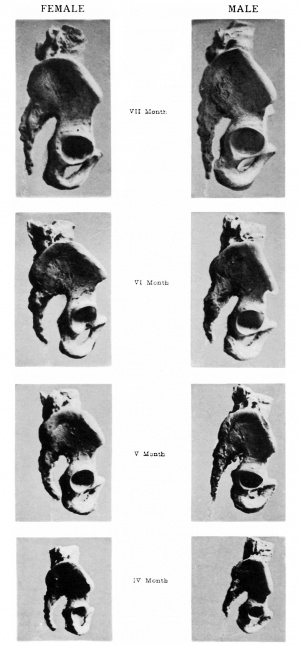
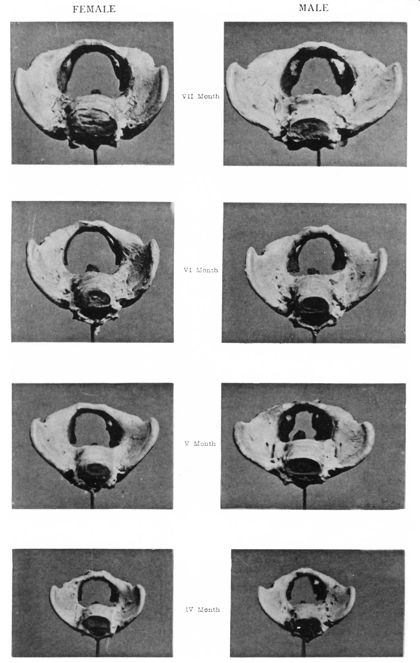
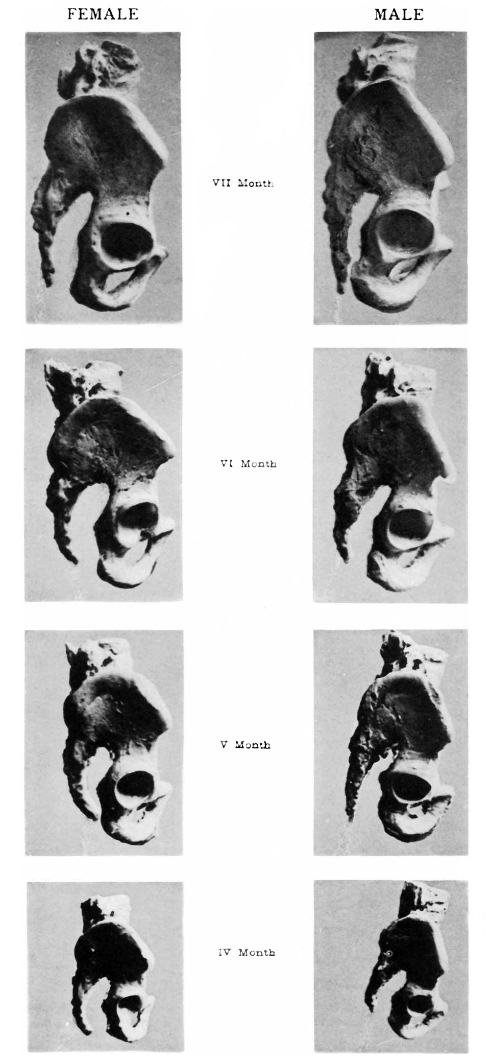
A study of the photographs shown on Plate XIV., as well as the diagrams therefrom constructed, will, I trust, demonstrate the fact that there is remarkable uniformity of type in the disposition of this part of the pelvic girdle in the two sexes.
As viewed from above, the male ilia are clearly seen to be more open and expanded, whilst the fore part of the female ilia tends to be inturned ; this, together with the lesser splay of the innominate bones in the female already referred to, combines to form a false pelvis, the sides of which are more vertical. Whilst the form of the false pelvis undergoes great individual variations in the two sexes, the reader cannot fail to be struck with the characteristic type displayed in the male and female series of foetal pelves. Further, the curve of the iliac crest reaches a higher level, and is more pronounced in the male than in the female, as happens also in the adult forms (see Plate XV.). I have estimated this difference in the adult in the following way: A line was drawn across the outer side of the ilium from the anterior superior iliac spine to the posterior superior iliac spine ; from this line the greatest perpendicular height of the iliac crest was measured, with the result that in four males the distance was found to average 66 mm., as contrasted with 58 mm., the mean of eight females.
It is, however, in the true pelvis that the appearances characteristic of sex are principally met with. These differences are so generally recognised in the adult that it is unnecessary here to dwell upon them,except to point out that in the foetus they are just
1 The Science and Practice of Midwifery, 6th edition. ‘3 The Science and Art of Midwifery. 3 Le Bassin clans les sexes dans les races, Paris, 1870.
as distinctive. The form of the inlet in the male is described as cordate, as contrasted with the more uniformly oval, elliptical, or reniform aperture in the female; this difference is due to the maximum transverse width in the male being placed in a plane posterior to the greatest transverse diameter in the female. The same characteristic forms are seen in the foetal pelvis liere figured, the shapes typical of the two sexes being readily recognisable as early as the fourth month. It has long been assumed, and the assumption has been based, no doubt, on the measurements of dried specimens, that the conjugate exceeded the transverse diameters in the foetal condition. According to Burns} whose figures have been extensively quoted, it is not till about the tenth year that the transverse diameter exceeds the conjugate. Hennig,2 in an elaborate paper on the child’s pelvis, has pointed out that such is not the case, and confirms Fehling’s. observations on the pelvis of the new-born child; yet, despitethese researches, there seems to be a strong tendency to cling tothe older accounts.
In the four female foetal pelves which I have measured, the average transverse diameter of the brim was found to be 208. mm., as compared with an average conjugate of 176 mm.; the four males yielded an average of 212 mm. for the transverse,. and 17"? for the conjugate diameters. Verneau gives for adult. females the following averages: conjugate 106, and transverse135; males, conjugate 104, transverse 130. Comparing these results, it will be seen that the differences between the conjugate and transverse diameters in the foetal pelves are very nearly proportional to the difference between the same measurements in the adult. Thus, in order to attain the adult proportions, there need only be an increase in width of 7 mm. in the female and 5 mm. in the male, supposing the foetal proportions are otherwisemaintained. The proportions of the conjugate diameters to the transverse in the series of foetal pelves has been estimated by means of the formula suggested by Zaaijer and employed by Sir W. Turner, Viz.,
Conjugate diameter X 100
, - elvic index. Transverse diameter P
1 Principles of Mz'dwz'fery. . 2 “ Das Kindliche Becken,” Archiv fur Anal. co. Entwiclcelungsgeschichte, 1880.
The results obtained are an index of 832 for the females and 862 for the males. They are therefore included in the
platypellic group, according to Turner’s classification. It is remarkable how close the agreement is between the indices of the foetal form and the adult. Quoting from Sir W. Turner’s
IV.
Females. Months. Males. FIG. °2.——Foetal pelves.
monograph} the indices have been estimated as follows by the subjoined authorities :—
p Males. Females.
J. J. Watt, . . . 87°9 87'3 Pelvic index. John Wood, . . . 85 86 ,,
Sir W. Turner, . . 77 79 ,, Verneau, . . . 80 78 ,, Gegenbaur . . . 84 85 ,,
Sir W. H. Flower, . . 81 78
77' 1 "Report on the Human Crania and other Bones of the Skeleton,” Challenger Reports-Zoology, vol. xvi.
“In the measurements made by J. Wood, Gregenbaur, and myself (Sir W. Turner), the brim index in the male pelvis is a little below the brim index in the female; but in those measured by J. J. Watt, Verneau, and Flower, the brim index of the males somewhat exceeded the females.” .In this respect, therefore, the indices of the foetal pelves are in agreement with the observations of the latter observers, for the male index, 86, exceeds the female index, 83.
The proportion which the sacrum bears to the pelvic inlet, as well as its size in relation to the pelvis as a whole, is a matter of some interest, in view of the statement so often repeated, that the shortness of the transverse diameter of the brim is due to the narrowness of the foetal sacrum.‘ Now, as a matter of fact, the foetal sacrum is larger in proportion to its surroundings than in the adult. When we consider that the sacrum forms part of the axial skeleton, whilst the innominate bones belong to the appendicular skeleton, and are necessarily associated with the develop ,ment of the lower limbs, this does not appear surprising. The facts, however, are best ascertained by comparing the proportion of the breadth'of the sacrum to the greatest pelvic breadth in both the adult and the foetus, taking Verneau’s figures, and comparing the greatest width between the iliac crests with the sacral width at the inlet, taking 100 to represent the former.
Sacral width at inlet X 100
_ , , — index, Greatest width between 1l1ac crests we obtain the following results :— Greatest Width Greatest Width between Iliac of Sacrum at . Index. Crests. Inlet. , Males, . . 255 mm. : 108 mm. : : 100 : 42°33 Females, . 245 ,, 2 109 ,, :: 100 : 444
Comparing with the above my own observations on the foetal pelves, we find—
Male, . . 5l'1 mm. : 227 mm. :2 100 : 445 Female, . 48 ,, : 22'6 ,, :: 100 : 47
1 Schauta, in Muller’s Hcmdbuch der (Jeburtshmfe; Playfair, The Science and Practice of Jlfidwvgfery ; Galabin, Manual of M2'dw1fe7*y ; Wood, article on Pelvis in Todd’s Oyclopcedia.
that proportions very nearly the same are observable in these specimens; and further, that the females, even in the foetal condition, possess a sacrum which bears a relatively greater proportion to the maximum pelvic width in the male, such as maintains in the adult.
Comparing now the sacral width with the transverse diameter of the inlet, supposing the latter to be 100, we find from Verneau the proportion works out as follows :—
Max. Trans. Breadth of
Dia. of Sacrum at Index. Inlet. Inlet. Males, . . 130 mm. : 108 mm. :: 100 : 83 Females, . 135 ,, : 109 ,, :: 100 : 80'7
whilst the foetal pelves yield the following results : Max. Trans. Dia. Max. Breadth
of Inlet. of Sacrum. Index‘ Males, . . 21'2 mm. : 22"? mm. : : 100 : 107 Females, . 20'8 ,, : 22°6 ,, : : 100 : lO8‘6
From this it will be seen that the sacrum in the foetus exceeds the width of the pelvic inlet; and here it is necessary to point out that the maximum width of the sacrum in the foetus lies above the pelvic inlet} whereas Verneau’s figure gives a greater transverse width for the sacrum at the level of the inlet than at the base. For this reason I have taken the larger of Verneau’s diameters to compare with the widest measurements in the foetus. From these facts it follows that the explanation so frequently offered of the mode of ‘ growth of ‘the female pelvis,’ z'.e., the remarkable increase in the diameters of the sacrum, is altogether wrong, and based on a misconception of the proportions of the bones in the foetal condition. As will be shown later, the increase in the size of the diameters of the inlet of the pelvis is due in large measure to the growth and development of that part of the iliac bone which overtops and forms the upper boundary of the great sacro-sciatic notch.
1 In this connection it is interesting to note that Sir W. Turner, in his “ Challenger” Monograph, has drawn attention to the fact that “Sometimes, however, in a male pelvis the sacral breadth was more than that of the transverse diameter of the inlet. This was the case in one Australian, two Negroes, a young Andaman Islander, and one Bushman.”
In regard to the length of the sacrum, and therefore also in regard to the proportion of the breadth to the length, as indicated by the sacral index, too much reliance must not be placed on the measurements of the foetal specimens. It was very difficult to measure accurately the length of the sacral segment of,the vertebral column without further dissection and such destruction of ligaments as would spoil the specimens for the purpose for which they were prepared. The estimates there given of the length of the sacrum must be taken only as approximate, and the index based upon this estimate -must not be accepted as absolutely accurate. -Quoting again from Sir W. Turner’s classic monograph, we find the following measurements and indices
_ given :— ”§?“§a§;%’i%'.‘h M°§‘E 3:3?” IndexVerneau—— Males, . . 105 mm. 118 mm. 1l2'4 Females, . . 101 ,, 116 ,, l14'8 Gortz-— Females, . . 1l8'9 Garson— Females, . . 101 mm. 1183 mm. 1168 Carl Martin? . . 100 ,, 105 ,, 105 The measurements of my foetal specimens .are as follows :— Males, . . 216 mm. 227 mm. 104 Females, . . 22'7 ,, 22'6 ,, 997
Too much reliance must not be placed on these figures. Still they are interesting, as showing that the proportions of the breadth to the length do not differ so much from what obtains in the adult, and that the narrowness of the foetal sacrum, so often referred to in works of reference, has no existence in fact. For all practical purposes, the female sacral index, although it works out at 997, may be regarded as 100, under which circumstances both male and female indices fall within the Platyhieric group of Turner, in which the breadth of the sacrum equals or exceeds the length. It is, however, curious to note that in the male foetuses the index is higher than in the female, indicating a proportionately greater Width of sacrum in that sex, a condition the converse of what holds good in the adult.
Another comparison of some interest is to contrast the relative proportions of the length of the sacrum with the maximum pelvic height. According to Verneau’s figures, the proportions in the adult are as follows, taking 100 as the pelvic height :—
Hei ht of Length of
Pe vis. Sacrum. Index‘ Males, . . 220 mm. : 105 mm. : : 100 : J 47 '7 Females, . 197 ,, : 101 ,, : : 100 : 5l'2
My own figures for the foetal pelves are : Males, . . 42 mm. : 2l'6 mm. : : 100 : 5l'4 Females, . 41 ,, : 22'7 ,, : : 100 : 553
In each case the length of the sacrum, as compared with the pelvic height, is proportionately longer in the female than in the male; but a comparison between the foetal and adult series will prove that during growth the relative proportions of these parts of the skeleton are little if at all disturbed. Further inquiry and special methods of measurement are, however, necessary before these observations can be accepted; none the less, the results are interesting, as they are based on measurements taken with all the accuracy possible under existing conditions.
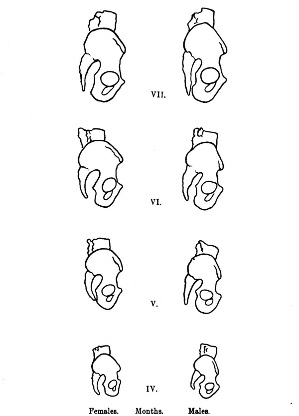
In passing to the consideration of the cavity of the true pelvis, the reader’s attention is directed to the photographs shown on P1. XIV., and to the diagrams on p. 369. He will see at a glance that even during the foetal condition the configuration of the parts is as pronounced as what we see in the adult. Apart from the differences in the outline of the inlet, to which attention has been already directed, he will notice that in the male the walls of the true pelvis encroach much more on the cavity. This is due to their greater obliquity, as shown in fig. 1, and also to the fact that the ischial spines are more inturned, and therefore brought closer together, thus giving to the entire pelvis a funnel-shaped appearance, as contrasted with the female. These observations are supported by a reference to the measurements, wherein the average width between the ischial spines in the female foetuses is 14 mm., as compared with 12 mm. in the males. Contrasting the width between the ischial spines and the transverse diameter of the brim, assuming the latter=100, we get from Verneau’s figures the following proportions : Trans. of Width between I d Brim. Ischial Spines. “ 9”’ Verneau— Males, . . 130 : 90 : : 100 : 69".? Females, . 135 p : 108 : : 100 : 80
Comparing the foetal pelves with the above, we find the proportions as follows :—
Males, . . 21-2 : 12 :; 100 ; 56-6 Females, . 20'8 : 14 :: 100 : 673 The difference between the male and female proportions is in the adults (Verneau) 10°8, whilst the foetal pelves yield an almost similar figure of 107. The relative width between the ischial spines is, however, greater in the adult than in the foetus, and this may be explained by the fact that, unlike most of the other spinous prominences on the innominate bone, the ischial spines are not provided with secondary epiphyses} so that we may assume that their growth ceases at a much earlier period, with the consequent effect of proportionably increasing the width between them in the adult.
The reaction of the slope of the pelvic walls on the outlet is also seen in the photographs and figures. For reasons already stated, I have disregarded the influence of the coccyx on the antero-posterior diameter, although in each figure it appears as it is shown in the photographs. As regards the contours of the anterior part of the pelvic outlet, the differences in form and size are well seen, and will be considered at a later stage, when the differences in the formation of the pubic arch are discussed; meanwhile, it may be pointed out that the width between the ischial tuberosities is greater in the female foetal pelvis, 20"? mm., than in the male, 196 mm.
Concerning the depth of the pelvic cavity, the measurement in the male foetus, 198 mm., exceeds that of the females, 188 mm., taken from the brim near the pectineal eminence to the most depending part of the ischial tuberosity.
The proportion of the depth of the pelvic cavity in the adult
- 1 I am aware that such an epiphysis has been described, but I have failed to ascertain its existence in all the specimens I have examined.
to the total. height of the pelvis, taking the latter = 100, as. deduced from Verneau’s measurements, is as follows : Total Height Depth of
Verneau‘ of Pelvis. True Pelvis. Index’ Males, . . 220 mm. : 107 mm. : : 100 : 48‘6 Females, . . 197 ,, ; 93 ,, :; 100 : 47-2'
Comparing this with my own measurements on the foetal pelvis, We find that—
Males, . . 42 mm. : 19-23 mm. :5 100 :.47-1 ‘Females, E . . .41 ,, : 18'8 ,, :: 100 : 458
thus proving that there is little disturbance of these proportions during subsequent growth.
A glance at the photographs and diagrams will enable the reader to realise that there is a marked difi"erence between the angle of the pubic arch in the male and female foetuses, despite the many statements to the contrar 31 and directly opposed to the opinion expressed by Schauta,2 that the pubic arch first assumes its characteristic form in the female about the age of 12 or 13. Owing to the small size of the foetal specimens, considerable difliculty was experienced in measuring this angle. It was found that it could be much better estimated from the photographs by the use of a projector; and whilst this method may be open to criticism, it may be pointed out that the same sources of error are common to both the male and female series and further, that the results obtained had been checked by reference to the wet preparations, and found sufficiently/aqgqgate for all practical purposes. The sexual difference, how tar", is : s',o.~ marked, that it can easily be recognised without w e,_.§ridC-° of} instruments when the male and female specimens are ‘laced side by side (see Pl. XIII. and fig. 1). The average sub-pu '°*'°‘“ "‘ / in the females is about 68°, as contrasted with 50° in the males. Verneau estimated the angle at 74° in the females and 60° in the males. Comparing these results, it will be evident that though there is an increase in the angle with the growthof the pelvis, the sexual difference is maintained throughout; for whilst -the difference between the average angles of the male and female foetal pelvis amount to 18°, the difference in the adult, as shown by Verneau’s figures, amounts to 14°. In this connection it is interesting to note the maxima and minima in both sexes. According to Verneau, the male angle ranged from 38° to 77°, the female from 56° to 104°; in my own specimens the extremes were 46° and 56° for the males, and 60° and 76° for the females, -—the female minimum exceeding the male maximum. Wewmust ‘therefore regard it as established that, in degree at least, the sexual differences of the sub-pubic arch are as well marked in the foetus as in the adult.
1 Wood, Todd’s Cyclopcedia. Lusk, Science and Art of Midwvfery. Galabin, Manual of Midwz_'fe7y. 3 Mii1ler’s Handbuch der Geburtshiil/‘e.
VOL. XXXIII. (N.S. VOL. x111.) 2 13
Another feature in which the adult female pelvis differs from the male is the form and size of the great sacro-sciatic notch. This in the female is usually wider and shallower than in the male. The form of the notch depends on the relation of the sacrum to the innominate bone, its shape being modified by the degree of curvature of the sacrum. The width of the notch necessarily depends on the degree of separation of the two bones which form its boundaries in front and behind, but it must be noted that this reacts on the shape of the ilium, the lower free border of which forms the summit of the notch. Here a characteristic difference is met with in the two sexes. If this part of the ilium be measured from the posterior inferior iliac spine, in correspondence with the external margin of the sacrum to the anterior margin of the great sacro-sciatic notch, the length in the females will, on an average, be found to exceed considerably the corresponding measurement in the males. The average length of this part of the ilium, which I measured in eight adult females, was 49 mm., as contrasted with 40 mm., the average which I obtained from five males. The nearest approach to this measurement which I can find in Verneau’s tables is the distance between the ischial spine and the posterior inferior iliac ‘spine :. this for the males averaged 50, whilst the females yielded a mean of 51. Obviously, however, this measurement is not the same as that to which I have already drawn attention, nor is it one which appears to me at all satisfactory, as the spine is liable to much individual variation, both as regards length and direction, nor does the slight difference shown between the figures for the two sexes at all invalidate the conclusions derived from my measurements, for they are in reality different sides of a ‘triangle, the angles of which are formed at points in correspondence with the posterior inferior iliac spine, the ischial spine, and the angle formed by the anterior confluence of the anterior margi of the great sacro-sciatic notch and its upper border.
Fig. 3. The increased widthof the hinder portion of the ilium is neces sarily correlated with a greater breadth of the sacro-sciatic notch.
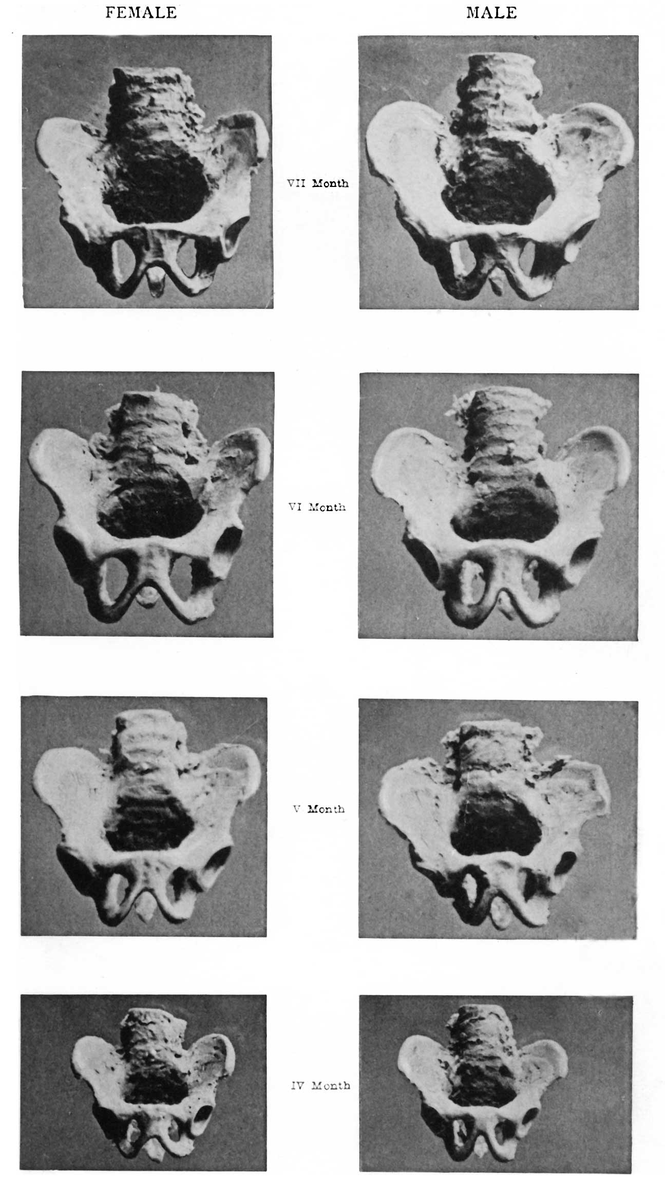
This, as Testut states, measures 72 mm. across in Women as: compared with 60 mm. in males, a result in harmony with the figures given above relating to the adult pelves I examined. I am aware that grave objection may be taken to any measurements of the Width of the sacro-sciatic notch in foetal pelves, on the grounds that the parts are so pliable that slight compression, may give rise to very considerable differences in the diametersFurther, it is necessary to remember that the measurements are so small that it is next to impossible to estimate the slight differences with accuracy. For these reasons I have not attempted to record this difference by figures, but will content myself with a reference to the photographs and diagrams carefully constructed therefrom (Pl. XV. and fig. 3). The reader will have no difficulty in recognising the fact that in the four female foetal pelves here represented the sacro-sciatic notch, and with it the hinder part of the ilium, is distinctly Wider than in the male.
I confess I Was hardly prepared to be able to demonstrate this sexual difference in the foetus, but it seems to me to have a very far-reaching influence on the subsequent development of the pelvis. If the forms of the male and female adult innominate bone be compared, it will be seen that in the female the Width of the posterior part of the ilium is greater than in the male ; unfortunately, it hardly seems possible to estimate this proportional difference in the adult for comparison with the same proportions in the foetus ; but the fact I wish to emphasise is, that this difi"erence exists even during foetal life, and that during subsequent growth it is maintained, and possibly emphasised; and that it is not to be attributed to the exercise of influences such as pres sure, muscular action, etc., which have been supposed in the past to act in some specially selective Way in the female pelvis, whilst, for some obscure reason, the males have been exempt. from the operation of these same forces.
From a consideration of the facts already stated, it has, I trust, been sufficiently pointed out that in many of its forms and proportions the foetal pelvis conforms very closely to the adult. The greatest difi°erence is met with in the innominate indices, and in the proportion of iliac width to pelvic height The increase in the pelvic diameter, especially those of the brim, have been in the past particularly ascribed to the rapid increase in width of the sacrum. It was assumed——without, so far as I know, any fact to support it—that the sacrum was relatively narrower in the foetal pelvis. Not only is this not the case, but I trust I have been able to show that its width is proportionately -greater to the rest of the pelvis than in the adult (see p. 371). Such being the case, the explanation that the increase in the transverse diameter is due to its great proportional increase in width necessarily falls to the ground. It is in the posterior part of the ilium that growth most rapidlyoccurs, thus leading to greater proportional increase in the antero-posterior diameters of the
pelvis, as well as augmenting its transverse width; but the fact that this difference in the form of the ilium is already clearly displayed in the foetus proves that during growth the relative proportions of the sexes are maintained throughout. Hubert and Valerius,[1] in discussing the growth of the pelvis, describe the "transverse diameter of the pelvic canal as increasing by the horizontal evolution of the sacrum and the hind parts of the ilwla, the antero-posterior by those of the iliac bones alone.
So far, I have confined myself to the description of those features of the foetal pelvis which are characteristic of sex. I have not discussed the changes which take place in the position of the sacrum, the development of the sacral promontory, and the obliquity of the pelvis. These lie outside the scope of the present inquiry, and doubtless are largely dependent on mechanical cause associated with the assumption of the erect position. But what I do desire to insist upon is, that these influences operate alike in the two sexes, and are in no way responsible for the remarkable differences met with in male and female adult forms, these being due to an inherent difference in the forms of the bones and cartilages.
Another feature of possible importance is suggested by a consideration of the photographs in Plate XV. As will be seen, the pelves have been placed for comparison in a position in which the anterior superior iliac spines and the symphysis pubis occupy the same vertical plane. Now, a comparison of the two seriesreveals the fact that in the females the lumbar part of the columna appears to curve upwards and backwards, whilst in the males it is more inclined forwards. This circumstance, which can hardly be considered accidental, appears to suggest that the mode of articulation of the ilium with the sacrum differs in the two sexes, a fact no doubt correlated with the difference in the form and size of the hinder portion of the ilium already alluded to, and would appear to indicate that even in foetal life therewwas a characteristic difference in the obliquity of the pelvis of thetwo sexes such as maintains in the adult. It would be unwise, however, to draw any conclusions from so small a number of specimens, though my examination of other foetal pelves appears to confirm this view.
To sum up, it appears from a consideration of the foregoing facts that during foetal life the essentially sexual characters are as well defined as they are in adult forms, and that any differences that occur during growth between the adult and foetal forms, due it may be to the influence of pressure or muscular traction, affect both sexes alike, and that such influences are in no way accountable, as has been maintained, for the characteristic features of the pelves of the female as contrasted with the male.
Note. — Since the foregoing was written, I have had an opportunity of consulting Fehling’s paper, but I have considered it advisable, under the circumstances, not to alter my communica tion, but to present it in its present form, as evidence confirma.tory in the main of Fehling’s views.
- ↑ Memovlres de l’Academie Royale de Médecine de Belgique, tome iv.
Plates
Plate XIII. Female Male
VII Month
VI Month
V Month
IV Month
Plate XIV. Female Male
VII Month
VI Month
V Month
IV Month
Plate XV. Female Male
VII Month
V1 Month
V Month
IV Month
Cite this page: Hill, M.A. (2024, April 23) Embryology Paper - The sexual differences of the fetal pelvis. Retrieved from https://embryology.med.unsw.edu.au/embryology/index.php/Paper_-_The_sexual_differences_of_the_fetal_pelvis
- © Dr Mark Hill 2024, UNSW Embryology ISBN: 978 0 7334 2609 4 - UNSW CRICOS Provider Code No. 00098G


