Paper - The morphogenesis of the systems of juxta-aortic tissues in human embryos
| Embryology - 27 Apr 2024 |
|---|
| Google Translate - select your language from the list shown below (this will open a new external page) |
|
العربية | català | 中文 | 中國傳統的 | français | Deutsche | עִברִית | हिंदी | bahasa Indonesia | italiano | 日本語 | 한국어 | မြန်မာ | Pilipino | Polskie | português | ਪੰਜਾਬੀ ਦੇ | Română | русский | Español | Swahili | Svensk | ไทย | Türkçe | اردو | ייִדיש | Tiếng Việt These external translations are automated and may not be accurate. (More? About Translations) |
Jimenez-Castellanos J. The morphogenesis of the systems of juxta-aortic tissues in human embryos. (1949) Q Bull Northwest Univ Med Sch. 23(4):428-31. PMID: 18148736
| Historic Disclaimer - information about historic embryology pages |
|---|
 |
The Morphogenesis of the Systems of Juxta-Aortic Tissues in Human Embryos
- Contribution no. 517 from the Department of Anatomy, Northwestern University Medical School. Received for publication, June 1. 1949.
Juan Jimenez-Castellanos
From the Department of Anatomy. University of Granada, Spain, and Northwestern University Medical School.
Introduction
The chromaffin system of tissues has been reviewed as to origin, morphogenesis, relationships and functions by Kohn (1), Zuckerkandl (2), Iwanow (3), and Hollinshead (4). Reconstructions of the juxta-aortic bodies have been made in various mammals. Kohn (1), Zuckerkandl (2), and Iwanow (3) have presented planographic reconstructions of this system in human embryos and fetuses of various ages, and Wrete (5) has presented models prepared from human embryos of 38 and 39 mm.
In order to contribute to the better understanding of the sympathochromaffin system in the human embryo, three stages in the morphogenesis of this system have been reconstructed planographically. The two older stages have also been reconstructed as wax models. The three embryos so utilized were of 9 mm. (greatest length), 14 mm. (greatest length) and 40 mm. (crown-rump length). It was judged that stages intermediate between 14 and 40 mm. did not offer sufficient differences to warrant separate consideration. The 9 mm. embryo was fixed in Zenker’s solution; the 14 mm. and 40 mm. specimens were fixed in 20 per cent formalin. All three embryos were embedded in paraffin, sectioned transversely at 10, 11 and 30 micra, respectively, and stained in hematoxylin and eosin. The original models and the planographic reconstructions were made at magnifications of 22.5 diameters (40 mm. embryo) and 62.2 diameters (14 mm. embryo). The original planographic reconstruction of the 9 mm. embryo was made at a magnification of 156 diameters.
This investigation was carried out in the laboratory of Prof.-Dr. José Escolar at the University of Granada, to whom I wish to express gratitude for many excellent suggestions. The author is also grateful to Dr. John Jacobs and Dr. Alberto Vaz Ferreira of Northwestern University Medical School for their kind assistance in preparing the original manuscript in English and to Professor L. B. Arey, who has recast that manuscript into its present form.
Observations
1. Embryo of 9 mm
At this stage the reconstruction (fig. 1) shows that the suprarenal glands have not yet appeared, the metanephroi are at the beginning of their development, the mesonephroi are fully developed and the gonads are prominent, elongate folds along the ventromedian surfaces of the mesonephroi. The ganglionated sympathetic cords descend dorsolateral to the aorta, and near the cranial ends of the mesonephroi gradually disappear as such and become continuous with paired juxtaaortic masses. These latter extend caudad ventrolateral to the aorta, and in their more caudal extents interconnect by three bridges ventral to the aorta.
In the sections the juxta-aortic tissue shows as masses that stain deeply with hematoxylin (fig. 4).
2. Embryo of 14 mm
As the reconstruction (fig. 2) shows, this stage is marked by the increasing prominence of the metanephroi and by the suprarenal glands, which not’ only have appeared since the previous stage but also have grown to relatively large size. The juxta-aortic substance differs from that in the previous stage by being dispersed into 31 discrete masses. These lie both in front of the aorta and on both sides of it; indeed, some that are not shown in figure 2 are even included in the interior of the suprarenals themselves. In the region located between the suprarenals and between the cranial halves of the metanephroi, there are numerous small masses, the more caudal ones tending to be larger than those at higher levels. Between the caudal halves of the metanephroi there are two masses, much larger than any others in the embryo. These are the forerunners of the aortic bodies of Zuckerkandl. The sympathetic cords continue caudad beyond the region reconstructed. The splanchnic nerves also descend to the level of the juxta—aortic masses and end in association with them; the nerve fibers, however, do not actually enter the cellular masses.

|

|

|
| Fig. 1. Planographic reconstruction of the juxtaaortic masses and the adjacent organs of a human embryo of 9 mm. :3 5.9. (1) metanephroi; (2) mesonephros; (3) gonad; (4) sympathetic cord; (5) juxta-aortic body; (6) post-cardinal vein; (7) coeliac axis; (8) superior mesenteric artery. | Fig. 2. Planographic reconstruction of the juxtaaortic masses and the adjacent organs of a human embryo of 14 mm. :c 47. (1) Aorta; (2) coeliac axis; (3) superior mesenteric artery; (4) middle sacral artery; (5) common iliac artery; (6) metanephros; (7) suprarenal gland; (8) mesonephros; (9) gonad; (10) sympathetic cord; (1 1) splanchnic nerve; aortic bodies (of Zuckerlcandl). | Fig. 3. Planographic reconstruction of the juxtaaortic masses and the adjacent organs of a human fetus of 40 mm. :13 8.5. (1), (2), Vertebral body and arch; (3) aorta; (4) common iliac artery; (5) renal hilus; (6'), (7'), suprarenal glands; (8) superior mesenteric artery; (9), inferior mesenteric artery; (10), (16), (24), aortic body; (11) middle sacral artery; (12) common iliac vein; (13) inferior vena cava; (14), (17), (20), renal vein; (15), (19) vein from aortic body; (18) left suprarenal vein; (21) splanchnic nerves; (2.?) right vagus nerve; (23) sympathetic cord. |
In sections (fig. 5) there is no particular tinctorial difference between the large inter-renal masses and the small masses located between the suprarenal glands. Both stain deeply with hematoxylin. Certain of the small masses can be observed in the process of being incorporated into the interior of the suprarenal glands. This, of course, is a normal initial stage in the development of the suprarenal medulla.
3. Embryo of 40 mm
The distribution or Juxta-aortic tissue is much as in the previous stage (fig. 3). Mostly lateral to the aorta, and roughly coincident with the extent of the suprarenal glands, are small masses composed of small cells that stain deeply with hematoxylin. In size and stainability, at least, they resemble closely the syrnpathoblasts in the ganglionated sympathetic chains. These latter cells, likewise, are not far along in the course of differentiation into ganglion cells. All of these small juxtaaortic masses that were examined for the purpose were proved to be in intimate relationship with the terminations of fibers from the splanchnic and right vagus nerves. It is possible, under high magnification, to observe the nerve fibers penetrating into the interior of such masses.
There are two large inter-renal masses which represent the future aortic bodies. These are elongate and irregularly lobed; they interconnect, ventral to the aorta, by two prominent bridges of tissue. A similar, smaller mass is displaced down the middle sacral artery. All three bodies stain more weakly than do the smaller masses at higher levels, already mentioned. Their paler color with hematoxylin is like that observed in the most central part of the suprarenal gland.

|

|

|
| Fig. 4. Photomicrograph through a human embryo of 9 mm. at the level indicated in fig. 1. :1: 59. (1) Aorta; (2) posterior cardinal vein; (3) mesonephros; (4) gonad; (5) liver; (6) juxtaaortic body; (7) body of vertebra. | [[[:File:Jimenez-Castellanos1949 fig05.jpg|Fig. 5]]. Photomicrograph through a human embryo of 14 mm. at the lower level indicated in figure 2. 1: 47. (1) Metanephros; (2) juxtaaortic body; (3) sympathetic cord; (4) aorta; (5) mesanephros; (6) body of vertebra. | [[[:File:Jimenez-Castellanos1949 fig06.jpg|Fig. 6]]. Photomicrograph through a human fetus of 40 mm. at the middle level indicated in fig. 3. ac 15. (1) Suprarenal gland; (2) aorta; (3) body of vertebra; (4) duodenum; (5) metanephros; (6) crus of diaphragm; (7) sympathetic cord; (8) right splanchnic nerve; (9) masses of sympathoblasts; (10) paraganglia; (11 ) renal vein; (12) inferior vena cava. |
Intermediate between the two levels already mentioned is a zone containing small masses that have a double structure (fig. 6). The main material of such a complex consists of the larger, pale type of cell; at the periphery are aggregations of the smaller, dark type of cell.
Discussion
This study sheds no light on the origin of the juxta-aortic tissue since at 9 mm. it is already present as paired, continuous masses. At this period this common substance is not visibly differentiated in its definitive directions: ganglion tissue of the coeliac plexus; suprarenal medulla; and the chromaffin tissue of the aortic paraganglia. This lack of differentiation is in accord with the observations of other investigators and is not only shown by routine methods of staining but also by the application of specific methods for chromaflin tissue and by silver procedures.
The 14 mm. stage likewise fails to provide information concerning divergent differentiation in the common juxtaaortic tissue. It does, nevertheless, illustrate the subdivision of the common primordia into multiple small masses superiorly and two large inter- renal masses (aortic bodies of Zuckerkandl) that lie at a more caudal level. Likewise, the invasion of some smaller masses (future suprarenal medulla) into the cortical primordium has begun.
At 40 mm. some specialization, as indicated by staining differences with hematoxylin, can be detected readily; Zuckerkandl (6) was the first to notice this divergent stainability. The sympathoblasts of the small, cranial masses are larger, paler elements. These are coming into relationship with nerve fibers, all of which are preganglionic elements (Hollins head, 4). The small masses at an intermediate level between the ganglionic primordia and the aortic bodies tend to show representations of both cell types, as do also the later paraganglia.
Summary
Planographic reconstructions have been made of the sympatho-chromaffin tissue in human embryos of 9, 14 and 40 mm. The two older stages were also modelled in wax.
The 9 mm. stage possesses two elongate, interconnecting juxta-aortic masses which consist of small, dark-staining cells.
At 14 mm. the suprarenal glands are beginning to be invaded by some of this tissue which also has subdivided into many small masses cranially and into two large masses located caudally between the metanephroi.
At 40 mm. the differentiation has taken three directions: (1) suprarenal medulla; (2) sympathetic ganglia; (3) paraganglia; (including the aortic bodies of Zuckerkandl). The cells of the future suprarenal medulla and paraganglia are large and stain palely. The small masses, located cranially (between the suprarenal glands), consist of small, dark-staining cells that are becoming similar to the sympathoblasts. In a location intermediate between these two extremes are compound, small masses containing both kinds of cells.
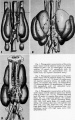
Fig 1-3
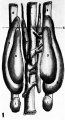
Fig 1
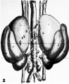
Fig 2
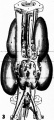
Fig 3
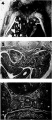
Fig 4-6
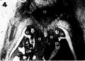
Fig 4
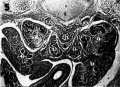
Fig 5
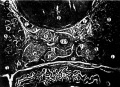
Fig 6


References
1.Kohn, S.: Die Paraganglien, Arch. f. mikrAnat., Bd. 62, S. 263-365, 1903.
2. Zuckerkandl Z. XV. The development of the chromaffin organs and of the suprarenal bodies pp157-179 in Keibel F. and Mall FP. Manual of Human Embryology II. (1912) J. B. Lippincott Company, Philadelphia.
3. Iwanow, G.: Das Chromafline and Interrenales System des Menschen, Ergeb. d. Anat., Bd. 29, S. 87-280, 1932.
4. Hollinshead, W. H.: Chromaflin Tissues and faigganglia, Quart. Rev. Biol., 152156-171,
5. Wrete, M.: Beitrag zur Kenntnis von der Entwicklung des Chromaffinen Gewebes der Bauchregion beim Menschen, Zeitschr. f. mikr.-anat. Forsch., Bd. 9, S. 79-98, 1927.
6. Zuckerkandl, E.: Ueber Nebensorgane des Sympathicus in Retroperitonealraum des Menschen, Anat. Anz. (Ergéinzrheft), Bd. 19, S. 95107, 1901.
Cite this page: Hill, M.A. (2024, April 27) Embryology Paper - The morphogenesis of the systems of juxta-aortic tissues in human embryos. Retrieved from https://embryology.med.unsw.edu.au/embryology/index.php/Paper_-_The_morphogenesis_of_the_systems_of_juxta-aortic_tissues_in_human_embryos
- © Dr Mark Hill 2024, UNSW Embryology ISBN: 978 0 7334 2609 4 - UNSW CRICOS Provider Code No. 00098G