Paper - The formation of the duodenal curve
| Embryology - 1 May 2024 |
|---|
| Google Translate - select your language from the list shown below (this will open a new external page) |
|
العربية | català | 中文 | 中國傳統的 | français | Deutsche | עִברִית | हिंदी | bahasa Indonesia | italiano | 日本語 | 한국어 | မြန်မာ | Pilipino | Polskie | português | ਪੰਜਾਬੀ ਦੇ | Română | русский | Español | Swahili | Svensk | ไทย | Türkçe | اردو | ייִדיש | Tiếng Việt These external translations are automated and may not be accurate. (More? About Translations) |
Frazer JE. The formation of the duodenal curve. J Anat. 1919 53(4):292-7. PMID 17103870
| Historic Disclaimer - information about historic embryology pages |
|---|
| Pages where the terms "Historic" (textbooks, papers, people, recommendations) appear on this site, and sections within pages where this disclaimer appears, indicate that the content and scientific understanding are specific to the time of publication. This means that while some scientific descriptions are still accurate, the terminology and interpretation of the developmental mechanisms reflect the understanding at the time of original publication and those of the preceding periods, these terms, interpretations and recommendations may not reflect our current scientific understanding. (More? Embryology History | Historic Embryology Papers) |
The Formation of the Duodenal Curve
BY J. Ernest Frazer, F.R.C.S., St M ary’s Hospital, Professor of Anatomy in the University of London
Introduction
In a paper dealing with intestinal rotation, and written a few years ago, Dr R. H. Robbins and I showed that the duodenum is not involved in this rotation, and we therefore treated the production of its curve very shortly. We pointed out that this portion of the intestine is a.t first very short, and that it is subsequently elongated in accordance with, and curved out by, the growth of the head of the pancreas. From an early stage it is attached to the dorsal wall by the mesoduodenum, in which the head of the gland grows, and the bowing out of the tube takes place round this base of fixation; we stated, however, that we were not certain whether there might not be some later secondary fixation of the extreme terminal part of the gut known as the duodenum. I propose now to give some further details bearing on the matter.
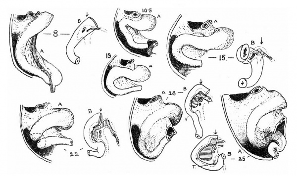
Fig. 1. The duodenum throughout the “first stage” of intestinal development. A, side view, B, front view. The numerals give the size of embryo in millimetres. See text.
In the figures I have illustrated the parts seen at several stages. The younger specimens were mostly drawn from models, the later ones directly.‘ The Formation of the Duodenal Curve 293
In the former the duodenum is shown from the left (.41.), but in some cases additional projections from the front are added (B.). Measurements (sittingheight) are given in numbers of millimetres.
At 8 mm. (fig. 1) the duodenum can be recognised as that part held by the mesoduodenum, and is a short and nearly straight tube directed caudally and dorsally, and likewise towards the right, as can be seen in the frontal projection. It is continuous below with the intra-abdominal portion (d'uodeno— umbilical loop) of the short U-loop of the gut.
The mesoduodenum may be defined for our present purposes as a thickened region of the common median dorsal mesentery, supporting that part of the tube immediately distal to the stomach. It occurs just below the pouch of the bursa omentalis, of the opening of which it forms the lower boundary or floor. Hence the lower part of the opening is seen in the drawing just above the mesoduodenum, and the vitelline vein, passing through the mesoduodenum behind and to the left of the gut, runs up to the liver (portal vein) in front of the bursal opening; that is, the upper “edge” of the mesoduodenum becomes continuous, dorsal to the line of the tube, with what is the free edge of the gastro-hepatic omentum. Caudally, the mesoduodenum stands out well, and a small recess (inter-mesenteric recess) exists between its rounded caudal border and the median mesentery; this recess persists to a late period.
In the frontal projection the position and attachment of the mesoduodenum is shown by a dotted line, and its continuity with the bursal floor is seen on the one hand, and with the general mesentery on the other, with the intermesenteric recess intervening. The duct openings are indicated by dots, with the small pancreatic growths associated with them; the body of the gland is seen to be already invading the bursal wall, but the head, very small, is in the mesoduodenum itself. The vitelline vein is not shown, but runs behind and to the left of the upper duct opening.
In the 10-5, 13 and 15 mm. specimens there does not seem to be much growth of the duodenum apart from the general increase in size of the parts, but there is a more pronounced dorsal direction. This seems to be due to growth of the stomach, and possibly of the liver; the frontal projection of the 15 mm. state appears to show this, for the gastric end is more to the right, and the duodenal direction seems to be horizontally to the dorsum and inwards. The mesoduodenum is apparently a little broader, but the head of the pancreas has not grown at the same rate as its body, for this last stands up well towards the floor of the bursal opening and is just apparent above and behind the duodenum.
The 22 mm. stage shows a definite duodenal curve, convex to the right, seen in the front view; an 18 mm. model, not drawn, shows a much less marked tendency toward the same curve. This curve is evidently associated with the decided increase in size of the head of the pancreas, which now occupies the greater part of the right portion of the mesoduodenum, extending down in it but not reaching its lower end. The duodenum now stands out to the right to some extent from its attachment, rolled out, so to speak, by the growth on its inner side, so that the duct openings are more definitely posterointernal than in previous stages. The tube has increased correspondingly in length; the first increase would seem to be mainly in its proximal part, but, as the head grows, the portion distal to the ducts extends more rapidly. Its distal end is turned in toward the mesentery and joins the duodeno-umbilical loop through a twisted U-shaped kink, which I have already shortly discussed in the previous paper and need not notice further.
The head is larger in the 28 mm. specimen, and the curve more pronounced in correspondence, but its effect is spoilt by the absence of the U-shaped kink distally; in the direction of the tube there is a decided indication (.21.) of the end of the duodenal region, and it is quite possible that there may have been a bend below this in the fresh state, but in the model the tube passes directly, at the site of the angled junction, into the duodeno-umbilical loop. The head of the pancreas is now for the first time apparent ventrally, and this is necessarily associated not only with the opening out of the duodenal curve but with lessening of the general dorsal direction of the tube.
A marked advance is seen at 35 mm. Here the duodenal curve would at first sight appear almost complete, and is associated with a large pancreatic head; as we shall see shortly, such reading of the appearance would not be correct, but at any rate it is not unlikely that the specimen exhibits, as an individual variation, a state rather more advanced than others at this stage. The 40 mm. specimen in fig. 2 shows a more angled curve, with a less wide head, but the depth of the head is well-marked. Treitz’ band (T.) is indicated in the frontal figure of 35 mm. I have not been able to recognise it definitely at an earlier stage. Its situation here shows the fallacy of appearances in this specimen, for it is evident that the hinder limb of the “kink,” as seen in the frontal draving, is not the ascending piece of the duodenum; this will have to be formed later and more proximally, and is indicated in the 40 mm. specimen.
In this, as in the others, the intermesenteric recess is evident, and its presence shows conclusively that the duodenum remains attached to, and only held by, the mesoduodenum; as it curves out, it stands out from the mesoduodenum, which is only continuous with it near its inner edge, except at its upper end. The curve is evidently due to the growth of the mesoduodenum and pancreas; the former enlarges ahead of the latter in its lower part, as may be seen in the figures, and the part as yet unoccupied by the pancreas contains a vascular plexus which is the forerunner of the lower pancreaticoduodenal loops, and quite distinct from the vascular supply to the next succeeding part of the gut.
We can see then that the duodenal curve is well advanced in formation before the bowel enters the abdomen and undergoes its “rotation” therein. This occurs about 40 mm.; in the 40 mm. specimen figured the gut was in the belly, a position which it had probably just taken, perhaps even during preparation, but manifestly the state of the duodenal curve has nothing to do with this. The model shows, however, that the duodenum as a whole had moved somewhat to the left, swinging on the dorsal attachment of the mesoduodenum.
This is easily understood and to be expected. The previous figures and those in the original paper show that the “kink” in the duodeno—jejunal region is pressed against the median mesentery (mesocolon). When this structure moves dorsally and to the left as a result of the entry of intestinal coils from the umbilicus, it not only permits the region of the “kink ” to follow in the same direction, but, through its continuity with the mesoduodenum, tends to swing this somewhat also to the left; hence the changed relation, especially of the distal part, to the middle line as indicated by the arrows in the frontal views.
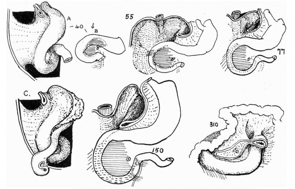
Fig. 2. Side and front views of 40mm. model. Front views of older specimens. 0, a semidiagrammatic drawing to show position of colon at end of second stage. In the later stages shown, pancreas is indicated by horizontal shading, adhesions by oblique shading and interrupted lines.
As in many other parts of the body, individual variations become more apparent as the specimens become older, and the later history of the duodenum, including its final fixation, is not so smoothly written in the foetus as the earlier stages seem to be in the embryo. I have put in fig. 2, however, a few drawings from the dissections I have made which will give, I hope, a fairly accurate if rather general idea of these later happenings.
The progressive enlargement of the duodenal curve goes on as before, with variations. The few stages shown illustrate this and the association between it and the pancreas and mesoduodenum. But the secondary attachments of this portion of the gut are still more variable in their occurrence; presumably they wait for their time till the curve is complete, and then find the governing factors variable — but what these factors may be, other than the teleological call for support mechanically, I have not been able to ascertain.
The commencement of the ultimate fixation occurs when the (originally umbilical) colon comes back across the “neck” of the intestinal mesentery and thus lies in front of the duodenum. It seems to be fairly constant (fig. 2, c.) in its position here, so far as my observations go, and is found running downwards and outwards, parallel to and just below the upper part of the duodenum, and then crossing it to come into relation with (caecum) the kidney. If this drawing is compared with that of the 40 mm. specimen above it, it is clear that the site of crossing is opposite the lower part of the mesoduo W denum as seen from the right. Here occurs the first secondary fusion affecting the duodenum, soldering it and colon and kidney together at this level. In all my specimens the colon has not come into position at a lower level, so that, depending on these, it seems justifiable to regard the original right free surface of the mesoduodenum as being visible in the adult above the level of the crossing colon. This surface extends up to the margin of the opening into the omental bursa, as seen in the drawing. The bursal opening is not the foramen of Winslow, but is marked, .so far as its floor is concerned, by the lower pancreatico-gastric fold[1]; in the embryonic state the hepatic artery lies in the lower edge of the opening and has the base of the intra-bursal liver process resting on it (papillary process of Spigelian lobe). The upper part of this surface of the mesoduodenum, then, is continued through the floor of the foramen of Winslow to the line of the hepatic artery. The foramen of Winslow is a late formation, probably due to the increase in size of the inferior cava, and possibly also of the liver and pancreas; this can be understood from the figures, and no further stress need be laid on it or on the simultaneous addition of a “vestibule” to the small sac, not derived from the omental bursa.
The lower aspect of the mesoduodenum, however, remains unattached behind the transverse part of the duodenum, which is fastened to its front and lower margin. This may be understood from the 55 mm. drawing, where the mesoduodenum evidently comes down to a level lower than that of the crossing colon, and an open groove or sulcus underlies the duodenum distal to this line. This is the same as saying that, beyond the level of the colon, the duodenum is free from attachment on its posterior surface.
Increasing areas of attachment are indicated in the 77 and 150 mm specimens, and, speaking generally, the adhesions seem to be developed first near the colon and to extend distally. One cannot recognise the intermcsenteric recess with certainty as soon as the adhesions affect the ascending portion of the tube, but before this occurs it is quite distinct (60-70 mm) and shows definitely that the duodenal curve is completed round the mesoduodenum and head of the pancreas. In the 77 mm drawing there is a recess of doubtful value; probably the true recess has been obliterated by the adhesions which have taken place behind the gut.
No useful purpose would be served by describing or figuring the various adhesions met with in the foetuses examined. The general conclusion arrived at was, as already intimated, that these were last formed and most variable in the region of the ascending part; two are shown in the 150 mm. specimen, overlying the inferior mesenteric vein, of which the immediate causation is not Very evident, although the mechanical call for their existence may be plain enough. The connection with the common folds and fossae in the neighbourhood is obvious. In the 310 mm. specimen there is a marked and extensive adhesion below the first half of the transverse part, which would become continuous[2] with the mesentery when this is attached to the front of the duodenum; a sulcus, unmodified by any adhesions, lies behind the rest of the duodenum.
This drawing is introduced, however, to show a secondary attachment of the highest part of the tube to the transverse mesocolon ; the true continuity between mesoduodenum, mesentery, and mesocolon lies to the right, where it is clearly seen. A small fossa (? Gruber’s fossa) separates the two folds. Evidently, in some cases at least, the adult duodenum may include, at its extreme end, a very short piece of gut which is not attached in the embryo by the mesoduodenum, and becomes fixed in position by means of secondarily acquired adhesions.
Enough has been said to show the possibility of explaining the occurrence and site of the different fossae—with the exception of the paraduodenal, about the origin of which I have nothing to advance—as mere variations in the adhesions formed in relation with the duodenum distal to the crossing of the colon. While the duodenum as a whole is curved out by the enlarging head of the pancreas and is primarily held only by the mesoduodenum, the lower part of it, below the crossing of the colon, is the part which projects most beyond the position of the base of attachment and is secondarily fixed by late adhesions. Immediately distal to the crossing of the colon these adhesions will, still later, be reinforced by the fixation of the mesentery across the face of the duodenum; this, as pointed out in the earlier paper, is really a secondary adhesion of the “mesocolon of the loop” — as is also in all probability the first adhesion at the level of the crossing. Thus, from the colon to the “line of attachment of the mesentery” across the duodenum, the gut is more firmly fastened down, and it is beyond this line that the variations in the secondary bands become apparent.
Footnotes
- ↑ I mention this fold in deference to the view that it marks the mouth of the bursa omenti majoris. My own View is that the band is produced later, and that the line of the hepatic artery is the only original and proper line to take.
- ↑ This fold in the 310 mm. foetus is probably adherent mesocolon and hence differs from the attachments shown in 150 mm. It would lie superficial to them and would not vary with them
Cite this page: Hill, M.A. (2024, May 1) Embryology Paper - The formation of the duodenal curve. Retrieved from https://embryology.med.unsw.edu.au/embryology/index.php/Paper_-_The_formation_of_the_duodenal_curve
- © Dr Mark Hill 2024, UNSW Embryology ISBN: 978 0 7334 2609 4 - UNSW CRICOS Provider Code No. 00098G


