Paper - The development of the seminal vesicles in man
| Embryology - 27 Apr 2024 |
|---|
| Google Translate - select your language from the list shown below (this will open a new external page) |
|
العربية | català | 中文 | 中國傳統的 | français | Deutsche | עִברִית | हिंदी | bahasa Indonesia | italiano | 日本語 | 한국어 | မြန်မာ | Pilipino | Polskie | português | ਪੰਜਾਬੀ ਦੇ | Română | русский | Español | Swahili | Svensk | ไทย | Türkçe | اردو | ייִדיש | Tiếng Việt These external translations are automated and may not be accurate. (More? About Translations) |
Watson EM. The development of the seminal vesicles in man. (1918) Amer. J Anat. 24(4): 395 - 439.
| Historic Disclaimer - information about historic embryology pages |
|---|
| Pages where the terms "Historic" (textbooks, papers, people, recommendations) appear on this site, and sections within pages where this disclaimer appears, indicate that the content and scientific understanding are specific to the time of publication. This means that while some scientific descriptions are still accurate, the terminology and interpretation of the developmental mechanisms reflect the understanding at the time of original publication and those of the preceding periods, these terms, interpretations and recommendations may not reflect our current scientific understanding. (More? Embryology History | Historic Embryology Papers) |
The Development of the Seminal Vesicles in Man
Ernest M. Watson
James Buchanan Brady Urological Institute, Johns Hopkins Hospital, and the Carnegie Laboratory of Embryology, Baltimore, Maryland
Twenty-Seven Figures
Introduction
In reviewing the literature on the embryology of the seminal vesicles one finds a surprising lack of any detailed or accurate study covering their course of development in foetal life. There are many references to the time of their initial appearance, but from that time until birth, or in fact until adult life, there are very few observations. The work of Pallin (’01) on the development of the prostate and seminal vesicles is unquestionably the most complete.
Kolliker (’79) stated that the Vesicles have their beginning in the third month of foetal life as offshoots from the proximal portion of the Wolffian ducts. Mihalkovics (’85) foundgthat at about the fifth month of intra-uterine life the evaginations from the deferent ducts, going to form the seminal vesicles, proceeded first in a horizontal, then in an upward curving direction. Minot (’92) stated that the Vesicles do not attain a length of 1 mm. until about the fifth month of foetal life. 0. Schultz C97) wrote that they appear at the end of the third month as outgrowths of the lower part of the Wolffian ducts, and are then simple, hollow, pear-shaped appendages about 1 mm. long. Heisler C99), in his text-book, stated that from the caudal end of the Wolfiian duct there arises a pouch-like evagination, which later becomes the seminal vesicle. Pallin (’01), reporting his studies on the development of the prostate and seminal vesicles in man and in the lower animals, found that they appear at the third month of foetal life. They are noted first as longitudinal folds constricted off ‘of the Wolffian ducts; in the fourth month they become bent in a ‘knee-fashion,’ with one portion horizontal and lateral, the other directed cranialward. He also found that the diverticula first appear at the end of the fourth month, and stated that the vesicles then have the general topography of the adult organ. The diverticula of the ampulla appear simultaneouslywith the diverticula of the vesicles, as longitudinal constricting folds. McMurrich (’02) said that at about the third month a lateral outpocketing appears from the wall of the vas deferens in the form of a longitudinal fold, which goes to form the seminal vesicle.
More recently Lowsley (’12), writing upon the development of the prostatic gland and briefly referring to the embryology of other structures about the neck of the bladder, stated that at the sixteenth Week of foetal life the seminal vesicles appear as tortuous tubes. He found them at this time surrounded by a thick muscular layer, their lumina connecting with each vas deferens by means of one small duct or opening directly under the internal sphincter. At birth each vesicle in its upper portion consists of five lumina with muscular walls as thick as those of the vasa deferentia. These lumina all communicate lower down, and the end duct finally communicates with the ejaculatory duct just below the point Where the musculature of the prostate gland is first encountered.
W. Felix (’12, Keibel and Mall’s Human Embryology, vol. 2, p. 938), states that the seminal Vesicles have their origin from the lower portion of the primary excretory duct which has become dilated to form the ampulla. They are formed by the constricting off, in a craniocaudal direction, of a simple epithelial tube. This constriction, which ends at the beginning of the horizontal portion, he first found in embryos of 60 mm. head-foot length.
The material for the present study Was obtained in part from the Embryological Collection of the Carnegie Institution and in part from the Obstetrical Service of the Johns Hopkins Hospital. The specimens were preserved in 4 per cent formalin, imbedded in paraffin and cut serially. The measurements recorded are crown rump length, and the ages of the specimens were determined according to the figures in the table of classification in Keibel and Mall’s Human Embryology. Before taking up the discussion of the various specimens it might be well to define the terms used to describe the direction of growth of the vesicles. Anterior refers to the cranial end of the body; ventral, to the region of the abdominal wall, and dorsal, to the back or vertebral aspect. The lateral measurements were taken by means of a micrometer eyepiece on a microscopic stage. The anterior posterior measurements Were made by counting the number of sections, which were all cut of a known thickness and every section saved for study. The reconstructed drawings of the eight specimens, showing their dorsoventral View in two diameters with varied configurations, were made from the superimposed serial sections. These sections were reproduced under a magnification of fifty diameters to insure greater accuracy, and transferred to millimeter paper from which the reconstructed diagrams were made. The drawings were then reduced to a size suitable for illustrations.
Fetuses about 80 mm long
This stage is represented by specimen no. 7680 of the Carnegie Collection, length 80.3 mm., and illustrated by figures 1, 2, and 3. In this specimen the beginning of the seminal vesicles and their relation to the ampullae of the deferent ducts, to the trigone, the venous plexus and the general musculature forming the neck of the bladder are first noted. Serial transverse sections were made of the specimen, each 6 p thick and stained with hematoxylin and eosin. Every section cut through the genitourinary tract, from the urachus to the anterior urethra, was mounted and studied separately.
At the level of the entrance of the ureters into the bladder wall the paired Wolfiian ducts are seen 011 either side of the midline, 1 mm. apart. At a distance of 2.4 mm. lateralward from these and slightly ventral are the openings of the ureters into the bladder Wall. The Wolffian ducts are well developed and oval-in shape, with the axis running from left to right. Their lumina measure each $x% mm., being twice as Wide in their lateral as in their dorsoventral diameters. The’ previously undifierentiated inesenchymatous tissue about the Wolflian ducts has at this stage become more specialized, and surrounds each duct in a more or less circular fashion at a distance of about 0.1 mm. from its lumen. In the beginning the different muscle layers are clearly defined, and are separated from the tissue entering into the formation of the outer coat of the bladder on the ventral side, and also from that going to form the peritoneum and fascial coverings of the ducts and vesicles on the dorsal side. The ducts are lined with mucous membrane composed of a layer of single cells with large nuclei situated at their base. Beneath this the basement membrane is distinctly formed, and is upheld by a single layer of mesenchymatous cells having a circular or ring—like formation, thus giving a semblance of their future form as the inner or circular muscular coat of the ducts. The ducts lie % mm. back of, or dorsal to the lumen of the bladder. At this site—the upper boundary of the trigone in the region of the interureteric ligament—the bladder wall is nearly twice as thick as at any other point and has a diameter of a trifle over % mm. The bladder at this stage of development is lined with mucous membrane in a double layer of cells and over the trigone with a triple layer, the cells having large, deeply staining centrally placed nuclei. The different muscle cells are now pretty well differentiated and the muscular coats can be well made out.
In their course down from the level of the upper limit of the trigone the Wolffian ducts gradually draw nearer together, each approaching the midline, until they come to lie about % mm. apart. At the same time they have noticeably increased in size, while their relative thickness has changed from one-half to two—thirds of their lateral diameter. In other words, they are now more nearly round. This increase, which amounts to an increase of nearly 40 per cent in the size of their lumen, corresponds to the future ampulla of the dcferent duct. At this stage the ampulla is a straight tube and, aside from this slightly increased dimension, corresponds in every Way to the deferent duct in other regions, there being no indication of any convolutions or diverticular formation. As the ducts approach each other at this site their surrounding mesenchymatous tissue, instead of being differentiated into two separate masses, each surrounding a duct, becomes fused into a single ovoid mass about the two duets, with a more deeply staining and closely packed ring of muscle cells about each duct. At this level the ducts, with their surrounding tissue, are well separated from the muscular coats of the bladder which are clearly differentiated into the several muscle layers. The presence, however, of a differentiation of the fascial coverings of the Wolfl’1an ducts, dorsally towards the peritoneum, cannot be seen, the undifferentiated mesenchyme extending well back to the peritoneum.
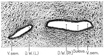
The earliest indication of the formation of the seminal vesicles is observed in this specimen on the left side (fig. 1), where there is a definite out-pushing of the Wolffian duct lateralward, forming a slight irregularity in the otherwise regular ovoid contour of the basement membrane about the lumen of the duct. This out-pushing is very small, measuring 51;, mm. in depth, and yet definite enough to cause a more close approximation of the adjacent mesenchymatous cells about the duct at this site than at other points. The evagination is lined with a single layer of epithelial cells, which is continuous with that lining the Wolffian duct. The outpocketing is traced on this side through a series of six continuous sections, which gives the longitudinal diameter of the vesicle a dimension of a little over 5% mm. and making its lateral width about four times as great as its depth.
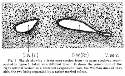
On the right side (fig. 2) the seminal vesicle is seen to be a little farther advanced in its development. Here it forms a pocket 7.5 mm deep, excluding its single cell layer of mucous membrane, and 3-13 mm in its dorsoventral diameter. The basement membrane of this evagination, together with its lining of mucous membrane, is continuous with similar structures of the Wolflian duct on this side. The budding is demonstrated through a series of twenty-nine continuous sections, Which gives a perpendicular dimension of a little over % mm for the vesicle at this stage, making the outpushing slightly oval in shape, its width being three-fourths of its length. The origin of the seminal vesicles in this earliest specimen, therefore, appears definitely as a lateral evagination of the deferent duct, situated in the lower region of that portion which in older stages is designated as the ampulla, and lies above the base or beginning of the tubules of the prostate gland. This budding, even at its earliest stage of recognition, is always wider in its lateral than in its perpendicular or anteroposterior dimension. Immediately as it reaches an appreciable size there appear two depressions or folds, in the nature of constrictions. These are situated opposite each other, one on the dorsal, the other on the ventral aspect of the vasevesicular junction, and form a definite double furrow of demarcation separating the vesicle from the duct, and making the intervening portion appear as a pedicle of attachment between the Vesicle and the deferent duct.
Fig. 1 Sketch showing a transverse section through the right and left Wolffian ducts in a human fetus 80.3 mm. crown-rump length, estimated age 13 weeks (Carnegie Collection, no. 768(3). Enlarged 100 diameters. The primordium of the left seminal vesicle can be seen extending as a lateral evagination from the Wolffian duct of that side. The primordium of the right seminal vesicle is also shown and is cut in a broader portion, its lumen being separated from that of the Wolffian duct by a slight ventral sulcus.
Abbreviations: A or Amp, ampulla ductus deferentis F., prostatic furrow D.D. or D. def., ductus deferens (vas L, left side deferens) Rt, right side D.E. or D. ejac., ductus ejaculatorius U., utriculus prostaticus D.W., ductus Wolfii V.S. or V. sem., vesicula serninalis GL., glandula V.U., vesica urinaria
After the area of vesicular budding is passed, the deferent ducts noticeably increase in size as they approach the urethra, par— ticularly in their lateral diameter, until they attain a width of 3} mm. being over three times as Wide laterally as they are dorso— ventrally, yet maintaining their oval contour. There is noted also at this level a more close approximation of mesenchymatous cells about the ducts, which gradually form a ring of four or five layers, eventually differentiating into muscle layers in the walls of the ducts. This enlargement of the Wolffian ducts marks the beginning of the common ejaculatory ducts and continues for a distance of a little over % mm. As the ejaculatory ducts enter the prostate gland they decrease in size and become nearly round, measuring about mm. in diameter at the site where the prostatic tubules are first noted, in the region of the middle lobe. It is here also that the prostatic utricle (the fused atrophying Mullerian ducts) is first seen, located in the midline between the two ejaculatory ducts. When first observed at the upper portion or base of the prostate it is very small, oval in shape, about one-fourth as wide in its long diameter as the ejaculatory ducts, and situated between the ducts with its long axis in a dorso-ventral direction.
Fig. 2 Sketch showing a transverse section from the same specimen represented in figure 1, taken at a different level. It shows the primordium of the right seminal vesicle as a flattened evagination from the Wolffian duct of that side, the two being separated by a rather marked sulcus.
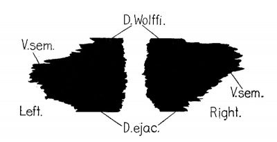
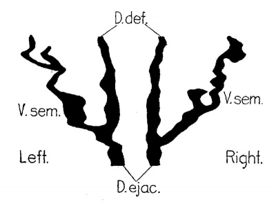
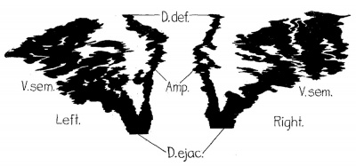
In tracing the ejaculatory ducts and utricle farther down towards the internal sphincter and neck of the bladder, the ducts decrease in size and become nearly round, until, as they reach the apex of the trigone, all three structures are about the same size, the utricle, however, retaining its oval form. In continuing through the prostate, and below the middle lobe, the ducts decrease still more in size and become perfectly round, their lumina measuring % mm. in diameter. The utricle, on the other hand, increases in size until its lumen is several times the diameter of the accompanying ejaculatory ducts. With the appearance of the tubules of the posterior lobe, which lie dorsal to the ejaculatory ducts, the ducts and the utricle begin their upward course towards the urethra. At first they lie just beneath the prostatic furrows, the .utricle being uppermost in the substance of the Verumontanum. As the ducts approach the surface of the urethra they come to lie nearer the midline, and finally open into the urethra simultaneously through the substance of the colliculus seminalis (verumontanum), midway between the top of the colliculus and the base of the prostatic furrows, one on each side of the midline. The prostatic utricle continues through the prostate and colliculus at this point, where it suddenly ends as a blind pouch below the surface of the mucous membrane and in the submucosa of the colliculus. The contour of the vesicular pouches is indicated in figure 3, which represents a graphic reconstruction of part of the Wolflian ducts.
Fig. 3 Graphic reconstruction of the lower end of the Wolffian ducts, showing the lateral evaginations which form the seminal vesicles. Human fetus 80.3 mm. crown-rump length, estimated age 13 weeks (Carnegie Collection, no. 768c). Enlarged 25 diameters.
Fetuses about 100 mm long
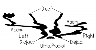
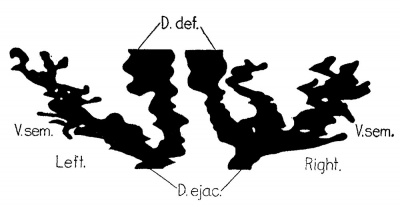
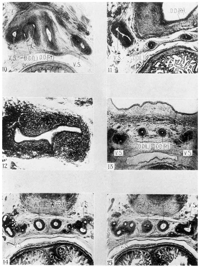
During the week following the first appearance of the seminal vesicles a very considerable growth takes place. The specimen used to represent this stage is a human fetus 105_II1II1. crownrump length (Carnegie Collection, no. 13589), having an estimated age of fourteen weeks. The pelvic region was prepared in transverse serial sections, and that portion from the upper border of the trigone down to and including the colliculus seminalis was studied. A graphic reconstruction of the deferent ducts and seminal vesicles is shown in figure 4, and photographs of typical sections are shown in figures 10 to 12.
Fig. 4 Graphic reconstruction showing the seminal vesicles and their relation to the deferent ducts in a human fetus 105 mm. crown-rump length, estimated age 14 weeks (Carnegie Collection, no. 1358e). Enlarged 25 diameters.
At the level where the ureters enter the bladder wall are seen the paired deferent ducts, symmetrically placed, each lying just medianward from the corresponding ureter, and posterior, or rather dorsal to the bladder wall. They are loosely placed, well apart from the muscle-fibers of the bladder, and only sparely attached to the subperitoneal fascia] planes, or even to the peritoneum itself, which at this point is Well developed and closely approximated to the posterior bladder wall and to the rectum. Each duct is here nearly round with a diameter approximately -3; mm., including the thickness of its wall, though the diameter of its lumen just beneath the mucous membrane lining is only 1% mm. The ducts are lined with a single layer of rather flat epithelium, as in the previous specimen, beneath which is a supporting basement membrane and a single coat of mesenchymatous cells, the latter surrounded by ten or twelve layers of closely packed undifferentiated mesenchymatous cells arranged in circular form, which later go to form "the muscle coats or walls of the ducts. The deferent ducts are here 2% mm. apart, each a little over 1 mm. from the midline, and about 3,; mm. dorsal to the lumen of the bladder in the region of the trigone. The trigone at this stage and in this region of the interureteric ligament is twice as thick as the other portion of the bladder wall, measuring nearly —§— mm. It is covered with a triple—layered mucous lining which, lateral to the ureteral orifices, becomes double—layered, and over the extreme lateral margins and ventral surface of the bladder, a single layer of small, rather flat epithelial cells with large, centrally placed nuclei.
In the course of the ducts along the dorsal surface of the bladder, beginning at the upper limit of the trigone down to the first indication of the appearance of the seminal vesicle, a distance of 1% mm. in this specimen, the size, contour, and general appearance of the deferent ducts remain unchanged. There is, however, about the lumen of the duct a little more marked separation‘ be tween the mesenchymatous tissue referred to above and that more loosely arranged, areolar—like tissue which forms the fascial layers about the vesicle beneath the peritoneal covering. Moreover, the position of the ducts at this level has noticeably changed. They now lie much nearer the midline, about 1 mm. apart, and also much nearer the lumen of the bladder, being separated from it by less than % mm. There are rather marked changes, too, in the bladder Wall in this region, its thickness at the trigone being no greater~in fact, even less than in some other areas. This relative increase of thickness in the other portions of the bladder wall is due entirely to an increase in amount of muscle fibers of the inner or circular layer. These are now distributed evenly, just beneath the mucosa, and unquestionably represent the beginning of the circular or sphincteric fibers about the neck of the bladder, which in the adult form a part of the internal sphincter.
In this series the right vesicle makes its appearance first, its tip being % mm. to the right of the Wolflian duct. From this point it stretches out as an oval-shaped mass of mesenchymatous cells measuring § xi mm., and is seen to contain the lumina of three ducts or pouches, each about -§— mm. in diameter, as shown in figures 11 and 12. On following their course downward these appear to be simply diverticula of a larger cavity measuring % x mm., the latter showing in addition the beginnings of two lateral diverticula similar to the three just mentioned. It also sends off one or more small diverticula posteriorly placed. All of these are lined with a single layer of epithelial cells, immediately beneath which is a basement membrane with several layers of undifferentiated mesenchymatous cells supported, in turn, by ten or twelve layers of mesenchymatous cells, all in one homogeneous oval mass. Leading from this large pouch like cavity is a small duct, similar in shape, size, and structure to the Wolflian duct. This in its course gradually approaches the midline, where it finally joins the deferent duct, the two forming the ejaculatory duct. Just before this fusion, however, there is noted a small pouch-like formation or irregularity in the shape of a median diverticulum from the vesicle, which is similar in all respects to those noted in its distal extremity, except that it buds forth from the vesicle itself, whereas those previously described occur as outgrowths from its sacculated end. The vesicle, on this side extends through a series of fifteen sections, which gives it an anteroposterior length of about -% mm. It extends from the beginning of the ejaculatory duct laterally for a distance of nearly 1 mm.
The left vesicle appears somewhat lower down than the right. It is noted first as an oval mass of closely attached mesenchymatous cells measuring a little over ix fi mm., and is situated % mm. from the deferent duct. Its lumen at the distal extremity is oval in outline, measuring ‘i x% mm. in diameter. From this distal end there arise two small evaginations which are constricted at their bases, one projecting laterally and measuring %x 1% mm., the second arising a little lower down and of about the same size, but projecting medianly. The duct, lined with its single layer of rather flat epithelial cells, its basement membrane immediately beneath and its supporting layers of mesenchymatous cells, descends somewhat medianward. At its lower end are seen two small out-pushings, one of which is 1-15 mm. deep and dorsally placed, the other & mm. deep and ventrally placed. These diverticula appear as evaginations of the wall of the duct, carrying with them the duct lining and pushing ahead all the coverings of mesenchymatous tissue. After these diverticula increase in size they become somewhat saeculated and their base or pedicle becomes constricted to allow for only a small lumen of communication with the main duct. In this specimen the left vesicle is smaller than the right and measures about % mm. in its anteroposterior diameter, by about % mm. in its lateral diameter. The duct of the vesicle, after the last-mentioned evagination, is seen toljoin the left deferent‘ duct and then to proceed with its fellow of the opposite side through the substance of the prostate gland, following a course above the posterior-lobe tubules and below those of the median lobe, until both ejaculatory ducts open on the floor of the urethra, coming out on either side of the colliculus seminalis just above the prostate furrows. As can be seen in figure 4, the deferent ducts are still observed as straight tubes, showing no attempt at convolution in the region of the ampullae.
Fetuses about 130 mm long
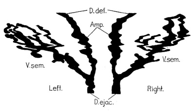
The intervening period between these two stages. of development of the vesicles is characterized by the growth of ducts and sacculations already begun, rather than by the appearance of new diverticula. The present stage is represented by a fetus 130 mm. crown-rump length (Carnegie Collection, no. 1018), and illustrated in figure 5, which is a graphic reconstruction of these structures, and in figure 13, which is a photograph of a typical transverse section.
At the upper level of the trigone the Wolffian ducts are seen as simple, straight, epithelium~lined tubes lying beneath the peritoneum and separated from it by only a few layers of areolar tissue. The trigone at this point is six times as thick as other portions of the bladder wall, measuring ;i- Inm., over half of it being made up of circular tissue, which is not quite so abundant in the other areas of the bladder wall. The mucous membrane of the trigone is here composed of four or five layers of epithelial cells, while that lining the remainder of the bladder cavity consists of only one layer. In their course beneath the trigone the Wolffian ducts gradually approach the midline until they lie only % mm. apart. The trigone maintains the same thickness as noted ‘above down to the point where the vesicles appear, when it thins out rather rapidly, the bladder wall, on the other hand, increasing to twice its thickness at the higher level, due to an increase in circular muscle fibers.
Fig. 5 Graphic reconstruction of the seminal vesicles showing their relation to the deferent ducts in a human fetus 130 mm. crown-rump length, estimated age 16 weeks (Carnegie Collection, no. 1018). Enlarged 25 diameters.
The vesicles in this series are first encountered at their distal extremity, 1% mm. below the upper level of the trigone and interureteric ligament, and lying close beneath the lower portion of the trigone. The right vesicle is noted as a somewhat oval mass of well-differentiated mesenchymatous cells lyiI1g % mm. lateral from the Wolfiian duct, and measuring % x ,1; mm. This cell group quickly enlarges to twice this size and shows a lumen somewhat crescent-shaped, and measuring % mm. in its longest diameter. In following the course of the vesicle down through the series of sections it is noted that there has been a lateral budding at the extremity, which has reverted and begun to grow backward along the course of the vesicle, so that for a short distance there are two parallel ducts—one the main duct of the vesicle, the other its distal diverticulum which has become bent to assume the proportions of a second duct. Both of these, as in the earlier specimens, are lined with mucous membrane supported by a well—defined basement-membrane surrounded by a homogeneous oval mass of mesenchymatous cells well differentiated from the surrounding areolar-like tissue (fig. 13). This diverticulum continues back along the course of the main duct for a distance of §-,mm., where it ends as a blind pocket, the only point of communication being at the distal extremity of the main duct from which the diverticulum buds off. As the blind pocket is reached there appears a small ventral outpocketing from the main duct of the vesicle in the shape of a second diverticulum. This last evagination is only about ;% mm. in "length, but is nevertheless the beginning of a definite sacculation near the extremity of the main duct. From this last diverticulum the main duct continues posteriorly and medianly as a straight tube for a distance of % mm. At this point it joins medianly a larger, definite sacculation which is several times larger than either the main cavity or the deferent duct-. This pouch continues as a straight tube for a distance of nearly %— mm., where it joins the right Wolflian duct to form the common ejaculatory duct beneath the apex of the trigone, before the appearance of the prostastic tubules. The larger sacculation, which forms the proximal end of the vesicle, is similar in structure to that of the deferent duct, and also to that of the distal extremity of the seminal vesicle. The right vesicle in this specimen measures a little over 1 mm. in its anteroposterior length and a trifle over i mm. in its lateral length from the ampulla of the deferent duct.
The left vesicle first appears as an oval mass of darkly staining mesenchymatous cells measuring about %x% mm. in diameter, and lies about i mm. lateral from the deferent duct. It soon increases to double this size when the lumen of the duct can be made out; the latter is somewhat crescent—shaped and measures % X 5}; mm. At this extremity it is quite irregular, and following it down the irregular extremity is seen to give off a ventral evagination somewhat similar to the one described at the tip of the right vesicle. It is much smaller, however, and a picture similar to that described at the distal end of the right vesicle, i.e., the main duct and a diverticulum running parallel,~—can be seen through a few sections. From this point the main duct continues as a straight tube for a distance of a little over % mm. Its course is posterior and median, and it gives off only one very tiny ventral out-pushing about i m. from the first one. After this straight uninterrupted course the chief tube or duct is seen to enter a medianly placed sacculation or duct, similar in all respects in form and outline to the one described on the right side, though somewhat smaller. This last pouch continues as a straight -tube, posteriorly and medianly, for a distance of ~§ mm., when it joins the deferent duct to form the ejaculatory duct immediately below the lower portion of the trigone, in a position corresponding to that of its fellow on the right side. The vesicle measures a little over 1 mm. in its anteroposterior length and almost § mm. in its lateral width.
The two ejaculatory ducts are formed in this specimen at almost the same level, and together they continue their course into the substance of the prostate, first pushing aside the laterallobe tubules and then taking the course above the few tubules of the posterior lobe. When the prostatic utricle (the fused Miillerian ducts) is reached it occupies a perpendicular position between the two ducts, and is about three times as large as its perpendicular diameter, and about equal in its lateral diameter to that of the almost cylindrical ejaculatory ducts. From this site all three structures, thestwo ducts and the prostatic utricle, continue their course up to and through the colliculus seminalis. The ducts end almost simultaneously on either side of the col1icuIus just above the prostatic furrows. The utricle continues a short distance beyond the opening of the ejaculatory ducts into the posterior urethra, and finally ends as a blind pouch just beneath the mucous membrane covering the top of the colliculus.
In this specimen the deferent ducts were studied from the upper limit of the trigone to their outlet, and gave no indication of any irregularities or dilatation. In fact, the region of the ampulla was in all respects the same as the duct in other places.
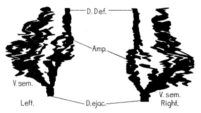
Fig. 6 Graphic reconstruction of the seminal vesicles showing their relation to the deferent ducts in a human fetus 171.4 mm. crown-rump length, estimated age 19 weeks (Carnegie collection, no. 1049). Enlarged 25 diameters.
Fetuses about 170 mm long
This stage is represented by a fetus 171.4 mm crown-rump length (Carnegie Collection, no. 1049), and illustrated in figure 6, which is a graphic reconstruction, and in figures 14' and 15, which are photographs of sections. The specimen was imbedded in paraflin, and sections 40 u thick were cut, arranged serially and stained in hematoxylin and eosin. Every section of the series, from the upper limits of the trigone down to and through the colliculus seminalis, was studied.
At the upper limit of the trigone the ureters are seen to enter the bladder cavity simultaneously. At this point the thickness of the trigone is five times as great as the ventral Wall of the bladder, measuring nearly 2 mm. On either side of the trigone, beyond the ureteral orifices, the bladder Wall gradually begins to thin out, and at the midpoint of either lateral Wall the diameter is reduced to something less than i~ mm. This thickness is maintained around the remainder of the bladder wall. The musculature of the trigone is nearly half made up of circular muscle, while in other areas this comprises only one—fifth of the thickness. The mucosa covering the trigone has five layers of epithelial cells, about the same number as are found in other portions of the bladder cavity. In most places four distinct layers can be counted.
The deferent ducts lie 2% mm. apart and 1:} mm. dorsal to the lumen of the bladder, just beyond the lateral limits of the trigone. They are separated from the peritoneum by several layers of areolar tissue, and from the musculature of the bladder by several planes of similarly loosely arranged tissue. Here the ducts are perfectly round with a diameter of % mm., and have a muscular coat 1% mm. thick and a lining of a single layer of epithelial cells. Around the ducts is a zone of connective tissue Well differentiated from the surrounding areolar tissue, and about this are noted several small groups of blood-vessels accompanying the ducts. These vessels are, for the most part, situated along the external or lateral aspect of each duct. As the ducts are traced along the dorsal aspect of the bladder their form gradually changes from round to oval, and at the same time they increase in size until, at a distance of 1% mm. below the upper limit of the trigone, they measure ~§—x% mm. in diameter, and have an oval lumen Whose greatest diameter extends laterally. In the meantime the ducts have gradually drawn nearer to the midline, and at the level of the first appearance of the seminal vesicles they lie only 1% mm. apart.
The right vesicle appears first in this series, as an oval mass composed of darkly staining cells and measuring %x§ mm. in diameter, with the greatest width extending laterally. This cell mass. lies § mm. to the right of the deferent duct and 2% mm. dorsal to the lumen of the bladder, and is surrounded by a network of areolar connective tissue. After the mass of cells is passed the succeeding sections show an increase in the tip of the vesicle until it measures % x i m. by the time its lumen appears as a slightly irregular oval cavity lined with a single layer of muscle‘ §, mm. deep. The cavity of the vesicle immediately becomes" constricted in its center at this level to form two distinct lumina. The vesicle continues in this manner with the two cavities enclosed in a single musculature for a distance of nearly % mm., when each becomes noticeably larger and somewhat ‘sacculated, and there appears ventrally a third small lumen closely‘ associated with the medianly placed lumen. In the next section there appear two additional cavities, very small, each in its own musculature and placed between the median portion of the ‘vesicle and the Wolflian duct. This arrangement continues for some distance, when the three small cavities about the larger dorsal lumen unite with it to form a larger irregular cavity. This leaves one small additional cavity placed between the vesicle and the deferent duct, as can be seen in figure 14.. The larger cavity next becomes constricted and gives off a small lateral pouch, while at the same time there appears a small ventral cavity, ‘thus making a four-cavity vesicle. These all become fused. into a single irregular pouch in the succeeding section, only to give ofi" again, farther on, two small pouches, one of which immediately disappears leaving a vesicle with a double lumen. Thiscondition persists for only a few sections, when the medianly placed cavity, which has persisted since the very first appearance of the lumen of the vesicle at its extreme tip, joins the lateral pouch to form one large irregular lumen. This finalsingle cavity continues for a distance of % Inm., gradually moving medianward, and having a somewhat irregular lumen. It finally joins the ampulla to form the common ejaculatory duct at a point dorsal to and medianward from the tubules of the lateral lobes of the prostate, before the appearance of any of the middle-lobe tubules. This vesicle measures 1% mm. in its anteroposterior length, and 1% mm. in its lateral width from the ampulla of the right deferent duct.
The left vesicle appears at a somewhat lower level than the right, and is first noted as a dumbbell—shaped mass of deeply staining cells, one of the group being slightly larger than the other and measuring i X % mm. in diameter. In the succeeding sections, as this cell mass enlarges in size, the lumina of two cavities appear, the number increasing to four in the next section, due to the appearance of two additional cell masses. The larger of these soon divides and the picture is one of la five-cavity organ at a distance less than i mm. from its extreme tip. The three larger cavities immediately become constricted to form three additional cavities, thus making eight pouches, two of which are each as large as the lumen of the deferent duct. About these are grouped the smaller pouches. The vesicle is here crescentric in form with the concave side pointing ventral. Four of the smaller pouches disappear in the succeeding sections, leaving two larger and two smaller lumina to the vesicle. The two small remaining cavities can be traced through only a few sections, when they, too, fuse with the larger ones. The two cavities persist for some distance, when the lateral one, which has gradually become smaller, finally unites with the larger to form a single, slightly irregular pouch. This single cavity continues posteriorly and medianly for a distance of % mm., when it joins the deferent duct to form the ejaculatory duct at a level corresponding with its fellow of _the opposite side. The left vesicle measures 1% mm. in its anteroposterior length, and 1% mm. in its lateral diameter from its junction with the ampulla. The ampullae are round tubes at the level of the first appearance of the vesicles, and have a lumen of % mm. in diameter. They soon enlarge, however, until, at a distance of § mm. below the tip of the vesicle, they have an elliptical lumen %x—{— mm. in diameter. The left deferent duct in this specimen shows, in addition to a dilatation in the region of the ampulla, a definite diverticulum formation which is the first observed in the course of this study, and is shown in figure 15. After the union‘ of the Vesicle and the ampulla the ejaculatory duct has an irregular lumen whose diameter is % mm.
The ejaculatory ducts, each surrounded by its individual musculature, begin their course through the prostate between the lateral-lobe tubules, and above them soon appear the few well-defined tubules of the middle lobe. They lie on either side of the midline % mm. apart, with the few blood-vessels which were observed higher up about the lateral margins of the deferent ducts, now all located between the ducts. At a distance of 1 mm. below the union of the vesicle and ampulla the prostatic utricle first appears as an irregular elliptical cavity with its wall approximated and measuring —§- em. in its greatest diameter, which extends dorsoventrally. The ejaculatory ducts have now become elliptical in shape and are situated on either side of the utricle. At this point appear a few tubules of the posterior lobe situated dorsal to the ejaculatory ducts and to the utricle. The ducts continue their course posteriorly and upward through the colliculus seminalis until they open simultaneously on the tip of -the colliculus, a distance of 1% mm. from the union of the vesicle and ampulla at the base of the prostate gland. The prostatic utricle here ends as a blind cavity at the same level as the openings of the ejaculatory ducts.
Fetuses about 180 mm long
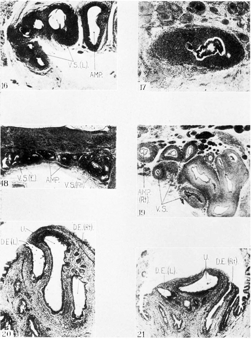
This state is represented by a fetus 178 mm. crown-rump length (Carnegie Collection, no. 1171), and illustrated by figure 7, which is a graphic reconstruction, and figures 16 and 17, which are photographs of sections. The specimen was cut in serial sections 40 in thick and stained in the usual manner with hematoxylin and eosin- Only those from the upper portion of the trigone and interureteric ligament down to and including the colliculus seminalis were studied in this series.
In the serial sections the right Vesicle appears first as an oval mass of well-differentiated mesenchymatous cells measuring % to % mm. in diameter, and situated % mm. to the right of the deferent duct. This cell group quickly enlarges in the succeeding sections to about twice the above size and is seen to contain the lumina of two diverticula, one probably being the chief duct. Each of these gives off another pouch, making four out-pocketings at the extreme distal end of the vesicle, and the succeeding sections show another very short dorsal budding from the largest pouch. The three smaller out-pocketings soon disappear, leaving a vesicle with two large pockets running parallel for some distance. The vesicle at this stage has assumed more nearly its adult form, being quite crescentic in shape and showing two lumina of considerable irregularity. At this site its dorsoventral diameter is a little over 1 mm. and its breadth nearly 1 mm., one lumen measuring about ;‘°;—x:i— mm. in diameter, the other lumen, nearer the deferent duct, is considerably smaller in diameter. Both cavities are lined with the characteristic single layer of epithelium, supported by a basement membrane and surrounded by a somewhat circular or oval mass of mesenchymatous cells.
Fig. 7 Graphic reconstruction of the seminal vesicles showing their relation to the deferent ducts in a human fetus 178 mm. crown-rump length, estimated age 21 weeks (Carnegie Collection, no. 1171). Enlarged 25 diameters.
The deferent ducts now show considerable dilatation in the region of the ampullae. They are oval in shape, and the diameter of their lumina is a little more than %x i mm. At this point the basement membrane about the ducts is supported by five or six layers of well-differentiated mesenchymatous cells, while the vesicles themselves have many more layers of supporting mesenchymatous cells, in many places numbering eighteen or twenty. The two parallel diverticula of the Vesicle, mentioned above, continue for some distance. The larger, which is situated more lateral, gives off in its course three small pouches, one lateral and two dorsal. The larger is quite sacculated and irregular, while the others, nearer the deferent duct, run as straight tubes. The larger sacculation soon joins the straight tube or duct which lies nearer the right deferent duct, or the ampulla of the deferent duct, as it may be called in this region, thereby making one larger irregular lumen to the vesicle. This continues posteriorly and medianly and soon unites with the ampulla of the deferent duct without further irregularities. This union takes place directly beneath the substance of the prostate gland, where the tubules of the two lateral lobes and several of the median lobe are seen to be well developed. The measurements of this Vesicle show it to be somewhat shorter in its anteroposterior diameter than in the preceding specimen, measuring about % mm. in length, while its lateral diameter is about 1 mm.
The left vesicle appears at a slightly lower level than the right. It is represented by an almost circular accumulation of mesenchymatous cells situated a trifle less than % mm. to the left of the deferent duct, and measures ixi mm. in diameter. This area almost immediately increases in size andthe lumina of two cavities appear, one lateral to the other, the larger, nearer the Wolffian duct, measuring %x i mm. in diameter. The smaller of these two diverticula constitutes only the distal extremity of the chief duct, which has become folded upon itself and is now running parallel back towards its origin. In the succeeding sections the smaller, more laterally placed pouch disappears, and there remains one larger, rather sacculated cavity which continues posteriorly and medianly “towards the deferent duct, without any marked sacculation except two small diverticula which branch out, one ventrally, the other laterally. These, however, soon disappear. The main duct continues for a short distance and then enters a larger, medianly situated duct or sacculation measuring % x :1— mm., which follows the usual course until it joins the ampulla of the deferent duct. This fusion takes place at a point where the deferent duct is slightly smaller than the proximal end of the vesicle, but still definitely larger than in the higher .region where the dilatation forming the am-4 pulla has begun. The vesicle joins the ampulla on this ‘side at a level somewhat higher than on the right, thus making the antero-posterior length considerably shorter than on the right side. This vesicle measures a little over % mm. in its anteroposterior length and % mm. in its lateral dimension.
Immediately after the Vesicles unite with their respective ampullae they begin their course through the prostate as the ejaculatory ducts, in which capacity they are of equal size, nearly round and with luJ:nina % mm. in diameter. In this re-’ gion the ducts are definitely surrounded by circular muscle about four cell layers deep, while beyond the mesenchymatous cells are less clearly diiferentiated. The ducts travel an upward course parallel to each other through the substance of the prostate gland, beneath, and in places seeming to almostpush upward the tubules of the middle lobe. They continue in this manner for a little over i mII1., when the prostatic utricle appears as a spherical lumen nearly 3; mm. in diameter immediately ‘above them. Their course is now divided, and they turn, each to its respective side of the prostatic utricle, and all three structures enter into the substance of the colliculus seminalis. Here they all become much smaller and elliptical in shape, their dorsoventral diameters remaining‘ always greater. ‘When the ducts reach the -middle portion of the colliculus they proceed upward to the surface of the mucous membrane covering it and open just above the prostatic furrow, one oneither side. The utrical is now elliptical iI1 shape, measuring % mm. in its dorsoventral diameter, but with its lateral surfaces approximated. This continues through several sections when the utricle ends as a blind pouch just beneath the surface of the mucous covering and in the substance of the colliculus.
Up to this stage the dilatations and irregularities of the ampullae have been only slightly developed. In fact, the changes in the ampullae made their first appearance in the previous specimen. At this later stage, however, the dilatation in the lower portion of the deferent ducts has become quite appreciable, as can be seen in figure 16. In this specimen there appear simultaneously in each ampullae definite folds which are the first steps in the later fully developed convolutions. These folds are two in number and appear i mm. above the junction of the vesicle and ampulla on either side. On the right they take the form, one of a. slight ventral, the other of a slight lateral fold, While on the left their positions are those of a slight ventral and a small median fold. This reduplication is too high up to be considered any part of the vesicle, which always joins the ampulla lower down, and hence can only be considered as the beginning of the tortuous course of the adult ampulla.
Fetuses about 220 mm long
This stage is represented by a fetus 221 mm crown-rump length (Carnegie Collection, no. 1172), and illustrated by figures 18 to 21, which are photographs of typical sections. The specimen was imbedded in celloidin and sections cut 15 11. thick, These were arranged serially, and every fifth one, from the urachus down to and including the colliculus seminalis, was mounted and stained in the usual manner with hematoxylin and eosin.
At the upper border of the trigone and interureteric ligament the deferent ducts are seen situated just inside the openings of the ureters into the bladder wall. Here they are round and of equal size, and each has a lumen % mm. in diameter and a definite muscular coat % mm. thick. They are lined with a single layer of mucous membrane with a basement membrane, as described in previous specimens. The ducts are situated close to the posterior wall of the bladder and are separated from the peritoneum by several planes of areolar tissue. The bladder wall is here well developedin its three muscle coats, the circular or inner coat being equal in thickness to the other twcr—the longitudinal and transverse. The mucous membrane over the trigone is composed of three or four layers of epithelial cells, while over the other areas of the bladder it consists of only two. The trigone in this region is one and a half times as thick as the other portions of the bladder wall, measuring 1% mm. At a distance of % mm. below the upper limits of the trigone the deferent ducts begin to show definite irregularities in their lumina and also to increase in size, particularly in their lateral diameters. These changes indicate the upper or anterior margin of the ampulla of the deferent ducts. A few sections farther on the ampulla is seen to have assumed nearly its adult form, and to be extremely irregular and convoluted. It is oval in contour, measuring 1% x 1 mm., and has a lumen %x-}— mm.
The right vesicle appears first as a round accumulation of cells measuring % mm. in diameter and situated % mm. to the right of the ampulla of the deferent duct. It almost immediately enlarges to 1% x% mm. and contains one large, irregular, sacculated cavity measuring %x % m. From this cavity are seen to arise, in the succeeding sections, numerous diverticula—— about eight or ten in number. These appear as irregular outpushings, all lined with the same sort of epithelium and all of the same structure as the original distal sacculation. Their architecture becomes still more complex as the sections are followed, and the diverticula increase to twelve in number, each one a definite epithelium—lined cavity with a basement membrane supported by eighteen to twenty layers of definite muscle cells which form a circular coat about the lumen of each cavity. The vesicle here has attained its greatest size, measuring 2% x 2 mm. in diameter. At this stage it is difficult to make out whether the diverticula arise from one central cavity or, ofttimes, from one another; but the impression is that they are all out-pusliings from the one distal sacculation described at the extreme tip of the vesicle. This sacculation, which continues throughout the slides, is larger than any of the diverticula and its lumen is extremely irregular. When it has reached its greatest circumference the vesicle immediately begins to diminish in size, and three of the twelve diverticula persist about the irregular cavity of the main sacculation throughout several sections. They become noticeably smaller, however, until the chief duct with the irregular lumen joins ‘the ampulla of the deferent duct on this side, within the substance of the prostate gland below the tubulesof the middle lobe and between‘ those of the two lateral lobes. For some distance within the structure of the prostate the ejaculatory duct still maintains its irregular outline, similar to that observed higher up in the region 0” the ampulla. The ducts are here oval in outline, measuring 33-): i mm. at their lumen, with their greatest diameter lateral. The general dimensions of the vesicle at this stage of development have changed noticeably from those of earlier specimens; the right vesicle measures % mm. in its anteroposterior length along the side of the ampulla and 3 mm. lateral from the junction forming the common ejaculatory duct.
The left vesicle arises in this specimen at the same level as the right, and is first observed as an oval mass of cells measuring %~x % mm., and situated tothe left of the ampulla of the deferent duct. It immediately increases in size through the succeeding sections, and, similar to the right, is seen to contain a large, irregular, sacculated cavity measuring %x% mm. in diameter. Surrounding this one soon encounters four definite pouches, epithelium—lined and similar to those on the opposite side. In succeeding sections their number is increased to nine, most of them being grouped medianly about the larger irregular sacculation seen at the end, such as was noted on the right, and which at this level persists as the chief duct. It is possible throughall these sections to identify this larger sacculation because of its irregularities. The diverticula at the lower level soon decrease to five in number and are grouped about two large sacculations, the larger of these still identified as the lower portion of the one noted first at the distal extremity, which has moved medianward and nearer the ‘left ampullae. In the course downward the remaining evaginations soon disappear, and there persists only the large chief duct, now a small, irregular—walled cavity, easily identified as the proximal end of the sacculation at the distal end of the vesicle. This soon joins the ampulla of the deferent duct, but at a somewhat lower level than the right, well within the substance of the prostate gland beneath the tubules of the middle lobe. The left vesicle measures % mm. in its antero— posterior length along the'ampulla of the deferent duct, and 2% mm. lateral from its junction with the common ejaculatory duct, being somewhat longer, but not so wide as the one on the opposite side.
The ejaculatory ducts, as they enter the substance of the prostate, lie side by side and are oval in shape with the long axis extending laterally. Their walls immediately become enveloped in a single oval musculature measuring 2 mm. across in its wide lateral diameter, the whole mass being clearly differentiated from the intertubular substance of the prostate itself. Their lumina still retain an irregular outline quite similar in character to the lumen of the ampulla. Their course through the prostate is an upward slant, running parallel on either side of the midline. They lie at first beneath the tubules of the middle lobe, and later the posterior lobe tubules appear beneath or dorsal to them, while the tubules of the lateral lobes are seen well to either side.
As the ducts approach the colliculus seminalis their shape changes rather abruptly and they appear oval, with the long axis directed dorsoventrally instead of laterally. This has come about in order to enable them to pass on either side of the prostatic utricle, which now appears in the central substance of the colliculus, as shown in figure 20. All three structures are now observed in their course; the ducts take a more upward direction than formerly, and are seen to open on either side of the colliculus, well up towards its tip, and on either side of the prostatic utricle, which continues for some distance beyond the openings of the ejaculatory ducts, finally ending as a blind pouch. In this specimenlthe utricle shows an upward evagination of its lumen in the form of a narrow pocket or duct, which is later to be its opening into the posterior urethra (fig. 21). The utricle runs through the colliculus for a distance of % mm. ; in its largest diameter——dorsoventral——it measures 1 x% mm.
Fetuses about 275 mm long
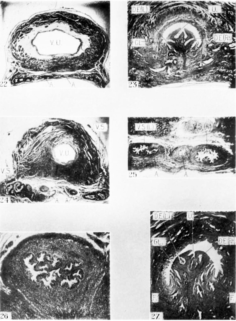
This stage is here represented by a fetus 276 mm. crown-rump length donated by Dr. Daniel Davis, Obstetrical Department, Johns Hopkins Hospital, and illustrated by figure 8, which is a graphic reconstruction of the structures, and figures 22 and 23, which are photographs of sections. The specimen was imbedded in paraffin and out in sections 40 1.: thick, which were arranged serially and stained in hematoxylin and eosin. Every section from the upper limits of the trigone and interureteric ligament, down through the colliculus seminalis, was studied. At the level where the ureters open into the bladder Wall the trigone has a thickness of 1% mm., approximately the same as in other parts of the bladder wall. Over half of it at this point is made up of circular muscle, which is not nearly so abundant in other areas. Over it the epithelial lining is composed of four or five layers of cells which, over the lateral and ventral walls of the bladder, become thinned out to two layerz and in some places to a single layer. The muscle coats of the bladder are very well developed, and the three layers are plainly delineated. The submucous layer is much thicker than in the earlier specimens and is of about the same thickness as the mucosa itself.
Fig. 8 Graphic reconstruction of the seminal vesicles showing their relation to the deferent ducts in a human fetus 276 mm. crown-rump length. estimated age 31 weeks. Enlarged 8 diameters.
The deferent ducts are seen at this level lying 2% mm. dorsal to the lumen of the bladder, 8% mm. apart. Each is situated a little over 4 mm. from the middle of the trigone and somewhat lateral to the openings of the ureters into the bladder Wall. The ducts have a well developed muscular coat measuring a little over 315 mm. in thickness. Their general shape is elliptical, with the long axis extending laterally. Their diameter is a little over fixi mm., and the entire duct measures nearly %x~}~ mm. They are surrounded by a network of blood-vessels, about sixteen to eighteen in number. Over half of these are as large in diameter as the lumen of the duct and form a venous plexus which completely surrounds each duct. The ducts lie close to the peritoneum, being separated by only two or three plenaes of areolar tissue, While between them and the outer bladder wall are four or five planes of areolar tissue with a few scattered bundles of longitudinal muscle fibers. As the ducts are traced down their course along the dorsal wall of the bladder they become nearly round, but remain the same distance apart. At the first appearance of the vesicles in their most distal portion the deferent ducts are still over 8 mm. apart.
In this series the tip of the right vesicle appears first, 2% mm. below the upper limit of the trigone and interureteric ligament. It is first noted as an oval mass of darkly staining cells situated 1% mm. to the right of the deferent duct. It measures i x% mm. in diameter, its lateral width being the greatest, and enlarges in the succeeding sections to a diameter of % X :1; mm. When this plane is reached the lumen of the duct of the vesicle is noted as an irregular, almost star—shaped opening with four buds or pouches arranged symmetrically, giving the outline of a four-pointed star. The dorsally situated evagination becomes elongated in the sections immediately following, and soon the star—shaped lumen disappears and the vesicle continues as a single oval pouch measuring 1%,» X =7; mm. in diameter. This single-lumen tube continues for about -1- mm., becoming gradually smaller, when there appear two oval cell masses, one situated dorsal, the other ventral to the lumen of the vesicle. These both increase in size until they reach a diameter of % mm., and both of their contained lumina are very irregular. These three evaginations soon unite to form a crescent—shaped vesicle with a continuous curved lumen. The single cavity continues through only a few sections when the vesicle divides into three nearly equal parts, each having a distinct lumen, and all arranged in the form of a crescent. After this division, the more dorsal portion, which has persisted from the extremity of the vesicle, immediately disappears, leaving two divisions, one ventral, the other lateral. The latter soon increases to almost twice its original size, measuring 1% x % mm. in diameter, and comes to lie nearer the midline in a position dorsal to the ventral pouch. This larger sacculation becomes constricted in its middle portion and soon has two distinct cavities instead of one. At this point all three cavities become more closely approximated and are supported in the same musculature. This condition persists through a few sections, when the more medianly placed sacculation, which comes from the division of the large pouch mentioned above, entirely disappears. Its place is soon taken by two‘ pouches with smaller lumina; these shortly unite, making three distinct cavities, triangularly placed, which form the vesicle. The lateral and dorsal sacculations soon join to form one large pocket; this almost immediately divides again, and two sacculations or pouches are sent off from it in the succeeding sections, making a four-pocketed vesicle with each sacculation a distinct cavity. The ventral and lateral pouches each give off another evagination, but the lateral one soon disappears, leaving a vesicle with five independent cavities at this site. This number is soon increased to six, after which the peripherally situated sacculations gradually become more lateral in arrangement. These gradually disappear, one by one in the succeeding sections, the more lateral going first; so that we have successive pictures of a five-, "four-, three-, and two-cavity vesicle, with the pouches arranged in a more or less crescentric form. Finally the two remaining cavities fuse and form a single irregular pouch measuring 1% x% mm. This finally unites with the ampulla to form the ejaculatory duct at the begirming of the prostate gland, Where the tubules of the lateral lobe are distinctly seen before any of the middle-lobe tubules appear. . This vesicle measures 5% mm. in its anteroposterior length, its greatest lateral width from the base of the ampulla being 2% mm.
The left vesicle appears at a somewhat lower level than the right, and is first noted as a nearly round mass of darkly staining cells measuring % x 1; mm. in diameter and situated % mm. to the left of the ampulla. This cell mass enlarges to a diameter of §x § mm., its greatest diameter extending laterally, when the lumen of the vesicle, as an irregular oval cavity, is first seen. This cavity increases in the succeeding sections until there appears a central constriction which divides the lumen into two distinct pouches situated dorsoventral. This form continues through several sections, when two additional pouches make their appearance, one placed just medianward from each cavity already present. These four sacculations continue for only a short distance, when the two ventrally situated pouches unite to form a single irregular cavity. The outer, more dorsal pocket, which was part of the cavity first observed, now disappears. The picture of a double-cavity vesicle soon changes to one where the more ventrally situated sacculation also becomes constricted to form two pouches. Another pocket, separate from the rest, is soon encountered to the extreme right of the above four cavities. These five pouches persist through only a few sections, when the more ventral diverticulum disappears, leaving a four—cavity vesicle with the pouches all enclosed in a continuous musculature. The more lateral pouch now becomes constricted to form an additional cavity, while the two ventral diverticula join to form a single irregular sacculation. At this point there appears medianward another separate branch containing two small lumina, which immediately fuse in the next section. The two lateral pouches now unite to form one, and the ventral pouch disappears entirely. At this point is encountered another small, medianly situated pocket, distinct from all the rest. The lateral diverticulum disappears and another ventral sacculation is brought to view. This soon enlarges to a dimension of 1% x 1% mm., and dorsal to this are grouped the four remaining pouches. The large sacculation immediately becomes constricted in two places to form a three-pocketed sac. Two of these pockets immediately unite, and there appear two additional cavities dorsal to the five already present. These two then join the two nearest to them and there appears medianly another small cavity besides the larger ventral division. This immediately disappears and leaves a crescent-shaped vesicle with seven cavities. This condition persists until the cavities disappear one by one, the more lateral ones ending first, until only two remain. This arrangement continues for some distance when the ventral sacculation disappears, after which the remaining cavity joins the ampulla of the deferent duct to form the ejaculatory duct. This union occurs within the substance of the prostate gland between the lateral-lobe tubules, but before any of the middle-lobe tubules have made their appearance. The left vesicle measures a little over 5% mm. in its anteroposterior diameter, and nearly 2% mm. in its lateral diameter from the ampulla of the deferent duct.
The ejaculatory ducts are formed in this specimen at the same level and appear as elliptical tubes, each with a "very irregular lumen measuring % x % mm. in diameter. In their course through the substance of the prostate gland they appear first between the tubules of the two lateral lobes. As they continue, their outlines become perfectly round and their dimensions much smaller. At the appearance of the middle-lobe tubules the ducts measure only % mm. in diameter. This occurs % mm. beyond the point of their formation.
The venous plexus, which was noted surrounding the deferent ducts along their course before they became united with the vesicles, has now entirely disappeared. The remains of it, some eight or ten vessels, occupy a position between the two duets, with no vessels along the outer, dorsal, or ventral aspects of the ducts.
A few sections beyond where the middle-lobe tubules appear the prostatic utricle is encountered as a round lumen % mm. in diameter, lying ventral between the two ejaculatory ducts. In its course it increases in size until it measures 1} x 1 mm. in diameter, the long axis extending from left to right. At this level the ejaculatory ducts have been pushed to either side of the utricle and the irregular character of their lumina has been lost. In following these structures into the substance of the colliculus seminalis, after they have taken an upward direction through the prostate gland and below the tubules of the middle lobe, the utricle becomes star—shaped. The ejaculatory duets again assume a position beneath or dorsal to the utricle, while all three cavities— —the ducts and the utricle——become much smaller. Toward the median portion of the colliculus the utI'icle becomes shaped like an inverted Y, as if two tubes (the M1"1llerian ducts) had become somewhat imperfectly fused at their lower ends, as seen in figure 23. At this level the ejaculatory ducts are situated more lateral and proceed through the substance of the colliculus to a position midway between its tip and the prostatic furrows. Here they open simultaneously, together with the prostatic utricle, into the posterior urethra at a distance 4 mm. below, or posterior to, the union of the ampulla and the vesicle. The utricle has a long, narrow, bifid opening, showing more clearly its relation to the separate Mfillerian ducts of the female.
The convolutions of the ampullae appear simultaneously in this specimen at a distance of 3% mm. below the upper limit of the trigone, and nearly 1 mm. below the upper level of the tips of the seminal vesicles. These convolutions become more extensive as the ampullae enlarge, and just above the union of the latter with the vesicles become so irregular asto frequently show two or more separate cavities in each ampulla at a given level. These sacculations have the same general contour and detail of arrangementaas the evaginations of the seminal vesicles. After the union of the ampulla and vesicle the ejaculatory duct loses its pouch-like character, but the irregularity of its lumen continues until the prostatic utricle is reached. These relations will be recognized in the graphic reconstruction shown in [figure 8.
Fetuses about 340 mm long
This stage of development is represented by a fetus at term 338 mm crown-rump length, from Dr. S. Bayne-Jones, Pathological Department of the Johns Hopkins Hospital, and is illustrated in figure 9, which is a graphic reconstruction, and figures 24 to 27 , which are photographs of typical sections. The specimen Was imbedded in paraflin and sectioned transversely, each section being 40 p. thick. These were arranged serially and stained in the usual manner with hematoxylin and eosin. Only those from the beginning of the seminal Vesicles down to and through the colliculus seminalis were studied.
Fig. 9 Grapnlc reconstruction of the seminal vesicles showing their relationto the deferent ducts in a new-born babe 338 mm. erown—rump length. Enlarged S diameters.
Just above the point Where the vesicles first appear the trigone is nearly twice as thick as the other portions of the bladder wall, measuring slightly over 1 mm. About half of it is made up of circular muscle, the other half of longitudinal and transverse fibers, and the mucosa is composed of four or five layers of epithelial cells. In the remaining areas of the bladder Wall the circular fibers are not nearly so abundant, and the mucosa is composed of only two or, in very occasional areas, three layers of epithelial cells. The submucosa is about the same thickness throughout the entire bladder cavity.
In this specimen the left vesicle is first noted as an oval mass of darkly staining cells measuring a little less than §x§ mm. in diameter, and lying about % mm. to the left of the deferent duct. The latter in this region is simply a straight oval tube with rather thick muscular walls and a very irregular lumen. It measures 1 xg mm. in diameter, and has a muscular coat % mm. in its greatest thickness. Both the duct and the vesicle are longest in their lateral diameters. The lumen of the vesicle soon makes its appearance as a small circular aperture situated at the extreme distal end of the cell mass. A large cavity with four smaller divisions is seen in the succeeding sections. The tip of the vesicle, with its five lumina, soon disappears, and in the sections following all five cavities unite to form a single cavity with a very irregular lumen which shows in places the beginning of the network of pockets of the cavity of the vesicle. Each strand of this network is composed of a single layer of epithelial cells supported by a basement membrane which has a frail fibrous tissue basc (fig. 26). The mucosa is similar in character to, and continuous with that lining the lateral wall of the vesicle. T his group of divisions is enclosed in a single unit of musculature. After the large irregular cavity is reached there appears, lateral to it, in a separate musculature, two additional cavities which finally unite to form a double-cavity vesicle, each cavity supported by its own individual musculature and lined similarly with a single layer of epithelial cells. The larger irregular cavity now divides, the two cavities being separated by a definite muscular partition covered with epithelial cells, forming a three-pocketed organ, each pocket supported by its own individual musculature, though all are grouped closely together. The more lateral of the cavities just formed again becomes constricted in two places, with definite muscle bands. The distally situated sacculation also divides to give at this level a sixcavity vesicle, all the cavities being separated by a definite muscular framework covered with a characteristic lining of epithelial cells. An additional lumen in a definite separate musculature now appears on the median side of the vesicle between it and the deferent duct. This last division immediately becomes reunited in the common musculature of the vesicle, and two of the cavities disappear by joining the two pockets adjacent to them. The five cavities now remaining are added to by another in its own separate musculature, situated between the vesicle and the deferent duct. .Most of the cavities at this stage are traversed by a network of divisions composed of an epithelial lining of single cells, a basement membrane and a fibrous tissue base. Two of the centrally situated cavities now unite, and those in the center of the vesicle gradually become smaller, until finally there remain only four cavities to the vesicle, hach in its separate musculature and each with a lumen quite irregular and traversed by a network of divisions. An additional pocket with a very irregular lumen now appears on the median side of the vesicle near the deferent duct, and this division unites, forming in all a four—pocketed vesicle, each division having a very irregular lumen. This numberiis soon increased to five pockets by a division of the larger middle sacculation. In the succeeding sections the pockets join each other to form three distinct irregular cavities, which again subdivide to form seven smaller cavities. The seven cavities persist with varying irregularities, until, all united in a common musculature, they begin gradually to disappear, those at either extremity of the Vesicle joining the ones nearer the center. In this manner we have successive pictures of a six—, five—, four-, three-, and two-cavity vesicle. Finally the remaining two cavities unite to form a pouch with a very irregular lumen, measuring with its musculature 1% X 1 mm. in diameter, the muscular walls being nearly %. mm. thick. This cavity, with its own individual musculature, persists for a distance of % mm., and in its course becomes gradually smaller and takes a position nearer the midline, retaining to a considerable degree, however, its irregularities. Finally its musculature unites with that of the ampulla at the site where the cavities are of nearly equal size. This fusion, which forms the ejaculatory duct, takes place beneath the tubules of the middle lobe of the prostate and at a site where the lateral lobe tubules are seen on either side of the common duct, all well within the substance of the prostate gland. The left vesicle measures 4% mm. in its anteroposterior length and 3% mm. in its greatest lateral width from the am.pulla of the deferent duct.
The right vesicle appears in this series at a level of % mm. below the extreme tip of the left. It is first noted as a round mass of darkly staining cells measuring i mm. in diameter, lying nearly 3 mm. to the right of the Wolffian duct and 4% mm. dorsal to the lumen of the bladder. The mass enlarges in the succeeding sections and contains a rounded irregular lumen traversed by a network of divisions. The cavity enlarges, losing eventually nearly all of its inner network of divisions until it becomes nearly twice its original size. At this point there appear two additional smaller cavities lateral to the pouch first noted, in an entirely separate musculature. The original pouch now divides to form altogether a five-pocketed vesicle at a distance of about % mm. below the first appearance of the vesicle. The vesicle continues in this manner for several sections, when the two more median cavities unite, and those remaining become more irregular. This group of cavities is now augmented by another small pouch situated between the vesicle and the deferent duct, which soon joi:ns thelarger irregular cavity adjacent to it. When this is accomplished the single cavity divides in another place to form a five-pocketed vesicle. The next picture shows a vesicle with six lumina, due to the appearance of another pocket between the complete vesicle and the deferent duct. l\Iost of the additional new cavities on this side are first observed medianward; that is, between the vesicle and the ampulla. The lastnot-ed cavity is very short and soon joins the larger one next to it, leaving a vesicle with five pockets, each having a very irregular lumen and well supported by its own musculature. As the vesicle is traced down two additional pockets appear, one in a separate musculature between the vesicle already present and the ampulla and the other formed by a division of the largest cavity already present midway between the lateral limits of the vesicle cavities. These seven cavities persist for only a few sections when the smaller ones disappear by becoming fused with the larger ones near them in the most lateral portion, and also the most median portion of the vesicle, until there are only two rather large, irregular cavities left, each supported by its own musculature. The laterally situated cavity now divides, and there appear three additional pockets in the musculature of the median pouch, all of which soon unite with the large irregular medianly situated pouch. In tracing the vesicle down through the next few sections the lateral pouch is seen to disappear entirely, leaving a single irregular sacculation with a complicated inner network of divisions to form the base of the vesicle. This cavity measures nearly 2 mm. in its lateral width and % mm. in its dorsoventral diameter, and has a muscular coat about i mm. thick. It proceeds through a few sections when another dorsally situated sacculation appears and the two unite; at the site of this fusion the vesicle and ampulla become supported in the same musculature. The single—cavity vesicle continues in this manner for a little over % min, when it unites with the defercnt duct to form the ejaculatory duct. This takes place a little lower down, or rather deeper within the substance of the prostate, than is the case on the opposite side. The union occurs well between the tubules of the two lateral lobes and beneath thc middlc—lobe tubules. The vesicle measures 4 mm. in its anteroposterior length and 4% mm. in its greatest lateral width from the ampulla.
At the level of the distal ends of the seminal vesicles the deferent ducts are seen as oval structures, each nearly 1% mm. in its lateral width and % mm. in its dorsoventral diameter. Their walls are nearly i mm. thick and the lumen of each is a single irregular cavity with walls nearly approximated in the dome ventral direction. Each duct lies 1% mm. on either side of the midline. In their course down along the dorsal surface of the bladder they maintain their dilated irregular character for practically the entire distance. As they approach the middle portion of the vesicle their irregularities become in places definite sacculations, and are frequently encountered as pockets which extend for some distance anteroposteriorly and form several dif ferent cavities to the ampulla. As the ampullae approach their union with the vesicles they become much smaller, and at this level have come to lie much nearer the midline until their musculatures are separated by only a few layers of fibrous tissue and their cavities are only % mm. apart. The lumina of the ampullae, while having Very irregular walls, show none of the network of inner-lining divisions that is so characteristic of the vesicles at this age. As in the previous specimen the bloodVessels which, higher up are lateral to the VVolffian ducts, here become centrally placed and lie as a definite plexus between the two common ducts.
The prostatic utricle first appears about % mm. below or posterior to the union of the Vesicle with the deferent ducts. It is observed within the substance of the verumontanurn as a circular lumen lined with a single layer of epithelial cells and measuring % mm. in diameter. It is surrounded on either side by three or four gland-like tubules similar to the prostatic tubules, but which appear to be more likely inherent glands of the colliculus seminalis (fig. 27).
The ejaculatory ducts pursue a course through the substance of the prostate gland below the tubules of the middle and lateral lobes, also above or distal to the tubules of the posterior lobeThey follow an upward, slanting direction until they come to lie just beneath, or dorsal to and slightly to either side of the prostatic utricle, which has now become materially increased in size. Here they are elliptical in shape, similar to the utricle, and all three structures are longest in their dorsoventral diame-1 ters. The ducts at this site begin a more lateral course on either side of the prostatic utricle, until both open into the prostatic urethra near the tip of the colliculus and over % mm. beyond or posterior to the opening of the utricle itself. The utricle extends only a few sections beyond the openings of the ejaculatory ducts, and finally ends within the substance of the colliculus. It has an_ anteroposterior diameter of a little over 3% mm., and at this age runs as a tube or duct-like structure which, at its opening, has a bifurcated lumen (fig. 27), showing its relation to the analogue in the female (Müllerian ducts). The ejaculatory ducts are of equal length in this specimen, and measure 33,3 mm. in their anteroposterior length.
Summary
- The seminal vesicles first make their appearance during the thirteenth week of foetal life (crown-rump length, 80 mm.) as lateral evaginations or out-pocketings from the lower portion of the Wolffian ducts. At this stage their lateral diameter is always greater than the anteroposterior length.
- The sacculations or diverticula of the vesicles are first noted in the fourteenth week (fetuses about 100 mm. crown-rump length) as evaginations of the walls of the vesicles. At this time three small but distinct sacculations may be counted at a given level in each vesicle.
- The dilatation of the Wolffian ducts in their lower portion, to form the ampullae of the deferent ducts, is first encountered in the nineteenth week (fetuses about 170 mm. crown—rump length). At this time is noted also the beginning of the saceulations of the ampullae in the form of slight but definite irregularities of their lumina. Each vesicle at this stage shows from three to eight distinct sacculations at a given level. The lateral and antero—posterior diameters are now about equal.
- At the twenty-first week (180 mm. crown-rump stage) the sacculations of the Wolflfian ducts have become well developed and the dilatation of the ducts to f« rm the ampullae have assumed very nearly their proportions at birth.
- At the twenty-fifth week (220 mm. crown—rump stage) the vesicles and ampullae have assumed practically their adult form as regards their general topography and arrangement of sacculations. At a single level may be counted from~nine to twelve separate diverticula.
- The prostatic utricle opens into the posterior urethra between the twenty—fifth and thirty—first week (fetuses 220 to 275 mm. crown—rump length) and at the latter date has a bifid lumen indicating the union of two partially fused tubes (the Mullerian ducts of the female). At this stage may be counted five to seven distinctsacculations in each vesicle at a given level.
- At birth the vesicles show at a given level seven distinct sacculations at one time. At this stage many of the diverticula are traversed by a network of fine trabeculae. After the twenty first week the anteroposterior length of the vesicle is usually greater than its lateral width. At this time the utricle still has a bifid lumen, as observed in the 276 mm. specimen.
I desire to express my thanks to Dr. Hugh H. Young, Director of the James Buchanan Brady Urological Institute, for his many kindnesses and keen interest throughout this work, to Dr. Franklin P. Mall for his cooperation in putting at my disposal the material of the Carnegie Embryological Collection, to Dr. George L. Streeter for his many helpful suggestions, and to Dr. S. Bayne-Jones and Dr. Daniel Davis for their kind donation of specimens.
Bibliography
Felix W. The development of the urinogenital organs. In Keibel F. and Mall FP. Manual of Human Embryology II. (1912) J. B. Lippincott Company, Philadelphia. pp 752-979.
Heisler JC. A text-book of embryology for students of medicine. 3rd Edn. (1907) W.B. Saunders Co. London.
KOLL1KER, A. 1879 Entwickelungsgeschichte des Menschen.
Lowsley OS. The development of the human prostate gland with reference to the development of other structures at the neck of the urinary bladder. (1912) Amer. J Anat. 13(3): 299-346.
MIHALKOVICS 1855 Untersuchungen über die Entwickelung des Harn und Goschleehtes apparatcs der Amnioten. Int-ernat. Monat-schr. f. Anat. u. Histol., 2.
Minot CS. Human Embryology. (1897) London: The Macmillan Company.
McMurrich JP. The Development Of The Human Body. (1914) P. Blakiston's Son & Co., Philadelphia, Pennsylvania.
PALLIN, GUSTAF 1901 Beitrag zur Anatomie und Embryologie der Prostata und des Samenblasen. Arch. f. Anat. u. Physiol., Anat. Abth., 135-176.
SCHULTZ, O. 1897 Grundriss der Entwickelungsgeschichtc des Menschen und der Siiugethiere. Leipzig.
Plates
Abbreviations
A or Amp., ampulla duetus deferentis
D.D. or D. dej'., ductus deferens (vas deferens)
D.E. or D. ejac., ductus ejaculatorius
GL., glandula
F., prostetie furrow
L, left side
RL, right side
U., utrieulus prostaticus
V.S. 07' V. sem., vesicula seminalis
V.U., vesica urinaria
Plate 1
10 to 12 are from photographs through the doferent ducts and seminal vesicles in a human fetus 105 mm. crown—rump length, estimated age 14 weeks (Carnegie Collection, 110. 1358e). A graphic reconstruction of these structures in the same fetus is shown in text figure 4. In figure 10 the left deferent duct is cut obliquely, so that its lumen is traversed twice. Figure 12 is an enlarged View of the same seminal vesicle represented in figure 11 and shows the first saeculations of the lumen of the vesicle.
13 Transverse section through the deferent ducts and seminal vesicles in 9,, human fetus 130 mm. crown-rump length, estimated age 16 weeks (Carnegie Colleetion, no. 1018). This is the same specimen shown in text figure 5.
14 and 15 Transverse sections taken at different levels through the deferent ducts and seminal vesicles in a human fetus 171 mm. crown-rump length, estimated age 19 weeks (Carnegie Collection, no. 1049). This is the same fetus shown in text-figure 6. In figure 15 it will be noted that the left ampulla is saeeulated.
Plate 2
16 and 17 Transverse sections through the region of the seminal vesicles in a human fetus 178 mm. crown-rump length, estimated age 21 weeks (Carnegie Collection, no. 1171), being the same fetus shown in text figure 7. In figure 16 is shown the left anipulla and seminal vesicle. In figure 17 is shown the right ampulla with two distinct evaginations.
18 to 21 Transverse sections through the seminal tract in a human fetus 221 mm. crown-rump length, estimated age 25 weeks (Carnegie Collection, no. 1172). Figure 19 shows an enlarged View of the same right seminal Vesicle shown in figure 18. Figure 20 passes through the prostatic utricle and shows the opening of the left ejaculatory duct. Figure 21 is at :1 slightly lower level near the opening; of the right ejaculatory duet.
Plate 3
22 Transverse section through the bla(lder, arnpullae, and seminal vesicles in a human fetus 276 mm. crown—rump length, estimated age 31 Weeks (series no. 7). This is the same specimen shown in text figure 8.
23 Transverse section through colliculus seminalis (veruinontanum) in the same specimen shown in figure 22. The terminal openings of the ejaculatory ducts can be seen. The prostatic utricle has a bifurcated opening, being a remnant of the two fused Mullerian duets.
24 to 27 Transverse sections through the seminal apparatus in a human fetus at birth, crown-rump length 338 min. (series no. 8). This is the same specimen shown in text figure 9. Figure 24 is through the main part of the vesicles. Figure 25 shows the junction of the left vesicle and ampulla, on the right side they have not yet united. In figure 26 is one of the sacculations of the left seminal vesicle under higher magnification, showing the trabeculae traversing its lumen. Figure 27 is a transverse section through the colliculus serninalis showing the bifurcated character of the prostatiu utricle and the openings of the ejaculatory ducts and the collicular glands.
Cite this page: Hill, M.A. (2024, April 27) Embryology Paper - The development of the seminal vesicles in man. Retrieved from https://embryology.med.unsw.edu.au/embryology/index.php/Paper_-_The_development_of_the_seminal_vesicles_in_man
- © Dr Mark Hill 2024, UNSW Embryology ISBN: 978 0 7334 2609 4 - UNSW CRICOS Provider Code No. 00098G


