Paper - Extra digits and digital reductions
| Embryology - 26 Feb 2026 |
|---|
| Google Translate - select your language from the list shown below (this will open a new external page) |
|
العربية | català | 中文 | 中國傳統的 | français | Deutsche | עִברִית | हिंदी | bahasa Indonesia | italiano | 日本語 | 한국어 | မြန်မာ | Pilipino | Polskie | português | ਪੰਜਾਬੀ ਦੇ | Română | русский | Español | Swahili | Svensk | ไทย | Türkçe | اردو | ייִדיש | Tiếng Việt These external translations are automated and may not be accurate. (More? About Translations) |
Prentiss CW. Extra digits and digital reductions. (1906) Popular Science Monthly. 336-448.
| Historic Disclaimer - information about historic embryology pages |
|---|
| Pages where the terms "Historic" (textbooks, papers, people, recommendations) appear on this site, and sections within pages where this disclaimer appears, indicate that the content and scientific understanding are specific to the time of publication. This means that while some scientific descriptions are still accurate, the terminology and interpretation of the developmental mechanisms reflect the understanding at the time of original publication and those of the preceding periods, these terms, interpretations and recommendations may not reflect our current scientific understanding. (More? Embryology History | Historic Embryology Papers) |
Extra Digits and Digital Reductions
By Dr. Charles W. Prentiss
University Of Washington
Although the mammalian extremities are nicely adapted by their structure to the functions they perform, the number of digits frequently varies from the normal. Moreover, different degrees of digital reduction may be observed in the extremities of animals whose habits are apparently identical. It is generally recognized that the digits of many mammals have been reduced to adapt the foot to rapid locomotion, but the evidence is chiefly circumstantial. In the present paper the writer will attempt to reconcile the various theories accounting for supernumerary digits, to call attention to certain evidences of reversion which may be daily observed, and to point out some little recognized factors concerned in the evolution of the mammalian foot.
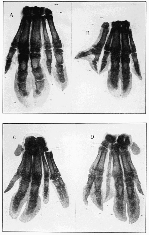
We may assume that the primitive and typical mammalian foot was pentadactyl, in spite of Bardeleben's contention that the progenitors of the mammalia possessed not five but seven digits. Bardeleben's assumption was based upon the observation that certain mammals, the whale, for example, have more than five digits; that among five-toed forms six and seven digits occasionally occur; and that in many species small cartilages are present on each side of the hand and foot. These cartilages Bardeleben regards as digital rudiments, and the occurrence of extra digits is explained by him as reversion, a 'turning back' through heredity, to ancestral conditions. Unfortunately, the facts do not support this beautiful theory. Paleontology tells us that the forerunners of the mammalia possessed only five toes. Embryology has shown that the sixth digit of the whale, and the cartilages which Bardeleben supposes to be digital rudiments, develop secondarily some time after the typical five digits have appeared. Finally, observations have proved that the extra digits which occur in polydactylism do not develop from Bardeleben's 'digital rudiments,' but originate in an entirely different manner. We may, therefore, assume that the primitive mammalian foot was pentadactyl, and this being so, the occurrence of six or seven digits on a foot normally five-toed can not be attributed to reversion, unless we assume with Albrecht that it is reversion to the many-rayed fins of the Elasmobranch fishes, an absurd supposition. Such cases of polydactylism are, nevertheless, of frequent occurrence on the appendages of man and the cat. They have been explained as due to bifurcations or duplications of one of the typical five digits. Dissections show that this is really the case, for, though the skeletal elements are often distinct from each other, muscle tendons and nerves are bifid, and in many cases the bones of the extra digit are more or less closely united to those of a normal toe. The question next arises as to whether these digital bifurcations are due to external influences or to internal variations of the germ plasm. Ahlfeld has observed that digital duplications may be caused 'in utero' by pressure from the thread-like outgrowths of the amnion. He attempted to make this explanation cover all cases of polydactvlism, but there are several serious objections. In the first place, the extra digits generally occur on both hands or on both feet, often on all four extremities (Fig. 1, A-D). The middle digits, moreover, arc not generally affected, but the duplication is chiefly of the first and fifth. Finally, and most important, the abnormality is strongly inherited and may increase in degree during successive generations. Thus Fackenheim cites the case of a woman born of normal parents. She had the little finger duplicated on each hand. Of two sons, one inherited the mother's extra fingers and the other had besides extra small-toes on both feet. Of eight children, three were normal, three had six toes and two had six fingers on both right and left extremities. In three succeeding generations the abnormality appeared, now on the hands, now on the feet, and in two cases on all four extremities; in two cases seven toes were present on both feet.
Similar observations have been made by Poulton and Torrey in families of cats. It is evident that extra digits produced by the chance pressure of amniotic threads would not be inherited, and that such chance pressure would certainly affect now one digit, now another; whereas, we have seen that the first and fifth digits are chiefly affected. Of twelve cases studied by the writer all were of the latter type.
It will be observed in Fig. 1, A-D, which represent the extremities of one child, that the fifth digit is affected differently in each case. In fact, it has been pointed out that occasionally no extra digit may be produced, that the first or fifth digit may simply be abnormally large. These facts, together with the frequent inheritance of the extra digits, show that we have to do here with variations of the germplasm. The first digit of man has been modified, and the fifth slightly reduced. Variation most often affects organs whose structure has been recently changed, and the variation or duplication of these digits might be naturally expected.
We are warranted, then, in assuming that the abnormal occurrence of six or seven digits on the five-toed extremity is not due to reversion. They are rather duplications of the normal digits, produced either by external influences or, more frequently, by germinal variation.
As the five-toed extremity is the primitive type of mammalian foot it is but natural to conclude that the appendage with less than five toes has lost some of the original number of evolutionary changes. The circumstantial proofs of such reductions are too well known to require more than a brief statement. Rudiments supposed to represent the absent first digit are found in the pes of the dog and the manus of the pig (Figs, 2, A, 3, C). The feet of sheep and cattle exhibit pairs of vestigial bones and hoofs, called the rudiments of digits 2 and 5; the splint bones of the horse are believed to be the vestiges of the second and fourth digits. These rudiments are often better developed in the embryo than in the adult. Thus of the dog's hallux only the upper part of the metatarsal bone remains. According to Bonnet, all the skeletal parts of this digit are formed in the embryo. The second and fifth digits of the sheep, represented by mere vestiges of the phalanges, are fully developed in the land). The foot of the adult horse shows only the metacarpals and metatarsals of digits 2 and 4; but in the embryo the writer has observed two cartilaginous phalanges on the metacarpal bones. Paleontology completes the ring of circumstantial evidence by showing us that the ancestors of the swine had five instead of four toes and that the forerunners of the ruminants and the Equidæ had three, four or five functional digits.

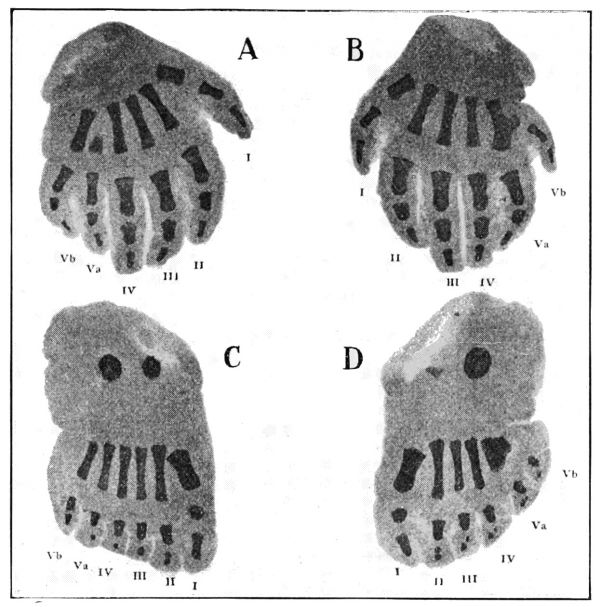
Fig. 1. X-ray photographs of a child's extremities showing duplication of the fifth digit in both hands and both feet. Va, Vb, the digits produced by duplication.
The question now arises as to whether the occurrence of extra digits on extremities normally possessing less than five toes is due to duplication, as in pentadactyl animals, or are the extra digits developed from the rudimentary structures we have described? If it can be shown that the supernumerary toes are due to reversion, we have no longer circumstantial evidence, but direct proof that the extremities of the ungulate have been derived by evolution from a five-toed type. This is an important point, but one about which investigators have been at variance. Bardeleben, Kollman, Marsh, Blanc and others recognize all cases of polydactylism as due to reversion. Gegenbaur warns us against such general conclusions, but admits that the extra digits sometimes found on the extremities of the horse are developed from the digital rudiments. Weismann, Bateson and Wilson ascribe all such abnormalities to germinal variation. But germinal variation may affect the rudimentary as well as the functional digits; if through such variation the supposed rudiment of a thumb develops into a digit with two phalanges, germinal variation and reversion are one and the same thing.

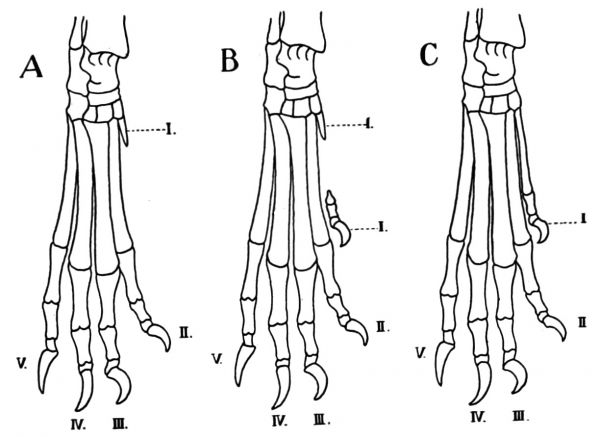
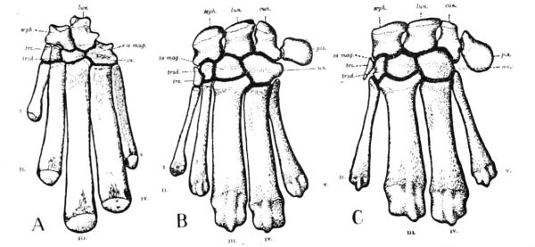
Fig. 2. A series showing the normal and polydactyl structure of the skeletal elements in the pes of the dog. A, normal pes with the rudimentary metatarsal bone of digit I.; B, a polydactyl pes, the hallux represented by two phalanges and the distal end of the metatarsal bone; C, polydactyl pes with hallux (I.) completely developed.
To attempt to reconcile the conflicting statements of various investigators the writer has made a comparative study of polydactylism in mammals normally possessing less than five toes. It was found that in the majority of cases the extra digits are developed from the so-called digital rudiments. This is most frequently observed in the pes of the dog. Normally the hallux, or great toe, is represented only by the proximal end of the metatarsal bone ( Fig. 2, A). Not infrequently, a claw and one or two phalangeal bones may appear at the point where the hallux should be; occasionally the distal end of the metatarsal bone is also represented, and sometimes a complete digit with all the bones and articulations of a functional hallux may be developed. Such cases, which may be regarded with certainty as reversions to the five-toed type of foot, occur not rarely on the pes of the Scotch collie, St. Bernard and Newfoundland (Fig. 2, B, C).
Fig. 3. A series to show the reversion of the pig's manus to the pentadactyl condition. A, carpals and metacarpals of the fossil Ancodus; B, of a polydactyl pig; C, of a normal pig. I.-V., first to fifth digits; trz., trapezium, the carpal element of the pollex.
In the foot of the pig the hallux is gone and the pollex is normally represented by a small carpal rudiment (Fig. 3, C). A small pollex was, however, present in the manus of Ancodus, one of the fossil swine (Fig. 3, A). It is, therefore, an interesting fact that in the polydactyl swine observed by the writer the extra digits were in every case located upon the manus, and in most instances were undoubtedly developed from the rudiment of the pollex; for the extra digit was attached to the carpal bone as a normal pollex would be, and careful dissections of muscles and nerves gave no evidence of duplications. This does not support Gegenbaur's assertion that the extra digits of swine were developed by the splitting of the second toe. His conclusion was based on the dissection of two ' pig's knuckles ' cut off below the carpus. Consequently he could not tell how the extra digit was attached. In any case this was scanty evidence on which to base a general conclusion. The writer was fortunate enough to obtain for study thirty-six perfect specimens. In one type observed, a small hoof, two phalanges and the distal end of the metacarpal bone were developed (Fig. 4, B) and in several cases a perfectly formed pollex was present (Fig. 4, C). In its general structure the manus of such polydactyl pigs resembles closely that of the fossil swine Ancodus, as may be seen by comparing A and B of Fig. 3. In other instances not a pollex, but a digit of three phalanges, was produced, and these in turn exhibited all stages of duplication up to the formation of two large extra toes. But in each case the two extra toes were developed by the variation of the rudimentary pollex (Fig. 5, A-D).

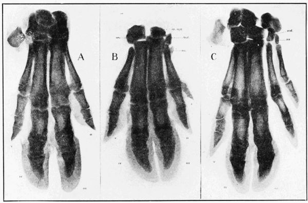
Fig. 4. X-ray photographs of the pig's manus showing normal structure and reversionary polydactylism. A, bones of normal manus; B, manus in which the pollex is represented by two phalanges and the distal end of the metacarpal bone (I.); C, manus with pollex completely developed; trz., trapezium.
Fig. 5. A series of four X-ray photographs showing variations and duplications of the pollex (I.) in the manus of the pig. A, a manus in which the pollex is represented by an abnormally large digit of three phalanges; B, the phalanges of the extra digit are duplicated: C, all the bones of the extra digit are duplicated, but both sets of phalanges are enclosed within a single hoof; D, two extra digits are present, articulating with a single trapezium (trz.).
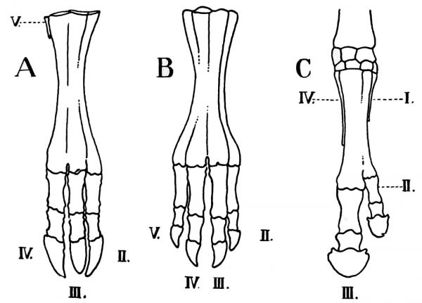
Fig. 6. A, calf's manus with digit II. fully developed (from X-ray photograph); B, manus or sheep with two extra digits (II. and V.) present (after Chauveau); C, manus of horse with two extra digits (I. and II.) (after Marsh).
The writer has also observed two cases in which the second digit of the ox was developed into a functional toe (Fig. 6, A), and in the foot of the sheep four complete digits sometimes occur (Fig. 6, B). As far back as Roman times the horse is known to have possessed extra toes. Suetonius alludes to a horse given to Julius Cæsar 'which had feet that were almost human, the hoofs being cleft like toes.' Two cases were described by Winter in 1703, and Marsh has since observed the development of an extra digit from one of the splint bones (Fig. 6, C); four or five digits may sometimes occur, but all of these are not completely developed.
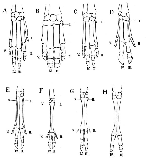
It is thus clear that the vestiges regarded as digital rudiments are really such, and that mammals possessing these vestiges must at one time have had a greater number of functional toes, some of which later became useless. It is a well-known theory that this reduction in the number of digits was in adaptation to some special function like that of locomotion. It has been carried to the extreme in the foot of the hoofed mammals; and of living forms, the swine and ruminants afford a beautiful series of digital reductions (Fig. 7, A-H). Even among living carnivora, forms like the cat and dog have the pollex reduced and the hallux absent, and, as we have seen, the forerunners of the swine had a reduced pollex on the manus (Fig. 7, A), and only four digits on the pes. The first digit is vestigial among the hippopotami; the second and fifth are slightly smaller than the third and fourth (Fig. 7, B). The difference in the size of the two pairs of digits is more marked in another fossil pig, but the small outer digits still articulate firmly at the wrists and ankle joints (Fig. 7, C). The third and fourth toes of the swine are relatively much larger and have taken unto themselves the whole articular surface of the carpus and tarsus (D).
Like the pig, the little water-deer (Dorcatherium) possesses four distinct functional toes, but in Tragulus, a closely related form, the outer toes are exceedingly slender and do not articulate proximally (E). The upper ends of these small digits have been reduced in the foot of the roebuck (Capreolus carca); in the extremities of the red deer (Cervus elaphus), these digits are represented only by the bones of the phalanges and vestiges of the metacarpals and metatarsals (F). In the foot of the sheep the outer digits are reduced to two small phalanges (G); these are absent in the foot of the ox and the antelope. Finally, the small hoofs, the only vestiges of the second and fifth digits of the ox, disappear in the extremities of the giraffe and the camel (H).
This series of extremities thus shows a reduction from five to two digits. The gradual atrophy of these three toes has been ascribed to the specialization of the foot as an organ of rapid locomotion. Primitive mammals were plantigrades, resting the whole surface of the foot upon the ground in running. This posture is not favorable for rapid locomotion, as instanced by the lumbering gait of the bear. It has been retained only by animals which, through burrowing, swimming, climbing or means other than speed are enabled to escape their enemies or obtain their food. But both to beasts of prey and to their quarry increased speed and leaping power would be of great advantage in the struggle for existence. To obtain this advantage they had recourse to the same expedient to which on occasion plantigrade man still resorts—they ran upon their toes. If by variation, the digitigrade position became gradually, or suddenly, the fixed posture of the foot in progression, the structure of the digits would soon be affected. Provided that the feet were used only in locomotion, the shorter digits would not reach the ground. Being useless, they might soon disappear. The reduction of the digits has, therefore, been ascribed simply to the adoption of the digitigrade posture. This is, indeed, the chief, but it is not the only factor. It does not explain why the hallux of the dog and cat has atrophied, while the pollex persists; why the pig and waterdeer have four digits, the giraffe and camel only two, though all are digitigrade.
There are evidently three factors upon which the degree of digital reduction depends: (1) the specialization of the extremity for locomotion; (2) the degree of perfection to which the digitigrade posture is carried; (3) the character of the ground which the animals traverse.
Fig. 7. A series of arterio-dactyl extremities showing successive reduction of the digits from five to two. A, Ancodus (fossil); B, Hippopotamus; C, Hyopotamus (fossil): D, Sus; E, Tragulus; F, Cervus; G, Ovis; H, Camelus.
The hallux of the dog and cat has been reduced, because the pes is used only for progression and in the digitigrade position. The pollex of these animals has been retained, as the claw is useful to the cats in climbing and in catching their prey—to the dogs and wolves in burrowing and in holding their prey. It is interesting to note that the pollex is no longer a functional organ in the manus of the hyena, an animal which feeds chiefly on carrion.
To the Carnivora, which are beasts of prey, a padded foot and sharp claws are necessary structures. We, therefore, find that the digitigrade posture is not developed to the extreme. These animals run upon the ball of the foot; whatever may be the character of the country traversed, all four toes are used in progression, and no further reductions have taken place.
To the herbivorous ungulates claws are useless structures, and in escaping from their foes noise is no drawback. Speed is their chief requirement and this is increased by leaping from the tips of the toes. This method of progression would blunt the claws, which would then be modified to protect the toes.
If the digitigrade position is not well developed (as is the case with the slow, heavy ungulates like the elephant, tapir and rhinoceros) all the toes, or all but the first, may reach the ground and function in locomotion. But if the digitigrade posture be extreme, the fate of the digits will depend upon the habitat of the animal. Should the region ordinarily traversed offer smooth, firm footing, an animal running upon the tips of its toes would use chiefly the longer third digit; this digit would naturally be strengthened and increased in size, while the other digits, being useless, might gradually disappear. Such changes have taken place in the foot of the horse, and in this way the perissodactyl, or odd-toed, type of foot has arisen. As is well known, the habitat of the horse family is the dry rolling plain, and what evidence we have goes to show -that the remote ancestors of the horse ranged a similar country. The fact that the digits of the horse were first reduced at their distal extremities points to the same conclusion. For, as we shall see, digits which do not support the weight of the body. may. if the animal frequents swampy regions, keep the foot from sinking too deeply. In such cases the reduction of the digits begins always at the upper or proximal end, just the reverse of conditions in the foot of the horse. We may conclude then that the odd-toed foot resulted from digitigrade locomotion over firm, comparatively level ground.
For rapid progression over swampy ground, the structure of the foot must conform to two requirements. It must be prevented from sinking too deeply and must be easily withdrawn. Any one who has attempted to walk across a mud flat can appreciate the importance of these two factors. It is in adaptation to these requirements that the artiodactyl, or even-toed, type of foot has evidently been developed. If an ungulate was of semi-aquatic habits, or attached to swampy places, all four digits, by spreading, would prevent the sinking of the foot; its withdrawal would be facilitated by making the foot occupy as little space as possible, and this could be accomplished by shifting the proximal articulations of the outer digits inward and posterior to the middle digits. The toes would then be arranged in pairs, the outer pair lying somewhat behind the other. Now in a semi-aquatic animal each pair of digits would be subjected to the same usage in walking or running through boggy ground; the digits of each pair would tend toward the same structure on this account. But as the middle digits would support the greater part of the strain brought to bear upon the foot, this pair of digits would naturally become larger and stronger than the other. Should our hypothetical ungulate change its habitat from the swamp to the firmer footing of the plain or upland, the outer pair of digits would not reach the ground, and unless they proved of use to the animal in some other way we should expect them to speedily disappear.
This theory as to the origin of the artiodactyl foot is supported when we examine into the habits of the even-toed ungulates. The more primitive forms are attached to the water. The amphibious hipppotamus possesses, as we should expect, four digits, arranged in pairs of equal size. The wild swine, although attached to water and to boggy ground, are nevertheless swift runners, and spend most of their time on more solid footing. The outer digits are retained because they are useful in keeping the foot from sinking in the mud, though they are not functional a good share of the time. The greater strains brought to bear upon the middle digits have resulted in their increased size (by variation) and their monopoly of the carpal and tarsal joints, firm articulation with which is no longer needed by the little-used outer toes. That these changes were advantageous to the swine is shown by the fact that related forms, less adaptive in this respect, have become extinct.
Of the deer family, the water chevrotain and its relatives are the only forms possessing four complete digits. Again we have to do with animals attached to swampy places, and the outer digits, though slender, are retained intact because of the extra support they offer in traversing boggy ground. The fusion of the metacarpal and metatarsal bones of the middle digits, which characterizes the foot of other ruminants, has evidently been prevented in the water deer by the spreading of the toes.
The red deer is one of the swiftest of runners and its usual habitat is the wooded plain and upland. It, however, readily takes to the water, as a large part of its food consists of aquatic plants. In roaming the more solid floor of the forest only the middle digits support the body. These have become relatively larger than those of swine, and are further strengthened hy the union of the metacarpal and metatarsal bones. The outer digits are perfectly useless in ordinary locomotion, but still perform two important functions: they serve to support the foot in yielding ground and give the deer a firm footing when running rapidly, especially down-hill. Any one may observe that in walking on fairly firm ground the foot of the deer leaves but two hoofprints, but that the foot of a running deer leaves four distinct marks. The performance of these functions has caused the retention of the lower portion of the outer toes. But as these digits no longer support any part of the weight of the animal, no proximal articulation is necessary and we find that the upper part of the metacarpal and metatarsal bones has atrophied.
Wild goats and sheep are mountain animals, feeding on rugged and precipitous slopes where the footing is precarious. Both sheep and goats are expert climbers and leapers, but in their ordinary habitat only the middle digits are used for supporting the weight of the body. The outer digits have, therefore, been reduced, but the hoofs and rudiments of two phalanges have been retained, because these small toes are used in climbing, and render the animals more sure-footed. No such function is performed by the second and fifth digits of the antelope, bison and ox; these animals roam the open plain and upland, and the outer toes would be of no use except for the occasional support of the foot when these animals enter the water to drink. We find, therefore, that only the small hoofs of the reduced toes persist. In the foot of the camel and giraffe even these vestiges have disappeared, as their habitat has long been the dry, sandy plain.
From these observations it seems plain that among ungulates the functions of the digits have been affected by the habitat of the various animals; and that there is a direct relation between the degree of digital reduction and the character of the country traversed. The use of the foot as an organ of locomotion alone, and the assumption of the digitigrade posture, were the primary factors producing reductions of the toes; the degree of such reductions and the type of foot produced have been dependent upon the habitat of the animals. The artiodactyl foot was formed in adaptation to semi-aquatic habits, and as the animals changed their habitat to 'terra firma' a further reduction of the digits resulted. The perissodactyl, or odd-toed, type of foot, began with the assumption of the digitigrade posture by animals which traversed solid ground, and the digits were further reduced as the digitigrade posture was developed to perfection.
It has been assumed that those digits which were useless would disappear. There is evidence that if they were not reduced they would be not only useless, but of distinct disadvantage to the animals. The writer has observed that the extra hallux rarely occurs on the pes of hunting dogs; when it does occur it is frequently injured and sometimes completely torn away. It is also noteworthy that the extra toe is most often found on the pes of the St. Bernard and Newfoundland, in which breeds it may be of some use for swimming, and walking through deep snows. It has been observed, too, that the small hoofs of the sheep and deer grow rapidly on the second and fifth digits, but are normally worn away by daily use. If these animals are kept in unnatural surroundings, as when sheep are deprived of rocky pasture, or deer kept in zoological gardens, the hoofs of the reduced digits will grow long, curved and twisted to such an extent as seriously to impede locomotion. We can readily see that should the wild deer or sheep change its habitat to the smooth footing of the open plains, the same abnormal growths might occur and hinder rapid locomotion. Variations tending toward the reduction of these digits would favor the survival of their possessors, and give rise to the type of foot found among the antelopes, cattle and giraffes.
In conclusion it may be of interest to speak briefly of the digital reductions which have taken place in the foot of the running birds (Ratitæ). The most primitive of the birds exhibited the digitigrade posture, but walked upon the ball of the foot. This may have caused the reduction of the fifth digit (which early disappeared), and certainly had to do with the reduction of the hallux. The hallux has, however, been retained by most flying birds because it is used in perching, in prehension and in swimming. As the legs of birds are set well forward on the body, they are more widely separated above than below. This position has thrown the greatest strains upon the outer or fourth digit, which is always longer than the second toe. Now in the foot of running birds like the emu, the digits are used only in locomotion and the hallux has disappeared, as it was useless in progression. But the digitigrade posture of the emu is the same as that of the flying birds; the ball of the foot touches the ground and the remaining three digits, being all functional, are well developed. The ostrich, however, has increased its swiftness by running (and walking) upon the tips of its toes. This posture would throw the weight of the body and the work of locomotion upon the longer third and fourth digits. As the foot of the ostrich is used only in locomotion, and as the birds traverse the smooth floor of the desert, the shorter second digit would fail to reach the ground and eventually disappear. As a result we find that the ostrich has only two functional digits.
The digits of birds, therefore, show structural changes which are exactly paralleled by those exhibited by various ungulates, and the digital reductions which have taken place may be attributed to the same factors in each case.
Cite this page: Hill, M.A. (2026, February 26) Embryology Paper - Extra digits and digital reductions. Retrieved from https://embryology.med.unsw.edu.au/embryology/index.php/Paper_-_Extra_digits_and_digital_reductions
- © Dr Mark Hill 2026, UNSW Embryology ISBN: 978 0 7334 2609 4 - UNSW CRICOS Provider Code No. 00098G


