Neural System - Fetal
| Embryology - 13 Jul 2026 |
|---|
| Google Translate - select your language from the list shown below (this will open a new external page) |
|
العربية | català | 中文 | 中國傳統的 | français | Deutsche | עִברִית | हिंदी | bahasa Indonesia | italiano | 日本語 | 한국어 | မြန်မာ | Pilipino | Polskie | português | ਪੰਜਾਬੀ ਦੇ | Română | русский | Español | Swahili | Svensk | ไทย | Türkçe | اردو | ייִדיש | Tiếng Việt These external translations are automated and may not be accurate. (More? About Translations) |
Introduction


During the fetal period there is ongoing growth in size, weight and surface area of the brain and spinal cord. Microscopically there is ongoing: cell migration, extension of processes, cell death and glial cell development.
Cortical maturation (sulcation and gyration) and vascularization of the lateral surface of the brain starts with the insular cortex (insula, insulary cortex or insular lobe) region occurs during the fetal period. This cerebral cortex region in the adult brain lies deep within the lateral sulcus between the temporal lobe and the parietal lobe.
This long development time generates the most complex structure within the embryo and the long time period of development means in utero insult during pregnancy may have consequences to development of the nervous system.
| neural crest | Sensory System Development | Second Trimester | Third Trimester | Category:Fetal
Some Recent Findings
|
| More recent papers |
|---|
 This table allows an automated computer search of the external PubMed database using the listed "Search term" text link.
More? References | Discussion Page | Journal Searches | 2019 References | 2020 References Search term: Fetal Neural Development <pubmed limit=5>Fetal Neural Development</pubmed> |
| Older papers |
|---|
|
Human Neural Timeline

Timeline of events in Normal Human Neural Development[7]
Fetal - Second Trimester

|
|
| Brain and Ventricular Development[1] | Brain Fissure Development[1] |

|

|

|
| Three months (median sagittal section) | Four months (inferior surface) | Five months (outer surface) |
PMID 3339373
At about 19 weeks (GA 21 weeks) neuronal migration ends and the radial glial cells that aided the migration now become transformed into astrocytes and astrocytic precursors.[8]
Fetal - Third Trimester
Three-dimensional magnetic resonance imaging and image-processing algorithms have been used to quantitate between 29-41 weeks volumes of: total brain, cerebral gray matter, unmyelinated white matter, myelinated, and cerebrospinal fluid (grey matter- mainly neuronal cell bodies; white matter- mainly neural processes and glia).
A study of 78 premature and mature newborns showed that total brain tissue volume increased linearly over this period at a rate of 22 ml/week. Total grey matter also showed a linear increase in relative intracranial volume of approximately 1.4% or 15 ml/week. The rapid increase in total grey matter is mainly due to a fourfold increase in cortical grey matter. Quantification of extracerebral and intraventricular CSF was found to change only minimally.[9]

Cortical surfaces for neonates at 28, 36 and 44 weeks PMA at scan with the labels overlaid.[10]
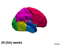
28 weeks PMA
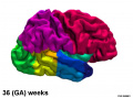
36 weeks PMA
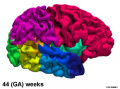
44 weeks PMA


Thyroid System and Neural Development

Timeline of human thyroid system and brain development from conception to birth.[11] (Estimation of neurogenesis adapted from Bayer et al.[12])
- Links: thyroid
Sulcation and Gyration
Cortical maturation (sulcation and gyration) and vascularization of the lateral surface of the brain starts with the insular cortex (insula, insulary cortex or insular lobe) region during the fetal period. This cerebral cortex region in the adult brain lies deep within the lateral sulcus between the temporal lobe and the parietal lobe.
- sulcation - The process of brain growth in the second to third trimester which forms sulci, grooves or folds visible on fetal brain surface as gyri grow (gyration). Abnormalities of these processes can lead to a smooth brain (lissencephaly).
- gyration - The development of surface folds on the brain (singular, gyrus)
Insular Gyral and Sulcal Development
- 13-17 gestational weeks - appearance of the first sulcus
- 18-19 gestational weeks - development of the periinsular sulci
- 20-22 gestational weeks - central sulci and opercularization of the insula
- 24-26 gestational weeks - covering of the posterior insula
- 27-28 gestational weeks - closure of the laeteral sulcus (Sylvian fissure or lateral fissure)
(Data from: Afif A, etal., 2007)
Three-dimensional magnetic resonance imaging and image-processing algorithms have been used to quantitate between 29-41 weeks volumes of: total brain, cerebral gray matter, unmyelinated white matter, myelinated, and cerebrospinal fluid (grey matter- mainly neuronal cell bodies; white matter- mainly neural processes and glia). A study of 78 premature and mature newborns showed that total brain tissue volume increased linearly over this period at a rate of 22 ml/week. Total grey matter also showed a linear increase in relative intracranial volume of approximately 1.4% or 15 ml/week. The rapid increase in total grey matter is mainly due to a fourfold increase in cortical grey matter. Quantification of extracerebral and intraventricular CSF was found to change only minimally.
(Text - modified from Huppi etal., (1998) Quantitative magnetic resonance imaging of brain development in premature and mature newborns. Ann Neurol 43(2):224-235.)
Neural development will continue after birth with substantial growth, death and reorganization occuring during the postnatal period.
References
- ↑ 1.0 1.1 1.2 Huang H, Xue R, Zhang J, Ren T, Richards LJ, Yarowsky P, Miller MI & Mori S. (2009). Anatomical characterization of human fetal brain development with diffusion tensor magnetic resonance imaging. J. Neurosci. , 29, 4263-73. PMID: 19339620 DOI.
- ↑ Matthews LG, Walsh BH, Knutsen C, Neil JJ, Smyser CD, Rogers CE & Inder TE. (2018). Brain growth in the NICU: critical periods of tissue-specific expansion. Pediatr. Res. , 83, 976-981. PMID: 29320484 DOI.
- ↑ Friedrichs-Maeder CL, Griffa A, Schneider J, Hüppi PS, Truttmann A & Hagmann P. (2017). Exploring the role of white matter connectivity in cortex maturation. PLoS ONE , 12, e0177466. PMID: 28545040 DOI.
- ↑ Goldstein IS, Erickson DJ, Sleeper LA, Haynes RL & Kinney HC. (2017). The Lateral Temporal Lobe in Early Human Life. J. Neuropathol. Exp. Neurol. , 76, 424-438. PMID: 28498956 DOI.
- ↑ Rajagopalan V, Scott J, Habas PA, Kim K, Corbett-Detig J, Rousseau F, Barkovich AJ, Glenn OA & Studholme C. (2011). Local tissue growth patterns underlying normal fetal human brain gyrification quantified in utero. J. Neurosci. , 31, 2878-87. PMID: 21414909 DOI.
- ↑ Rolo LC, Araujo Júnior E, Araujo E, Nardozza LM, de Oliveira PS, Ajzen SA & Moron AF. (2011). Development of fetal brain sulci and gyri: assessment through two and three-dimensional ultrasound and magnetic resonance imaging. Arch. Gynecol. Obstet. , 283, 149-58. PMID: 20878170 DOI.
- ↑ Report of the Workshop on Acute Perinatal Asphyxia in Term Infants, U.S. Department of Health and Human Services, Public Health Service, National Institutes of Health, National Institute of Child Health and Human Development, NIH Publication No. 96-3823, March 1996.
- ↑ Kadhim HJ, Gadisseux JF & Evrard P. (1988). Topographical and cytological evolution of the glial phase during prenatal development of the human brain: histochemical and electron microscopic study. J. Neuropathol. Exp. Neurol. , 47, 166-88. PMID: 3339373
- ↑ Hüppi PS, Warfield S, Kikinis R, Barnes PD, Zientara GP, Jolesz FA, Tsuji MK & Volpe JJ. (1998). Quantitative magnetic resonance imaging of brain development in premature and mature newborns. Ann. Neurol. , 43, 224-35. PMID: 9485064 DOI.
- ↑ Makropoulos A, Aljabar P, Wright R, Hüning B, Merchant N, Arichi T, Tusor N, Hajnal JV, Edwards AD, Counsell SJ & Rueckert D. (2016). Regional growth and atlasing of the developing human brain. Neuroimage , 125, 456-478. PMID: 26499811 DOI.
- ↑ Howdeshell KL. (2002). A model of the development of the brain as a construct of the thyroid system. Environ. Health Perspect. , 110 Suppl 3, 337-48. PMID: 12060827
- ↑ Bayer SA, Altman J, Russo RJ & Zhang X. (1993). Timetables of neurogenesis in the human brain based on experimentally determined patterns in the rat. Neurotoxicology , 14, 83-144. PMID: 8361683
Journals
- Neural Development Browse contents
- Developmental Brain Research Content Listing
- Neural Development Welcome to Neural Development | Pubmed Central Volume 1 2006 | Pubmed Central Volume 2 2007 |
- International Journal for Developmental Neuroscience Official Journal of the International Society for Developmental Neuroscience |
- Developmental Neuroscience Journal Homepage | Hippocampal Development | Vol. 29, No. 3, 2007 |
- Neuroscience Official journal of The International Brain Research Organisation (IBRO)
- Neuron Neuroscience journal published by Cell press
Online Textbooks
Developmental Biology (6th ed) Gilbert, Scott F. Sunderland (MA): Sinauer Associates, Inc.; c2000. Formation of the Neural Tube | Differentiation of the Neural Tube | Tissue Architecture of the Central Nervous System | Neuronal Types | Snapshot Summary: Central Nervous System and Epidermis
Neuroscience Purves, Dale; Augustine, George J.; Fitzpatrick, David; Katz, Lawrence C.; LaMantia, Anthony-Samuel; McNamara, James O.; Williams, S. Mark. Sunderland (MA): Sinauer Associates, Inc. ; c2001 Early Brain Development | Construction of Neural Circuits | Modification of Brain Circuits as a Result of Experience
Molecular Biology of the Cell (4th Edn) Alberts, Bruce; Johnson, Alexander; Lewis, Julian; Raff, Martin; Roberts, Keith; Walter, Peter. New York: Garland Publishing; 2002. Neural Development | The three phases of neural development
Health Services/Technology Assessment Text (HSTAT) Bethesda (MD): National Library of Medicine (US), 2003 Oct. Developmental Disorders Associated with Failure to Thrive
Search NLM Online Textbooks- "neural development" : Developmental Biology | The Cell- A molecular Approach | Molecular Biology of the Cell | Endocrinology
Reviews
Götz M & Huttner WB. (2005). The cell biology of neurogenesis. Nat. Rev. Mol. Cell Biol. , 6, 777-88. PMID: 16314867 DOI.
Greene ND & Copp AJ. (2009). Development of the vertebrate central nervous system: formation of the neural tube. Prenat. Diagn. , 29, 303-11. PMID: 19206138 DOI.
Articles
Saitsu H & Shiota K. (2008). Involvement of the axially condensed tail bud mesenchyme in normal and abnormal human posterior neural tube development. Congenit Anom (Kyoto) , 48, 1-6. PMID: 18230116 DOI.
Search PubMed
Search Pubmed: Fetal Brain Development | Fetal Spinal Cord Development | Fetal Neural Development
External Links
External Links Notice - The dynamic nature of the internet may mean that some of these listed links may no longer function. If the link no longer works search the web with the link text or name. Links to any external commercial sites are provided for information purposes only and should never be considered an endorsement. UNSW Embryology is provided as an educational resource with no clinical information or commercial affiliation.
- BrainSpan - Reference Atlases 15 pcw - Whole Brain, 21 pcw - Cerebrum, 21 pcw - Brainstem and 34 yrs - Whole Brain.
Glossary Links
- Glossary: A | B | C | D | E | F | G | H | I | J | K | L | M | N | O | P | Q | R | S | T | U | V | W | X | Y | Z | Numbers | Symbols | Term Link
Cite this page: Hill, M.A. (2026, July 13) Embryology Neural System - Fetal. Retrieved from https://embryology.med.unsw.edu.au/embryology/index.php/Neural_System_-_Fetal
- © Dr Mark Hill 2026, UNSW Embryology ISBN: 978 0 7334 2609 4 - UNSW CRICOS Provider Code No. 00098G