Neural System - Carnegie Stage 22
Introduction
| There individual serial slices have also been incorporated into a 3D model of this embryo. | |
| Central Nervous |
| Section | Name | Description |
|---|---|---|
|
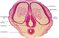
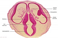
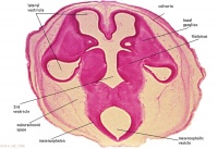
A1L | A1 - A4 In the human embryo identify the large telencephalic vesicles and the choroid plexus. The cavity in these (the lateral ventricles) communicate with the ventricle of the diencephalon (3rd ventricle) through the interventricular foramen.
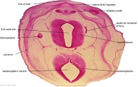
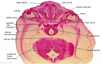
A1-A7, B1 The diencephalon. In these levels the brain comes into section twice, because of the cephalic flexure, but in A2-4 the two parts are connected. A1-5 Ventral to the thalamus you can identify the hypothalamus. |
|
A2L | Distinguish the wall of the forebrain. |
|
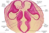
A3L | A3 - A4The basal part of the telencephalon forms the basal ganglia, a solid mass. Posteromedially these basal ganglia are in contact with the diencephalon. The large masses in either side of the diencephalon form the thalami. |
|
A4L | Description |
|
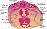
A5L | Description |
|
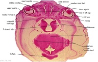
A6L | A6-A7, B1 The hypothalamus.
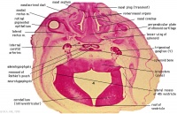
The junction between the mesencephalon and metencephalon is called the isthmus. The main metencephalic derivatives are the pons and cerebellum. The ventricular lumen of the hind brain is the 4th ventricle (A7L, B1-7, C1-3). The roof of the metencephalic part of the 4th ventricle is formed by the developing cerebellum (B2-3), beyond which the ventricle forms two large lateral recesses (B2L-B3L). In more caudal sections the roof of the ventricle is seen as a thin membrane only, bearing choroid plexus. |
|
A7L | Description |
|
B1L | Description |
|
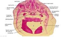
B2L | B2-B3 Further caudally the pituitary gland is seen, the neural part of which is a derivative of the diencephalon.
B2-B3 The trigeminal ganglion (CNV or CN5) and the trigeminal nerve. |
The myelencephalic part of the hind brain will form the medulla oblongata, the embryonic appearance of which is hardly different from the fully developed structure. The vagus nerve and its ganglion are seen to leave the base of the brain in B7, through the jugular foramen.
At all levels where the brain is present notice the meninges enveloping it and creating the large subarachnoid space which is filled with cerebrospinal fluid.
Study the histological appearance of the spinal cord. Note the alar and basal laminae, the dorsal root ganglia and the sympathetic trunk.
All Sections

|

|

|

|

|

|

|
| A1L | A2L | A3L | A4L | A5L | A6L | A7L |

|

|

|

|

|

|

|
| B1L | B2L | B3L | B4L | B5L | B6L | B7L |

|

|

|

|

|

|

|
| C1L | C2L | C3L | C4L | C5L | C6L | C7L |

|

|

|

|

|

|

|
| D1L | D2L | D3L | D4L | D5L | D6L | D7L |

|

|

|

|

|

|

|
| E1L | E2L | E3L | E4L | E5L | E6L | E7L |

|

|

|

|

|

|

|
| F1L | F2L | F3L | F4L | F5L | F6L | F7L |

|

|

|

|

|

|

|
| G1L | G2L | G3L | G4L | G5L | G6L | G7L |
Glossary Links
- Glossary: A | B | C | D | E | F | G | H | I | J | K | L | M | N | O | P | Q | R | S | T | U | V | W | X | Y | Z | Numbers | Symbols | Term Link
Cite this page: Hill, M.A. (2024, April 27) Embryology Neural System - Carnegie Stage 22. Retrieved from https://embryology.med.unsw.edu.au/embryology/index.php/Neural_System_-_Carnegie_Stage_22
- © Dr Mark Hill 2024, UNSW Embryology ISBN: 978 0 7334 2609 4 - UNSW CRICOS Provider Code No. 00098G
