Category:Carnegie Embryo 576
This Embryology category includes pages and images that relate to the Carnegie Collection Embryo No. 576.
| Serial No. | Size (mm) | Grade | Fixative | Embedding Medium | Plane | Thinness (µm) | Stain | Score | Sex | Year | Notes |
|---|---|---|---|---|---|---|---|---|---|---|---|
| 576 | E. 17 Ch, 60x40 | Good | Formol | P | Sagittal | 15, 20 | (Stain - Haematoxylin Eosin) | 14.5 | d | 1912 | Tubal |
References
Streeter GL. The factors involved in the excavation of the cavities in the cartilaginous capsule of the ear in the human embryo. (1917) Amer. J Anat. 22: 1–25.
Kunitomo K. The development and reduction of the tail and of the caudal end of the spinal cord (1920) Contrib. Embryol., Carnegie Inst. Wash. Publ. 272, 9: 163-198.
| Carnegie Collection - Stage 19 | |||||||||||
|---|---|---|---|---|---|---|---|---|---|---|---|
| Serial No. | Size (mm) | Grade | Fixative | Embedding Medium | Plane | Thinness (µm) | Stain | Score | Sex | Year | Notes |
| 17 | E, 18 Ch, 40x30x20 | Poor | Alc. | P | 50, 100 | Al. carm. | 16.5 | Male | 1894 | ||
| 43 | E, 16 | Good | Alc. | P | 50 | Al. coch. | 10 | Male | 1894 | ||
| 293 | E, 19 | Poor | Ale. | P | Sagittal | 50 | Coch. | 16.5 | S | 1905 | |
| 390 | E, 19 | Good | Formol? | P | Sagittal | 20, | (Stain - Haematoxylin Eosin) | 11.5 | Male | 1906 | Tubal Injected
50 |
| 409 | E.18 Ch, 50x40x40 | Good | Formalin | P | Transverse | 20 | Copper, iron H. & erythrosin | 14.5 | Male | 1907 | |
| 432 | E..18.5 Ch , 45x35x20 | Good | Formalin | P | Sagittal | 20 | H. & Congo red | 13.5 | Male | 1910 | Tubal |
| 576 | E. 17 Ch, 60x40 | Good | Formalin | P | Sagittal | 15, 20 | (Stain - Haematoxylin Eosin) | 14.5 | d | 1912 | Tubal |
| 626 | E., 21.5 Ch., 40x30x21 | Good | Formalin | P | Transverse | 100 | Al. coch. | 14_5 | 6 | 1913 | |
| 6??8 | E, 20 Ch, ca. 30 | Poor | Formalin | P | Sagittal | 50 | Al. coch. | 12 | 9 | 1913 | Head damaged |
| 709 | E, 19 Ch. 40x35x25 | Poor | Alc. | P | Coronal | 40 | Al. coch, Lyons blue | 15 | 49 | 1913 | |
| 837 | E. 21 Ch. 65x45x | Good | Formalin | P | Sagittal | 40 | Al. coch. | 14.5 | P | 1914 | |
| 1324 | E., 18 50x30x18 | Good | Formalin | C | Coronal | 40 | (Stain - Haematoxylin Eosin), aur, or. G | 125 | 79 | 1915 | |
| 1332 | E., 19 Ch., 40x43x22 | Poor | Formalin | C | Coronal | 40 | (Stain - Haematoxylin Eosin) aur, or. G. | 15 | Male | 1915 | |
| 1390 | E., 18 Ch, 40x38x15 | Good | Formalin | P | Sagittal | 20 | Al. coch. | 10_5 | Male | 1915 | Tubal |
| 1534 | E., 13 Ch.,35x31x25 | Poor | Formalin | P | 53% | 50 | Al. coch. | 13.5 | F | 1916 | Protruding midbrain |
| 2114 | E., 19.3 Ch., 49x42x33 | Good | Formol | P | Transverse | 40 | A1. coch. | 12 | M | 1918 | |
| 4405 | E., 15.5 | Good | Formalin | P | Transverse | 10 | Coch, Mallory | 13.5 | <3 | 1923 | Midbrain injured |
| 4501 | E, 18 | Exc. | Bouin | P | Transverse | 15 | Coch, or. G. | 14.6 | 1924 | Cystic left kidney | |
| 5609 | E., 18 | Exc. | Formalin | P | Coronal | 25 | A1. coch. | 13.5 | Male | ||
| 6150 | E., 17 Ch., 40x39x30 | Good | Bouin | C-P | Transverse | 15 | (Stain - Haematoxylin Eosin) | 16.5 | Male | 1930 | Tubal |
| 6824 | E., 18.5 Ch., 45x40x25 | Good | Formalin | C-P | Sagittal | 12 | (Stain - Haematoxylin Eosin) | 14.5 | Female | 1933 | |
| 7900 | E., 16.5 | Good | Bouin | C-P | Sagittal | 20 | (Stain - Haematoxylin Eosin), phlox. | 11.5 | . . | 1941 | Tubal |
| 8092 | E., 16.3 Ch., 52 x 47 | Exc. | Bouin | C-P | Transverse | 20 | (Stain - Haematoxylin Eosin), phlox. | 13 | Male | 1942 | |
| 8913 | E.,? Ch, 34 | Poor | Formalin | p | Transverse | 10 | Alan . | 7 | 1951 | rubella. Medical abortion. Isolated head damaged | |
| 8965 | E, 19.1 Ch, 42x32x19 | Good | Formol—Zenker | C-P | Transverse | 10 | Borax, carm, or. G. | 1952 | Univ. Chicago No. H 173 | ||
| 9097 | E, 21 | Exc. | Formol—glucose | C-P | Coronal | 10 | Azan ? . | ? | 1930 | Univ. Chicago No H 1380 | |
| 9113 | E, 185 Ch, 24 | Exc. | Formalin | C-P | Transverse | 10 | Alan > 6 | 1953 | Rubella. Medical abortion | ||
| 9325 | E, 17.0 Ch, 32x28x20 | Good | Formalin | —acetic p | Transverse | 15& 8-10 | Azan & Ag | ? | - | 1955 | Tubal |
Abbreviations
| |||||||||||
Embryo No. 576, 17 mm Crown-Rump Length
Kunitomo K. The development and reduction of the tail and of the caudal end of the spinal cord (1920) Contrib. Embryol., Carnegie Inst. Wash. Publ. 272, 9: 163-198.
This embryo has 35 cartilaginous vertebrae, the last consisting of three small pieces fused together. The demarcation between these pieces can be more clearly recognized in the lateral portions of the column than in the median plane; so in determining the composition of the last segment one nuist study carefully the more lateral line of sections. A profile reconstruction of the embryo is shown in figure 3<S, and a more diagrammatic sketch in figure 10. The tail, with the caudal end of the .spinal cord, is bent shari)ly dor.salward. The spinal cord narrows suddenly at the thirty-second vertebra anil from this point down the central canal, which extends the entire length of the cord, becomes much smaller and rounder, while in the more cranial portion a transverse section of it would form an elongated oval. The ventral wall of the canal in the atrophic jiortion presents several transverse folds, as seems to be usually the case at this stage of reduction. The caudal portion of the chorda dorsalis is convoluted and its end sharply retracted.
Cite this page: Hill, M.A. (2026, April 8) Embryology Carnegie Embryo 576. Retrieved from https://embryology.med.unsw.edu.au/embryology/index.php/Category:Carnegie_Embryo_576
- © Dr Mark Hill 2026, UNSW Embryology ISBN: 978 0 7334 2609 4 - UNSW CRICOS Provider Code No. 00098G
Pages in category 'Carnegie Embryo 576'
The following 2 pages are in this category, out of 2 total.
Media in category 'Carnegie Embryo 576'
The following 3 files are in this category, out of 3 total.
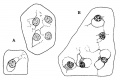 Streeter1917 fig05.jpg 800 × 531; 57 KB
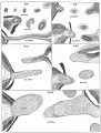 Streeter1957 fig05.jpg 1,280 × 1,690; 546 KB
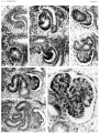 Streeter1957 plate04.jpg 1,500 × 1,986; 736 KB


