Book - Manual of Human Embryology 10
Pinkus F. X. The development of the integument. in Keibel F. and Mall FP. Manual of Human Embryology I. (1910) J. B. Lippincott Company, Philadelphia.
| Historic Disclaimer - information about historic embryology pages |
|---|
| Pages where the terms "Historic" (textbooks, papers, people, recommendations) appear on this site, and sections within pages where this disclaimer appears, indicate that the content and scientific understanding are specific to the time of publication. This means that while some scientific descriptions are still accurate, the terminology and interpretation of the developmental mechanisms reflect the understanding at the time of original publication and those of the preceding periods, these terms, interpretations and recommendations may not reflect our current scientific understanding. (More? Embryology History | Historic Embryology Papers) |
X. The Development of the Integument
By Felix Pinkus of Berlin.
A. The Epidermis
From the beginning of development the epidermis forms the outermost investment of the body. It consists of a uniform two layered sheet, the upper layer forming a sort of hard coveringlayer while the lower one remains soft and gives rise to new cells and to all the epidermal appendages of the integument. This twolayered stage persists over most portions of the body until into the fourth month, but even at the end of the second month it is not altogether unmodified.
The regions which show the first signs of further development are all upon the ventral surface of the body, the skull and back remaining covered by an unaltered, two-layered, indifferent epidermis. In an embryo of 15 mm Kallius found the first indications of the milk ridge, and Tandler observed it later in one of the 9.75 mm But the modifications of the epidermis are not confined to the regions of the milk ridges at the sides of the body; also on the ventral surface, anteriorly over the branchial arches and posteriorly as far as the tail, changes occur which indicate a strong formative tendency. In somewhat older stages (32 mm, 40 mm) an increased tendency towards development shows itself, especially over the facial region, on the anterior surface of the face and neck by the height and regularity of the basal columnar cells, and in the region of the eyebrows, the upper lip, and chin by the distinct commencement of hair formation.
a. Early Stages
Where its formation is most simple the epidermis consists of:
- A superficial layer of flat cells, the epitrichium, or, better, the periderm (W. Krause, 1902).
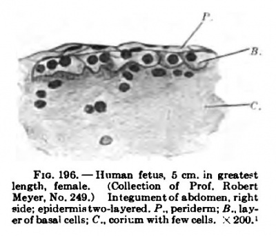
- A layer of cells greater both in height and breadth, the stratum germinativum (see Fig. 196).
Beneath the latter and sharply marked off from it is the fibrous and very cellular connective tissue.
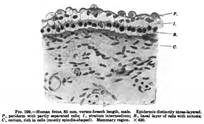
1. The periderm is the outermost layer of the epidermis. It consists, for the most part, of flat cells, which in transverse sections of the integument appear to be spindle-shaped with deeply staining, thin nuclei, while from above they appear as a layer of large polygonal cells with large roundish nuclei. Even in very early stages the peripheral portions of the cells flatten out, so that only the central portions containing the nuclei remain thick (Fig. 199). Gradually they become quite flat and unusually large (Minot, 1894). Frequently one finds some of these cells separated from the rest, so that they are seen from the surface in transverse sections, in which cases they appear as slightly irregular roundish disks with centrally placed nuclei, which are either still round or have become irregular. Around the nuclei there are frequently a large number of roundish cavities, which ^ve to the central portions of the cell the appearance of a coarse network. These are the cells which Rosenstadt (1897) found in the beak of an embryo chick, where they were full of large keratohyalin granules by whose solution the cavities are formed. Zander (1886) described them in the skin of all fingers and toes, where they were also observed by Kolliker (vesicular cells) and by Okamura (1900). As the outermost layer of the epidermis the periderm cells have the function of the later-formed corneous layer, and they actually form an investment of a horny character (as shown by their reactions: indigestibility, Unna., 1889; yel
In man the periderm is not a layer which requires to be especially distinguished as the oldest or specifically embryonic investing layer. It is only the outer layer of epidermis, whose cells are no longer turgid and have become firmer and incapable of reproduction. It merely occupies the place of the later homy layer and receives additions from the subjacent germinative layer, just as throughout life all the more superficial layers are recruited from the deepest layer, the stratum cylindricum.
Fig. 196-198 were drawn without use of a camera and consequently the enlargements cannot be given with certainty. The remaining: figures were drawn with a Zeiss-Abbe camera.
Fig. 196. — Human fetus. S cm. in greatest i o+nininc^ wifh r»ici-ic flciH length, female. (Collection of Prof. Roben '"W SiainiUg WlIU pitriC aClU, Meyer. No. 249.) in.e,™entofaixiom™ right Codercreutz, 1907). lu the more side; epidermis two-layered. P.. pendemi; 5.. lav, erol cells; C..cori.;mwithfewcells. \200.i deVeloped portions of the epidermis (as on the forehead) these cells become heaped up in two or several layers, and may even form distinct elevations, as at the nostril and mouth openings, in which cases the cells are especially large (Fig. 198).
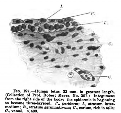
Fig. 197. Human Fetus 32 mm, in greatest lenfth (Collection of Prof. Robert Meyer, No. 307.) Measument from the right aids of the body; the epidermis is becinning to become three-layered. P., periderm; /„ sUstum bter. medium; if., etratum germinativum; C, corium, rich la eelLs;
That the periderm is added to is shown 1. By the desquamation of its cells. 2. By the arrangement of its cells in a regular layer, notwithstanding the increased growth of the skin surface. 3. By the local heaping up of layers of completely and similarly formed periderm cells in the course of development. Each of these three phenomena indicates an increase in the number of periderm cells. 2. The deeper layer, thestratumgerminativum, is the reproducing layer of the epidermis. Its cells are at first low, the breadth being equal to or even greater than the height; their nuclei are round or slightly oval, stain beautifully with a distinct chromatin network, and are very large in proportion to the entire volume of the cells. The basal surfaces of the cells, turned towards the connective tissue, are flat or slightly concave, and at first are but slightly connected with the corium, so that they readily separate in spots after the death of the fetus (maceration) or as the result of preparation. The lateral walls are variously curved, but in general but slightly, in correspondence with the pavementlike apposition of the essentially cubical cells. The outer surfaces are for the most part more or less convex. No special contents can be distinguished in their protoplasm by ordinary methods of preparation.
In those regions which already, in these early stages, show an advance in development, the cells of the deep layer become higher, and finally columnar; the nuclei are closer together and form a quite regular layer, parallel with the lower surfaces of the cells, as may be recognized by weak magnification of not too thin {15 um) sections. They stain distinctly darker and are round or oval, the long axis being perpendicular to the surface. The ttells are arranged palisade-like, close together, with perpendicular side walls ; and their upper surfaces are rather straight, forming a slightly wavy line beneath the stratum intermedium. Their lower surfaces are no longer smooth as in the first stage, but are drawn out into small projecting feet. The cell bodies are much clearer than those of the superposed layers ; they are homogeneous, without any granular contents. Since the nuclei all lie in the outer portions of the cells, the lower portions appear as a clear band between the row of dark nuclei and the dense mass of nuclei which occupies the most superficial portions of the corium.
b. Further Development
Very early tiiere appears between the periderm and the stratum germinativum a middle layer of cells, the stratum intermedium. Previous to its appearance the cells of the stratum germinativum become higher and more closely approximated, and their nuclei become round and large. First individual cells appear between the two primary layers (Fig. 197), and then a complete row of them (Fig. 199), their nuclei being small and transversely oval and the cell bodies smaller than those of the basal cells, and they take the nuclear stain (carmine) somewhat.
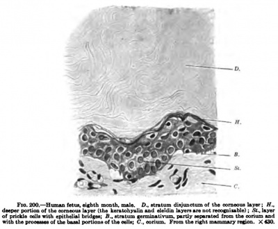
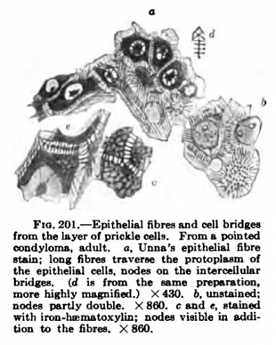
These simple conditions occur from the youngest up to rather advanced stages of development (end of the fourth month), where the Integument has not yet formed any special organs. In those places where a modification occurs, as, for example, in the region of the mouth and nose, the epithelium assumes quite early a very considerable thickness. Toward the end of fetal life tiie layer (layer of prickle cells) situated between the stratum germinativum and the corneous layer becomes the principal constituent of the epidermis. It is a solid layer, varying in thickness in different regions of the body, and its under surface forms an irregular network of ridges and convexities, which increase its surface of contact with the corium from which it is nourished (rete Malpighi). In vertical sections of the skin these ridges appear as the so-called rete papillie. The layer of prickle cells is composed of a mosaic of closely apposed and regularly spaced large cells; their nuclei are large, they have a polygonal outline in section, and are variable in form and size within narrow limits (Fig. 200). Between the layers of the twoor three-layered epidermis epithelial bridges cannot yet be made out with certainty; but as the epidermis increases in thickness, or in early stages where it has already thickened, they become distinct. With ordinary stains or when unstained they appear as prickles (Riffel, Max Schultze; filaments d'union, Ranvier), but with specific stains {Kromayer, Unna) they appear as epithelial fibres, which extend throughout a whole series of cells.
Fig. 199. Human Fetus BS mm. verMi-bmsh lenitb, male. Epidamii dirtinoUy (brae-Uyared. P., pcridenn with partly sBpantsd csIIb: /., stntum intenQedium: B„ ban] laygr of oelli with mitonij C, ooriiun, rioh in celli (moatly ipi]id]»4hap«d). Mammary refioti. X 430.
Fig. 200. Human Fetus month, male. D., stratum disjunctum of the comsous l&yer; H,. dsepsr portioD of ihe iwnieoiu Uy«r (the kentohyaliu uid eliidiu Isyen an not raccfuiuible) : St.. Uyer of prickle oella with epitheliBi bridiei; B., Btralum (ensiiiKtivum. parUy sepanled from the cxHium and ■idi theprooeaaaa of the bual portioDsof the Bella; C., oorium. From the nght mammary recioa. Xt30.
These epithelial fibres form only in the peripberal portions of the cells; these become denser and are distinguished as exopla^m from the endoplaem which contains the nucleus (Studnicka, 1903). The epithelial bridges arise by the formation of vacuoles at the boundaries between cells; the fibres differentiate from the exoplasm. In cell division the entire cell divides and both daughter cells again form on their contact surfaces a new eaoplasm layer eontaioin^ vacuoles. In a similar manner Ide (1889) regards the outer layer of the epidermis cells as a membrane, the prickles being formed by a process of drawing out, as is especially evident after division when an intervening wall is formed between the two young
Almost the same idea, that the outer parts of the epithelial cells are a membrane, is espressed by Unna (1903). according to his view the epithelial cells are in close contact, the apparent clear intervals between them (readily visible in the case of cells rich in protoplasm) not being intercellular spaces, but the outer layer of the cells, which stains with difficulty and is practically a membrane. The epithelial libres are not empty spaces or spaces merely filled with intercellular fluid; empty spaces have a very different appearance, as may be seen where the protoplasm has retracted from around material (leucocytes ) which has penetrated it. The limits between the cells are at the so-called nodes of Bizzostero, situated approximately
Fig. 201. —Epithelial fibres and cellbridew at the middle of the epithelial bridges. from the layer of Brickie cell?. From s poinied
These nodes lie in the verv narrow clefts between the cells and appear as nodes on the epitheiiii cells, nodes on the intercellular account of differences in refraction on coniification it is only this nodes partly double. X 860. c and e, iiained membrane like exoplasin layer that bewith iron-hainiBtoxyliii: nodes visi
We in addition to the fibres, X 880. , eomes comified, and the remains of the
nodes are retained on its surface.
The question whether the nodes are actually form elements or merely the result of light interference by superposed networks, is not yet detinitely settled ; it would appear that the cell walls traversed by fibres may be confused with nodes in the thin (unstained) section, for nodes are frequently seen to be united by a narrow streak parallel to the cell wall {see Fig. 201, a.d, e).
That the epithelial fibres arise from the exoplasm is generally admitted. They do not merely unite neighboring ceils, but may extend through a whole series of cells. according to Schridde (1906) certain regular fibre systems may be recognized: in the deepest layers of the epidermis they form perpendicularly placed ovals, which are found also in higher layers; nearer the surface they form circles; and at the surface horizontally placed ellipses. The form of the fibre arrangement consequently follows that of the cells, which nearer the corium are columnar, while those higher up are equal in all their diameters, and, finally, those at the surface are flattened.
c. Formation of the Stratum Corneum
Those regions in which the epidermis consists of many layers show a comification, but also in other regions of the body there early appear indications of it. These are distinctly visible in the second month, and in the third month the entire skin is undergoing comification.
Cederereutz (1907), using the method of Zilliacus, obtained the following colorations in a fetus 3.5 cm. in length:
- Yellow: the face, especially in the region around the mouth and nose (most marked in the epithelial plugs of the nostrils) and in front of the pinna.
- Yellowish: the lateral portions of the back, especially in the lower part, and also the lateral portions of the abdomen. The arrangement of the yellow spots on the body was rather distinctly symmetrical.
- Bluish-violet: the umbilical cord, the pinna, and the fingers and toes. In all other regions the skin assumed a dirty bluish-brownish green color.
In a fetus 5 cm. in length the entire body was distinctly colored yellow. The pavement epithelium, which becomes yellow with this stain, picric sublimate, and haemalum, must be regarded as comified. Bjorkenheim (1906) has shown that the same regions that stain yellow also resist pepsin and trypsin digestion — a peculiarity which in the skin and mucous membranes is associated only with comified epithelium.
The comification of the periderm, however, does not pass through the same stages as are to be seen in the formation of the definitive corneous layer. This is shown by the observation of Ernst (1896), who found that in the fourth month comification could nowhere be observed in the hand or foot, except in the nails, by the Gram method of staining, and that a uniform corneous layer first appeared on the toes in the sixth month. In young embryos whose cells of the corneous layer still retain their nuclei, keratohyalin and eleidin, which are later the constant by-products of comification, are completely wanting.
Keratohyalin and eleidin (parakeratose) are also lacking in the adult skin in places which comify (pathologically) in such a manner that the nuclei of the corneous cells remain colorable with nuclear stains (haematoxylin, methylene blue).
These substances first appear at the end of the third month in places where the epithelial cells are arranged in many layers. In fetuses 10 cm. in length keratohyalin granules are rather abundant in the face, but at this stage they are to be found elsewhere on the body only in places where longer outgrowths of the epidermis occur, as, for example, at the mouths of the long epithelial appendages around the nipples, in the anlage of the mammary gland, and, especially, in the epidermis of the nail bed. At the beginning of the fifth month keratohyalin is still lacking in the skin in general (Stohr, 1903) ; but trichohyalin has formed in the hairs and keratohyalin in the epidermis of the hair follicles. With the continued development of the skin the process of comification takes an entirely different course. The same substance that had already appeared in the early fetal stages, although in much smaller quantities, keratin, seems to result from the process ; but at the surface of the skin in later stages the cornification is usually accompanied by the formation of the by-products already mentioned. As a consequence the corneous layer, on account of characteristic refractive properties and staining peculiarities, may be divided from below upwards into certain readily distinguishable layers.
These are:
1. The stratum granulosum, with keratohyalin granules. The keratohyalin (Waldeyer), in the form of round or irregularly shaped granules (clumps, threads, occasionally bent at an angle or branched), is situated between the nuclei and the fibrillar layer of the epithelial cells, the exoplasma always forming an external investment of the cells free from granules. From it and its fibrillae the keratin is formed (Unna) ; the keratohyalin forms in the rest of the protoplasm. That the nucleus is concerned in its formation does not seem to be definitely shown by the similar staining properties of the keratohyalin and the nuclear chromatin, by the similar non-polarizing refraction of the nucleus and stratum granulosum (the layer of prickle cells, on the other hand, being doubly refractive, as well as the superficial corneous layers), and by the diminution of the nucleus as the keratohyalin increases. Arcangeli (1908), it is true, claims to have directly observed (in the oesophagus of the guinea-pig) the extrusion of keratohyalin granules from the nucleus; and Cone (1907) has seen the same outpouchings and expulsions of chromatin from the nucleus in human skin which had been kept for eighteen to twenty-four hours in a thermostat The keratohyalin is often situated at first at the periphery of the protoplasm (Weidenreich, 1901; Apolant, 1901), but collects later and preferably in the neighborhood of the nucleus. It is insoluble in our hardening fluids, and especially in water, alcohol, and ether ; but is soluble in alkalies and acids. In contrast to keratin it is soluble in hydrochloric-pepsin. It stains deeply with nuclear and acid stains (methyleosin. Zander, 1886; Rosenstadt, 1897; acid fuchsin), but not with osmic acid or Sudan (Rabl, 1902). Unna believes that with its appearance the glassy transparency of the skin of the young fetus disappears, since the keratohyalin (and eleidin), on account of its high refractive index, would render the skin opaque white.
2. The stratum lucidum (Oehl's layer) with eleidin drops. Immediately over the keratohyalin stratum is a thin layer which also remains unstained when treated with osmic acid, but after such treatment stains red with picrocarmine (Unna^s basal corneous layer, Ranvier's stratum intermedium). Upon this there follows a thicker layer, that blackens with osmic acid (Unna's superbasal corneous layer). This layer contains the eleidin, a name proposed by Eanvier for both keratohyalin and eleidin, the latter having been distinguished from keratohyalin by Unna and Buzzi in 1889. Eleidin is soluble in water and, in the fresh condition or after hardening, in strong alcohol; it stains with picrocarmine and nigrosin (Buzzi). Babl (1902) regards it as softened keratohyalin, the softening giving it the consistency of a fatty oil (keratoeleidin). according to the microchemical investigations of Ciliano (1908) it is an albumin. It does not appear in the form of granules, but either as a thickish fluid extending throughout the entire layer, or else in large drops or pools. Dreysel and Oppler (1895) could not detect it in a five-months fetus; it was abundant in the skin of one of eight months. With ordinary stains or when examined unstained in glycerin the stratum lucidum appears as a clear band. In this layer the cell membranes are already composed of keratin.
3. The stratum corneum forms the outermost portion of the skin. In its cells only the exoplasmic portion and the thickened fibrillar layer are comified (Unna; Eanvier; Weidenreich, 1901), and on their surfaces remains of the prickles can still be recognized. In the cells themselves a fat which reduces osmic acid collects and becomes very distinct in sections on treatment with osmic acid after fixation in Miiller's fluid (Rabl, 1902) ; according to Eanvier it has some resemblance to beeswax. The cells which contain this substance are flat and thin, but possess the property of swelling after an infiltrating injection of fluid into the skin. Probably the fat (pareleidin, Weidenreich) is formed in some way from eleidin (Rabl). Apolant (1901) assumes that the eleidin must be expelled from the flat corneous scales which represent completely cornified cells. The most superficial portion of the corneous layer stains diffusely with osmic acid. It has been termed by Eanvier the stratum disjunctum and by Unna (1883) the superficial corneous layer.
In the horny substance there are two kinds of substances which react differently to chemical reagents (Unna and Golodetz, 1908) : (1) those which are not digested by hydrochloric-pepsin and which color red with Millon's reagent (presence of tyrosin), but are insoluble in fuming nitric acid or in sulphuric acid + hydrogen peroxide (keratin A) ; and (2) those which also are undigested by hydrochloric-pepsin and stain red with Millon's reagent, but are soluble in the strong acids mentioned (keratin B).
d. Granule Inclusions of the Epidermal Cells
While the cells, as they pass toward the surface, become cornified and in so doing form keratohyalin, eleidin, a wax-like substance, and tyrosin, the deeper layers contain other substances which are not found nearer the surface.
1. Pigment
Pigment (melanin) occurs only in the deepest layers of the epidermis and develops chiefly only after birth. In a six months fetus Dreysel (1895) found no pigment; the granules which blackened with osmic acid did so also after previous treatment with chromic acid, a reaction which indicates fat but always destroys melanin. The extra-uterine development of pigment is much more distinct in the dark races than in Europeans. Negro children are quite light-colored at birth; they become brownish yellow on the second day and thereafter darken rapidly (Wieting and Hamdy, 1907), so that in six weeks they have reached the normal degree of darkness (Falkenstein; at four months according to Frederic, 1905). The children of the Australian aborigines are pale yellow with the exception of some black lines around the mouth, eyes, and nails — but by the broadening of these lines they become black in a few days (Gunn, according to Merkel). The pigment of the epidermis is much more abundant than that which occurs in the corium; indeed, the latter may be entirely lacking. The epidermal pigment seems to be formed in situ. That it may be formed there is shown by experiments on the adult (exposure to Finsen light rays for two hours, accompanied by cooling and deprivation of blood, after which treatment excised portions of skin, examined immediately, show an increase in the amount of pigment), by the pigmentation of skin from a cadaver (in incubator, Meirowsky, 1906, 1908), and by the pigmentation of vitiligo spots in the absence of pigment in the corium (Buschke, 1907). I'urthermore, the preferential presence of pigment in the skin, chorioid, and ependjma is an indication of a special pigment-forming faculty of the ectoderm ( chromatophoroma of the central nervous system).
In the epidermis the pigment granules occur:
- a. In the ordinary epithelial cells, at first surrounding the nucleus (Grund, 1905; Meirowsky, 1906, who regards the pigment as a transformation of the nucleolar substance), but later mostly in a cap-like mass on its outer surface.
- b. In stellate cells (chromatophores), which resemble the Langerhans cells. Whether these apparently stellate cells are really branched or whether the processes extending out from them are not streams of pigment granules passing out into the spaces between the small contact surfaces of adjacent ordinary rete cells (Rabl), is not as yet determined. according to Meirowsky 's (1908) observations these cells develop from ordinary epidermis cells.
The similarity of the chromatophores to the greatly branched Langerhans cells is striking, these latter cells, as well as the fonner, being demonstrable with especial distinctness and in large numbers by the gold and silver methods (Ramon y Cajal's and Levaditi's silver method), so much so that on the basis of this reaction the Langerhans cells have already and again recently been actually termed colorless pigment cells (Schreiber and Schneider, 190S; Bizzozero, 1908). The questions in discussion turn upon whether the epithelium always forms pigment in situ (Post, 1893, the development of feathers; Jarisch, 1892, no pigment in the hair papillee; Sehwalbe, 1893, the change of hair coat in the ermine, in which the white winter coat contains no pigment, while the summer coat makes its appearance pigmented; Rosentadt, 1897), whether the epithelium also forms the spider-like cells (melanoblasts) (Grund, 1905; L. Loeb, 1898; Wieting and Hamdy, 1904, the gradual pigmentation of the nose in new-bom dogs), which then may wander from the epidermis into the cutis (pigment stored up in the lymph-nodes, shown by Jadassohn, 1892, in pityriasis rubra and by Schmorl, 1893, in the negro), or whether the chromatophores are originally pigmented connective-tissue cells which wander into the epidermis and supply its cells with pigment (Ehrmann, 1896; in the unpigmented egg of the triton there is formed in the mesenchyme, when the development of the blood begins, a series of dark cells, melanoblasts, which later become rich in pigment granules and give rise to all the pigment in the body. No pigment is formed in the epithelium, it is carried there by the melanoblasts; they appear about the hair anlagen at an early period, even before the formation of the papillae). according to all recent works the idea that pigment is formed in the epidermis itself seems to be well founded.
2. Glycogen
Glycogen is abundantly present in the embryonic epidermis (S. H. Gage, 1906). After the sixth month it diminishes in quantity and is finally to be found only in the cells in which it occurs in the adult (Lombardo, 1907). The epithelium of the sudoriparous glands, especially, regularly contains glycogen, the more the more actively they are secreting; furthermore, the outer root sheath of the growing hair, from the bulb to the insertion of the muscle, contains it, while in that of the bulb hairs it is wanting (Brunner, 1906; Lombardo).
3. Fat
Fat is especially evident in the basal columnar cells and in the stratum granulosum even from the fifth month. The layer of prickle cells which intervenes between these is practically fat-free, its fat having presumably been used up during the divisions of the cells. As evidence for the absence of fat in the prickle cells it may be stated that post mortem no fat appears in them when they are placed in a thermostat, while in twenty-four to seventy-two hours the quantity of it in the stratum cylindricum and stratum granulosum is greatly increased (Cone, 1907). In the layer of columnar cells the fat, made evident by fettponceau, surrounds the nudeus in the form of granules of varying size (up to one-quarter the size of the nucleus) and streams out toward the periphery of the cells. In the stratum granulosum it is equally distributed from the cell periphery to the edge of the nuclear cavity, some loops lying even within this (according to Unna it is completely filled by a fat drop). Much fat is also present in the stratum lucidum, where it is more irregular both in form and distribution than in the stratum granulosum. In the actual stratum comeum it lies especially between the cells. In between the epithelial cells processes of branched connective-tissue cells filled with fat granules (lipophores, Albreeht) extend from below.
B. Corium
The connective-tissue portion of the skin, the corium, is developed from the most superficial portions of the somites. Ventrally it arises from the outer part of the dermomuscular plate and is applied to the myotomes so that, together with the epidermis, it sinks down into the furrows between suCoessive myotomes (sub-epithelial segments, Osc. Schultze, 1897). The case is similar dorsally, where the segmental furrows are continued between the sclerotomes which surround the spinal cord. The segmental furrows, however, soon disappear. The corium or dermis is at first very cellular, and the two portions into which it later divides, the corium proper and the subcutaneous fascia (tela subcutanea), are not distinguishable in the second month. Its oval cells elongate and begin to form fibrils (observed by Spalteholz, 1906, in the fifth week) in their own protoplasm, and these soon anastomose and form a delicate, somewhat irregular network, which, according to Spalteholz, remains throughout life unsheathed by the protoplasm mass and in connection with its original cells. A regular arrangement of the bundles of fibrils makes its appearance at about the end of the third month. In embryos 7-8 cm. in length the bundles arrange themselves in the lower part of the body in parallel bands running around the body (a result of the stretching of the skin by the growing liver). A little later the parallel arrangement of the bundles appears in the remaining portions of the skin as a result of the tension produced in it by growth (Otto Burkard, 1903). The regularity of the fibrils is interfered with by the development of the hairs (from the fourth month onwards), since the bundles separate around the down-growing hairs and form meshes, which after certain modifications are transformed into Langer's (1861) rhomboidal meshes.
In the later embryonic period the superficial layer of the corium separates into the papillary bodies and the stratum reticulare. The papillary bodies form a superficial layer whose fibres — collagenous connective tissue — stain reddish with Van Gieson's stain (picric acid and acid fuchsin) and are arranged horizontally, but with many vertical fibres that extend to the boundary of the epidermis.
The stratum reticulare consists of coarse and fine bundles of connective-tissue fibres, which, interwoven, run more or less parallel to the surface of the skin and take the picric acid of Van Gieson's stain somewhat more strongly. The varieties of fibres which can be clearly distinguished microchemically diflferentiate rather early in certain regions of the body. according to earlier accounts the elastic fibres of the corium first appear in the seventh or eighth month, but according to Spalteholz (1906) they are present in the truncus arteriosus of the chick even in the third day, in pig embryos of 9.2 mm., and in calf embryos (in the ligamentmn nuehae) of 35 nam. They arise intracellularly, either directly as fibres without any granular prophases in the protoplasm of the cells (Spalteholz; Gemmill, 1906, in tendons; Schiffmann, 1903), or as rows of granules (Jones, 1907, in the epicardium of chick embryos ; Teuff el, in the fetal lung ; Nakai, 1905, in the vessels of chick embryos). The elastic substance can, apparently, form in. all fibroblasts; special elastoblasts (Passarge, 1894; Loisel, 1897) are not recognizable.
Pigment cells occur in the corium in varying numbers; they are partly small, like the epithelial chromatophores, partly especially large and deeply seated. These latter appear during the fourth fetal month. according to Grimm (1895) and Adachi (1902) they are the cells which produce the blue gluteal spots (Mongolian spots). The corium pigment appears to be formed in the connective-tissue cells themselves, as the result of activities by which pigment is produced from uncolored constituents (according to Meirowsky, 1908, the cell nuclei are concerned in the process ; extruded nucleoli are transformed into pigment).
The young, richly cellular corium contains wide blood-vessels with a distinct endothelium, but without any of the other constituents of the wall. Gradually the abundant and specialized vascular supply of the skin of the child develops, with its superficial vascular network and a deeper one parallel with the surface of the skin and connected with the superficial network by vertical branches, and with the vascular supply to the glands and hairs and some superficial fat islands.
Beneath the corium is a looser tissue characterized by the formation of fat islands. according to Toldt these begin to form at definite places, so that the fat lobe is a special organ, with a peculiar blood-supply — a view with which, according to Rabl (1902), the sharp delimitation of the fat lobes is in agreement. More generally accepted is the idea, first suggested by Czajewicz (1866) and later more definitely by Hemming (1876), that in the subcutaneous tissue every cell may become a fat cell, even although there are constant areas in which fat develops by preference (Unna, 1881). The cells are at first branched and contain the fat in the form of small droplets, from which larger drops are formed, while the processes of the cells become less distinct. Gradually a large fat drop is formed, surrounded by a thin layer of protoplasm which contains the nucleus. The fat cells lie in groups, surrounded by ordinary connective tissue and richly supplied with bloodvessels. The masses of fat assume various forms, such as lobes into which the principal blood-vessel enters from below, cords extending along the blood-vessels, or islands standing isolated on the blood-vessels of the hair follicles and lacking a special vessel.
With the modem fat stains (fettponceau) it is possible to find even in the adult skin branched fat cells packed with small and large drops, whose processes extend into the epidermis (near to which they usually lie). These are Albrecht's lipophores (Cone, 1907). Fat granules may also be detected by staining in cells with pigment granules and in those with enzyme granules.
C. The Connection Between the Corium and Epidermis
The two-layered epidermis lies flat upon the corium throughout the greater portion of the body. In those places where the basal layer consists of high colunmar cells, even in early stages (30 mm.) a closer connection occurs, the bases of the epidermal cells being divided into fine processes which fit into corresponding depressions of the corium. With increasing growth this connection becomes continually more intimate, and in fetuses of 10 cm. the basal portions of the columnar cells consist of a finely fibred protoplasm, which stains deeply and represents the rooting feet of the epidermis cells, firmly connected with the especially cellular surface of the dermis. This condition becomes more marked with increasing growth. In the adult skin the central parts especially of the cell bases seem to have formed fibrous processes. By maceration in 10 per cent, salt solution (Merk, 1904), in pyroligneous acid (Loewy), or in weak acetic acid (Blaschko, 1888) the epidermis separates from the corium without losing its own continuity, so that it seems that a change in the arrangement of the protoplasm of the foot portions of the epidermal cells has occurred (Merk), a change which is also indicated by their different staining properties (bright red with eosin; reddish yellow with picric-acid-fuchsin, in contrast with the yellow color of the rest of the cell, a darker brown than the epithelial cells with saffranin after fixation in Flemming's fluid ; a brown or grey color, the epithelium remaining uncolored, with the elastic fibril stain of Unna and Weigert, Rabl, 1902). Quite as delicate as the fibre structure of the epithelial root feet is that of the subjacent superficial layers of the corium. But while the former are arranged perpendicular to the surface, in correspondence with the arrangement of their fibrils (epithelial fibres), the delicate fibrillation of the corium is, in general, parallel with the surface, only the finest of connective-tissue and elastic fibres ascending vertically towards the epithelium boundary. A penetration of corium fibres into the epidermis cannot be made out, but whether basal epithelial cells separate from the epidermis and wander into the corium or not is less certain. according to Kromayer (1905, dermoplasia), and especially according to Retterer (1904), the superficial portion of the corium is of epithelial origin, being foi-med by cells separated from the epidermis; and recently Krauss (1906), as the result of the employment of an especially distinctive staining method, ha3 regarded a portion of the corium in the Reptilia as derived from the epidermis. On account of the irregularity of the contact surface between the epidermis and the corium, even the thinnest sections are more or less oblique and give the appearance of an interstratification of the deepest portions of the epithelium and the connective-tissue fibres (the elastic fibres, namely), and so of the penetration of the fibres into the epithelium. So extensive a migration of epithelial cells and their conversion into connectivetissue cells has certainly not been demonstrated. A certain mingling in the course of development of living epithelial elements, such as touch corpuscles, pigment cells, and perhaps also unpigmented naevus cells, with corium constituents has been observed by many authors. In pathological processes separated epithelial cells (as in lichen ruber and vesicular eruptions) or epithelial islands or appendages (as in tuberculosis and trauma) usually gradually degenerate and only rarely find opportunities for further growth ; when they do so the growth is always of an epithelial form (traumatic epithelial cysts, milia).
The connection of the epidermis and corium, notwithstanding the ease with which they may be separated by maceration, is exceptionally intimate and is not broken by the displacements of the integument which occur in the course of normal development. The epidermis follows every outgrowth of the corium and the latter yields to every epithelial projection, closely surrounding it on all sides. Since the epidermis and superficial corium (in later stages separable into the papillary bodies and the subpapillary layer) constitute anatomically a single tissue-mass, and also are exposed in common to all changes, physiological and pathological, Kromayer (1899) has united them under the term parenchyma skin.
D. Dermal Ridges and Folds
Dermal Ridges Produced by Surface Growth, Growth Folds
For a long time the under surface of the epidermis remains smooth or slightly and irregularly wavy. With the development of hairs on the eyebrows and lips in the second month its first deep ingrowths into the corium occur. Much later, when the rest of ihe hair has begun to form everj^where, the lower surface of the epidermis begins to increase in certain regions, the rete ridges begin to form on the palms of the hands and the soles of the feet, and some time after these the more delicate outgrowths which form the papillary bodies and the rete Malpighi appear. The rete ridges make their appearance on the previously formed touch balls (see Figs. 73, 76, 77, and 78, p. 87-89). Each terminal phalanx of the fingers and toes bears one of these, four occupy the interdigital spaces between the heads of the metacarpal and metatarsal bones, and one corresponds to each of the lateral swellings of the hand and one to the side of the little toe. These are recognizable after the sixth week and reach their greatest relative development in the fifteenth week. After that they begin to disappear and their place is taken by corresponding systems of papillary ridges (papillary ridge patterns, which have a triradiate form, consisting of three lines arranged in the form of a triangle, with diverging lines from each single, Schlaginhaufen, 1905; Whipple, 1904). In the eighth week (Evatt, 1907), in fetuses of 3-4 cm. (Wilson, 1880), the epidermis lies flat on the corium and is separable by slight maceration; no trace of ridges is visible. In the eleventh week (Evatt), in fetuses of 9 cm. (Wilson), the skin appears streaked when seen from the surface, showing alternating light and dark lines, the beginnings of the formation of the rete ridges.
While the outer surface of the epidermis is still smooth, without any pattern, there arise on its lower surface simple ridges, that are triangular in section with the apex directed downwards. They produce the striated appearance of the skin and are Blaschko's gland ridges, the sudoriparous glands forming later on their lower angles. The outer surface is not raised into corresponding ridges ; rather it may show grooves (W. Krause, 1902). Only in the eighteenth week do ridgelike elevations of the outer surface appear, one corresponding to each of those on the lower surface. The development of the ridges begins at the tips of the fingers and toes and proceeds proximally over the entire surface (not radially from a centre), at the same time producing the ridge patterns with their whorls and spirals. These patterns even in this early stage show the same individual differences as may be observed in adults, and tliis is so even before the ridges of the outer surface are formed. First the gland ridges are formed and then the sudoriparous glands begin to form from them. At about the same time the same process takes place in the palm of the hand. In the cases of the gland ridges (papillary ridges, Hepburn; epidermic ridges^ Whipple, 1904) an epithelial elevation of the outer surface (crista epidermidis superficialis, Heidenhain, 1906) corresponds to an epidermal ridge of the under surface (crista epidermidis profunda intermedia, Heidenhain; subdermal ridge, Evatt, 1907). At regular intervals sudoriparous glands arise from them, their ducts later traversing them in a cork-screw fashion to reach the exterior. Then a second series of lower ridges, destitute of glands, is interposed between the gland ridges, forming Blaschko's folds (crista epidermidis profundae limitantes, Heidenhain) ; and finally delicate low transverse bridges are formed between the two series. Corresponding to the folds on the outer surface-, and therefore corresponding to the grooves between the ridges, are depressions of the epidermis (sulci superficiales, Heidenhain).
according to Whipple (1904) the gland ridges are formed by the union of isolated epithelial papillae, each of which is traversed by a sudoriparous gland ; and the transition of a ridge into a series of such papillae with few or but one gland duct is also to be seen in certain regions of the adult skin, constantly on the radial sides of the fingers and especially of the index finger. The development of the ridges on the palms and soles is completed at the end of the fifth month. From them there arise as secondary outgrowths the rete papillae, between which the true papillary bodies occur. In extra-uterine life the ridges lose much of their regularity, since new irregularly arranged outgrowths make their appearance.
Simultaneously with the formation of the rete ridges the outer surface of the corium develops elevations and papillae, as a negative, as it were, of the epidermis ; for, on account of the continuity of the entire parenchyma skin, an elevation of the epidermis must produce a practically corresponding depression of the corium. The dermal ridges of the hands and feet, so far as these are not movement folds but are the result of the enlargement of the epidermis surface (Lewinski, 1883), have their arrangement determined by a series of mechanical conditions. In general, they are arranged transversely to the direction of the limb in grasping or progressing (friction skin, Whipple) and, consequently, at right angles to the directions of the most delicate sensations for the substratum (Schlaginhaufen; their distally overlapping, imbricated arrangement at the tips of the fingers, pointed out by Kidd, 1905, also serves to increase sensation) ; they constitute neutral curves, so that they are neither stretched nor compressed by tensions of the surface, and, consequently, the touch sensation is not disturbed by the sensation of a surface tension (Kolosoff and Pankul, 1906).
In the regions of the body where hair occurs, longer or shorter meshed networks form between the hairs ; the rete papillae become much lower in the vicinity of the hairs, so that these, when fully developed, occupy the centre of a star of low rete papillae, which extend more or less distinctly upon the hair follicle (Philippson, 1906).
As a result of the formation of the papillae of the corium and of the ridges and papillae of the epidermis, the under surface of the latter becomes greatly increased in comparison with its earlier smooth condition, and a very much greater nutritive and functional surface is thus secured for the epidermis. The rete ridges follow in general the tension lines of the skin, so that a correspondence exists between the tension lines and the development of the ridges.
Tension Folds
In addition to the folds produced by the development of the skin — the growth folds — a second variety occurs, produced by the innumerable repeated bendings and foldings of the skin during movements of the parts (Lewinsky, 1883; Blaschko, 1888; Loewj^), or by the strains resulting from these movements (Philippson, 1889; distention folds, Charpy, 1905). In the palm of the hand, where the system of growth folds is most marked, some of the strongest flexion folds have the same direction; but elsewhere a contrast between the two systems seems to exist in that, in parts with strong flexion folds (joints, hands, nape), these cross the lines of tension of the connective-tissue bundles at right angles. The tension of the tissue is the cause of Langer's lines, the connections between the slit-like clefts formed when the skin of the cadaver is pierced by a round instrument. With these also correspond, in addition to the rete ridges, the arrangement of the hairs. The investigation of the tension lines shows that in the fetus great displacements of the skin occur during the course of development.
At first no directions of cleavage can be distinguished in the skin, the tensions to which it is subjected being equal in all directions ; but with the commencement of the parallel arrangement of the connective-tissue bundles there arises a definite cleavage (Otto Burkard, 1903). The cleavage lines pass transversely from the dorsum ventrally, diverging, accordingly, ventrally; in the extremities they run longitudinally. This cleavage arises gradually, those portions of the skin that are more strongly stretched by the growth of the deeper parts showing it at an earlier period than those parts in which the conditions of tension are indifferent. It begins in the third month and lasts until the end of the fourth or the beginning of the fifth. By the formation of the hair in the fourth month, the originally parallel bundles of connective tissue become arranged so as to form rhombic meshes, and then suddenly rearrange themselves as a result of the hair development in the fifth month, so that their new arrangement is at right angles to the original one and the cleavage lines become longitudinal in the trunk and circular on the arms though less so on the legs. The gluteal region now shows cleavage for the first time, the lines running circularly and diverging from the gluteal cleft. In the neck they run horizontally around and remain thus until the adult condition, in which they run horizontally from the occipital region to the thorax. Transitions between the courses of the first cleavage lines and the second do not occur. It is not a question of a gradual development as the result of growth processes, but of a very rapid rearrangement of the originally transversely directed rhombic meshes into longitudinally directed ones, with a brief intervening stage in which the meshes are quadrate and in which no definite lines of cleavage occur.
These second cleavage lines vanish in the fifth and the beginning of the sixth month and gradually become horizontal, returning to the primary direction and being at right angles to the secondary one. This change is the result of gradual growth displacements (growth in length) and from it there gradually develop the oblique cleavage lines of the adult, directed from above downwards. The cleavage lines of the head and face change but little during development. (See representation by Otto Burkard, 1903.)
E. The Metamerism of the Skin
The segmental plan upon which the human body is constructed suggests that a metameric arrangement exists also in the skin (Blaschko, 1888). In some mammals (mouse, pig, rabbit), and also in man, the skin in early stages of development follows the metameric arrangement of the underlying structures with which it is connected (the myotomes ventrally and the sclerotomes dorsally, 0. Schultze, 1897) ; this is the subepithelial segmentation.
In fishes the segmental arrangement of pigment cells (Bolk, 1906) indicates such a metamerism of the skin. In mammals none of the well-known metameric markings (such as those of young animals, the zebra and tiger), nor yet the metameric arrangement of the hair (trichomerism, Haacke), nor the circular arrangement of scales on the trunk and tail, can with certainty be referred to a primary metamerism of the skin. That this may exist, however, although invisible to our eyes, is indicated by pathological conditions, which are only to be explained as due to a certain predisposition to disease of growth zones or their boundaries (naBvi, linear inflammations). The method of investigation that represents to us the development of skin segments is the comparison of the course of development of the peripheral nerves with that of the skin areas which they supply. No other tissue, neither the skeleton nor the musculature, corresponds with the segmental structure of the skin. Even the blood-vessels are less satisfactory in this respect; for the blood follows the most convenient paths it can find. The blood-vessels accompany the nerves and frequently confuse, by their anastomoses, the distinct metameric course maintained by the nerves.
The nerve connection between the skin and the spinal cord is established in early stages, and it may be supposed that it remains unchanged until the completion of development in spite of all displacements and modifications dependent on growth. Those portions of the skin which are supplied, for instance, by the sixth and seventh thoracic nerves, and which Grosser and Frohlich (1902) have followed throughout the development from an embryo of 14 mm. onwards, are to be regarded as identical in both the fetus and the adult.
The territory which is supplied by a definite segmental spinal nerve remains the same from the beginning to the end and is known as a dermatome. At first the dermatomes form rings around the body, narrower ventrally and broader dorsally, in correspondence with the curvature of the embryo, in which the thorax and abdomen are much shorter than the back. The spinal cord at first grows more rapidly than the skin, and the lagging behind of the latter is shown by the fact that the cutaneous nerve branches pass proximally from, for example, an intercostal trunk, in order to reach their cutaneous areas. They are held back because their areas of distribution are of slower growth than the spinal cord. Later on the spinal cord lags behind, and the skin, with the outgrowths of the arms and legs, grows more quickly, drawing the nerves peripherally with it. The growth of the extremities draws the skin out, and the more distant cutaneous areas of the trimk must follow towards the region from which the limbs arise. The dissection of the nerves (Voigt, 1857) shows the situation of the corresponding skin areas, wliich are to be regarded as skin metameres. In spite of all displacements, such as occur, for example, in the extremities, the common supply of an area of skin by the branches of the metameric nerve indicates its individuality (Head, 1898; Sherrington, 1893; Seiflfer, 1898); and comparative studies (Grosser and Frohlich, 1902, 1904) show that it is to be regarded as a genetic anlage (dermatome).
The conditions are much the clearest in the trimk, although even in this region great displacements occur. To the skin pass :
- Twigs of the ramus posterior of each spinal nerve, the medial twigs in the upper regions and the lateral in the lower. Both pass downwards from the intravertebral foramen on their way to the skin, the medial (upper) ones to a lesser extent than the lower (lateral), which occasionally descend rapidly over three to four rib regions, since their cutaneous areas have been drawn downwards to this extent by the development of the lower limbs.
- The rami laterales and the rami mediales are carried out of their original course, parallel to the ribs, to a much smaller extent. On the ventral surface of the bodv the skin follows more regularly the growth of the deeper parts ; it is drawn downwards to a certain extent, but at the same time the ribs and the greatly enlarged abdominal wall are also displaced caudally. The skin and the deeper parts have a common growth and retain their relative positions.
F. The Hair
The development of the hair begins on the eyebrows, the upper lip, and chin at the end of the second month. On the eyebrows it has been seen in fetuses of 27 mm. (Keibel and Elze, Normentafel," No. 80, 1908), but the number of anlagen was still very small. In a fetus 32 mm. in its greatest length (No. 307 of the collection of Professor Robert Meyer) I found on the eyebrows 89 anlagen on the left and 81 on the right; on the upper lip 73 anlagen on the left and 57 on the right; on the chin only one of which I could be certain and several uncertain ones.
In another fetus 30 mm. in length (No. 310 of the same collection) the anlagen were somewhat more numerous and to a certain extent further advanced in development. At this time hair anlagen are present in no other regions.
The general hair coat begins to form at the beginning of the fourth month, and at this time only the earliest anlagen occur, except in the face region, where they already show a more advanced development. On the body the anlagen are quite closely placed, on the head they are somewhat further apart, and on the face they are especially close, being arranged laterally on the upper lip in a row which at places is unbroken. The hairs arise singly, but in many regions another appears early on each side of the first formed hair, so that groups of three are formed. Stohr (1907) saw on the back of the neck two additional hair follicles sprouting out beside a regularly arranged hair group, but the nature of these could not be determined on account of their slight development.
The phylogenetic origin of the hairs has not yet been definitely ascertained. Certain facts in connection with their structure and arrangement have, however, been taken as the basis for theories whereby the mode of formation of the hair might be explained. Of these theories the two most importaiit are the following:
1. Maurer's theory of the origin of hairs from the epithelial sense organs (lateral-line organs) of the Amphibia (Stegocephali) is based upon the similarity of these organs to the first epithelial hair anlagen (hair germs) and upon the comparable arrangement of the sensory hairs (sinus hairs) of the mammals and the lateral-line organs of the head in fish and Amphibia. It explains, according to Maurer, all the conditions of the evolution of the hairs. The transverse rows of hair groups, which are of the greatest importance for the following theory, constitute merely a topographic arrangement, and are independent of the arrangement of the scutes of the Stegocephali.
2. Weber's theory of the identity of the scutes of mammals with those of their reptilian ancestors (1893), a theory which has been further worked out by Reh (1896) and De Meijere (1894). As the type of the hair arrangement rows of hairs standing in groups of three have been taken, the groups in successive rows frequently alternating regularly (quincunxial arrangement). This plan is shown in the development of the hairs at the posterior margin of regularly arranged scutes (supposed to represent the scutes of the Promammalia). The coat of the most thickly covered mammals is denved from the groups-of -three arrangement by secondary hair formations, which are usually recognizable in the embryo, or by the arrangement of the hair muscles and the sudoriparous glands. The liair groups belong genetically to the scutes, behind (De Meijere), or, better, upon (Reh), which they arise. This theory receives support from my discovery of the hair disks, in case these may be identified with the touch spots of the reptilian scutes. Just as the reptilian and stegocephalan scutes are bilaterally symmetrical structures, whose planes of symmetiy correspond with the longitudinal axes of the trunk or the extremities, so, too, each skin area which is regarded as belonging to a group of three hairs is to be regarded as a bilaterally symmetrical structure from its first beginning, the middle hair of each group of three being situated in its longitudinal axis and being flanked on either side by the two additional hairs, or by all the additional hairs when the group consists of more than three; on the posterior surface of the hairs it has associated with it the sebaceous gland, the sudoriparous gland, the muscle, and the hair plate, the whole fonning a hair territory which must be regarded as coiTesponding to a scute of the promammaliau integument. according lo this theory the hairs are to be regarded as Dew acquisitions by the Mammalia, if the entire scute did not in much earlier periods surround a lateral-line organ. Such a condition would be analogous with what occurs in the fishes, in which the scales of the lateral line also bear the lateral-line organs, and it would render possible a union of the present theory with that of Maurer.
As the first aniage of a hair, in small areas of the three-layered epidermis the nuclei of the columnar cells become higher and more closely packed, so that more cells rest upon a given area of the corium surface than is the case elsewhere (Fig. :i02, primary hair germ). In addition, there may perhaps be a very slight downward convexity of the germ, but the nuclei of the corium usually do not show any increase in number. These structures are rather uncommon ; they seem to rejjresent a very transitory stage, and only when they occur among more developed hair anlngen can they be recognized as the first stage of these. A similar appearance is presented by marginal sections through structures which are distinctly hair -t human fetus. 8.6 genos (Stohr, 1903) and they XMribS™ppm'^ must not be confounded with ^"'nVumb^Ts"nd thcsc. They correspond in size the B™iinoinjre«Mofthe™ii«tive-u89ueceii.of the ^jth the hair gcrms, measuring 45-6(1 ft in diameter. The hair germ (Fig. 203) differs from them in the distinct bulging out of the layer of columnar cells towards the corium. The nuclei of the columnar cells are arranged radially to a somewhat distant centre, and are frequently slightly curved so as to be concave toward the centre of the structure (Maurer, 1895, the kiln-like arrangement of the hair cells). At the point toward which the cells converge there is occasionally a roundish opening. Even in this early stage the germs do not possess a radial structure, but are bilaterally symmetrical (Figs. 204 and 205). On one side (the side of the later acute angle between the hair follicle and the under surface of the epidermis, the anterior surface of the hair) the columnar cells come quite up to the hair germ; on the other side (the posterior surface of the hair) lower cells occur in its neighborhood (Stohr's anlage of the hair canal). The kilnshaped anlage is covered by some cells placed horizontally. The corium beneath the columnar cells, which are increasing in height, usually contains more nuclei than are present in yet undifferentiated regions, and beneath many hair anlagen the increase of the nuclei is especially pronounced (anlage of the papilla). No differences seem to exist between the early formed hair anlagen of the face and head and those of other portions of the body.
Fig. 204. Longitudinal section of a hair (emi of SDOther S.A<»d, human fetus, more advanral t1 H.-Kan.. hair canal cells (Siflhr). Spaces exist amoDi the oeUa of tbe lerm. V.. auterior surface; posterior surface of the hair germ. X 430.
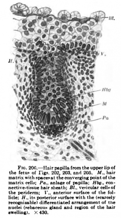
With the further development of the hair germ Stohr's stage of the hair papilla is reached, in which the hair anlage, while retaining approximately its original diameter, gradually grows downward into the corium (Fig. 206). The hair papilla projects downward from the stratum cylindrieum; it consists of an outer layer of cylinder cells, which are already beginning to differentiate in the deeper parts of the follicle, and of polygonal cells, arranged more irregularly, which fill the space bounded by the columnar layer. The latter, when the length of the hair papilla is about three times its breadth, shows on the posterior surface two low outgrowths with outwardly diverging, regularly J arranged nuclei. The upper of these is the aniage of the sebaceous gland, which in rare instances may be double (one above the other, Diem, liK)7) ; the lower one is that of the sweltiug (hair bed, Unna, 1883), which, usually, becoming lower, extends around to the anterior surface of the follicle. The lowest portion of the hair papilla consists of high columnar cells arranged in a kiln-like manner and forming a convexity which matrix converge, a roundish cavity (Fig. 206). The under surface of the hair papilla is flattened or even somewhat concave. The epidermis in front of the hair is unmodified and three-layered, consisting of periderm, stratum intermedium, and stratum cylindricum; the posterior surface consists of proliferated, flatter, and elongated cells (the hair-canal anlage), which in part already show a beginning cornification (Stohr). The hair papilla is enclosed laterally within a layer of connective tissue with numerous cells. On the under surface this is continued into a mass of cells with perpendicularly arranged, concave nuclei, the concavity looking upwards, which are immediately adjacent to the matrix (connective-tissue papilla). All these peculiarities of the hair papilla may be more or less pronounced. Thus a very distinct swelling may occur on one which does not show any marked apical flattening; or on a very short epidermal papilla there may be a distinct connective-tissue one, or vice versa. Further, in these early stages there may be opposite the swelling a marked aggregation of nuclei, which is to be regarded as the anlage of the rnuscidus arrector pili (Stohr). The hair anlagen, almost two months old, on the brows and lips have not advanced beyond this stage when the formation of the anlagen of the general hair coat begins. Some similar structures (gland anlagen) are aJone larger; these will be considered in connection with the perimammillary epithelial appendages.
Fig. 205. the fetus of rig?. 202 203. unfl 205. «.. hnir 1" Illglier lUaU Uldl Ol UlC bllUl m»irixwiih>ti««-.oiti,««,nver^nBpointo(ihf j^rly arranged cells of the hair miitnx «1U; Pa., anlngc of pBpiIln: Hbg.. ron•' . i ant V nective-tL-™eh>irBh«th;B;..ve'icui«r(Kii,ot germ (nuitrix plate, Gaicia, 1891). *ute;'*H!^rt?|i».(«VioTsurfa«™IIhThe'<™»"^ There is also occasionally to be n^'^^"'a^D"^X'n^>^oTii^'hl!^ found in this region, at the point nrdimj?. >: 430. toward whicli the cells of the
nto the bulb papilli. Pa., biiIbcp ot lir canal wlln: //.-K„ luigential MCet HiHtolov«, Fig. 300; EntwickluDS
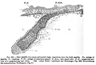
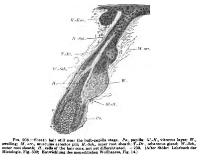
In the further course of development the layers of the hair and its sheath begin to form (Fig. 208). The epithelial papilla grows longer and thicker below (the bulb-papilla stage, Stohr). Its anterior surface abuts upon thin unaltered epidermis, while the posterior is continuous with the anlage of the hair canal, already recognizable in the two yoimger stages. This assumes the form of an elongated, partly comified mass of flat cells, projecting from the under surface of the epidermis behind the hair. The epithelial papilla is still surrounded by a high columnar epithelium, which at two regions on the posterior surface, that of the sebaceous gland above and that of the hair swelling below, projects more markedly than formerly. The anlage of the sebaceous gland does not always lie exactly in the posterior surface of the hair ; occasionally it lies more laterally and later may surround the entire periphery of the hair or come to be principally lateral or anterior to it. The specific fat formation begins at an early period in the central cells. The under surface of the epidermal papilla or bulb is at first only slightly concave for the reception of the connective-tissue papilla, but later (apparently very quickly, Stohr) it becomes deeply concave. The high columnar cells which line the concavity, the matrix plate, become the source of an upwardly projecting, conical, pointed mass of cells (the hair cone), which extends upwards into the still rather irregularly arranged cell material in the interior of the follicle. The outer boundary of this cone is formed by a layer of ceils {the anlage of Henle's sheath), which extends from the point where the outer surface of the follicle bends into the under surface to the summit of the cone. It is the first formed and outermost layer of the cone and arises from the most peripheral cells of the matrix plate ; it also cornifies sooner than any of the other layers of the hair. The inner sheaths and the hair are formed later from the middle cells of the matrix plate. Above the apex of the hair cone the cells arrange themselves to form an axial column, which eventually comes into relation with hair canal ceils abutting upon the superficial epidermis and indicates the path along which the hair sheaths, and with them the hair, must grow. The connectivetissue portions of the hair become more distinct. The concavity on the under surface of the bulb becomes filled by the large connective-tissue papilla, which consists of abundant, transversely arranged cells and which as yet shows no neck-like constriction. It seems to be covered by a very thin continuation of the vitreous layer and to be separated by this from the epithelium. From the richly cellular connective tissue of the hair follicle a denser homogeneous layer separates, especially in the region of the swelling; this is the vitreous layer. The musculis arrector has at first the form of elongated cells, which show the oblique course from the bulb to the epidermis that is characteristic for the muscle; it becomes more distinct in this stage.
Fig. 206. pepilla; (ll.-H., vitroDui Uyer; W„ i; T.-Dr.. aebafwous gland: W.-Sch., 230. (Afwr SWhr:
Fig. 207.
The follicle, gradually enlarging, grows obliquely downwards, and all its constituent parts undergo further development until it becomes the anlage of the actual hair. The sebaceous gland and the swelling assume noticeable dimensions and the connective tissue papilla increases in height in correspondence with a deepening of the concavity of the matrix plate. While the bulb forces its way downwards the cornified hair sheaths which arise from it are pushed towards the surface.
As the last stage, that may be taken in which all constituents of the hair are laid down (the sheath hair, Stohr). The hair elongates, especially in that part which lies below the hair bed. The upper part, with the swelling, sebaceous gland, and orifice, grows somewhat with the further development, but in general retains the same proportions. And, furthermore, it is the portion of the hair follicle intervening between the surface of the skin and the hair bed which remains unchanged throughout life, while the processes connected with hair change and the subsequent death of the hair take place only in the portions of the follicle below the hair bed. The outer root sheath, except in its lowest portion, from which the hair and its sheaths are developed, consists of two or three layers of cells, the innermost of which is flattened agajnst the inner root sheath and later is united with it (Stohr). The outer layer consists of more or less high columnar cells, which, in younger follicles, are all directed outwards and downwards (Fig. 209), probably in correspondence with the downwardly directed growth pressure of the follicle; later they become arranged perpendicularly to the axis of the hair. Towards the completion of development, when the hair change begins, the columnar cells become very high and their nuclei round or hemispherical with the flat surface directed outwards ; they lie in the inner portion of the cells, while the outer portions, which rest upon the vitreous layer, are clear and unstainable. On the outer surface of these high columnar cells a homogeneous layer is secreted. The portions of this layer, at first separated, fuse together to form the inner vitreous lamella and unite intimately with the outer vitreous laver, which is formed by the innermost layer of the connective-tissue portion of the follicle. Later the two vitreous lamellae become closely connected together. The columnar cells stand in small transverse grooves of the vitreous layer, these grooves corresponding in width with the cell bases and being readily recognizable in microscopic sections, in which the vitreous layer is easily separated from the follicle epithelium, as slight elevations between the rows of cylinder cells. The vitreous layer terminates at the swelling, and above its fibres lies close against the epithelium and extends upward as far as the sebaceous gland. In H-i the swelling the epithelium becomes many-layered, more so on the anterior surface of the follicle than on the posterior, which, from the beginning, shows the strongest development of the swelling. At this ^^ point the arrector muscle, surrounded from below upwards with elastic fibres, is usually inserted. Above the swelling the epithelium again becomes thinner and its cells lower, and it is especialIy thin in the region of the sebaceous gland, at w h o s e orifice the hair follicle has its -a. smallest diameter (the isthmus). At this point "^' the funnel of the follicle p. begins, not being formed by a depression from alx)ve, but by the cornification, accompanied by the formation of keratohyalin, of the central cells of the follicle and of the hair canal cells, whereby a long streak of comified cells, which extends far into the epidermis, is formed. This indicates the path which the hair will shortly take, and, after it has broken through, the funnel becomes usually much widened and its walls strongly comified.
1 upper edge an especially strong layer of elastic
Fig. 208. — Sheath hair still near the bulb-papilla stage
++++++++++++++++++++++++++
The lowermost part of the outer root sheath encloses the elements of growth for the hair. It has already been seen that in younger stages Henle's sheath could be followed down to the matrix and was the most external and at the same time the oldest of all the structures which arise from the matrix. It also cornifies the earliest and the most strongly of all the hair sheaths. Somewhere about the level of the connective-tissue papilla (according to Gavazzeni, 1908, even in the matrix cells) there is formed in it a ring of cells containing granules ; the nuclei of these cells do not become smaller, as is nearly always the case in the stratum granulosum, and their granules differ from keratohyalin in both their chemical and staining reactions (staining with eosin and fuchsin). It would seem that these granules, as also those of Huxley's layer and of the hair itself, are not keratohyalin, but a different chemical substance (trichohyalinf Vomer, 1903). The granules of Henle's layer became converted into elongated rods and soon disappear, the layer itself staining diffusely with eosin and becoming completely comified.
Henle's layer covers all the central structures and extends to the region of the hair canal, where it breaks up and is perforated by the hair in its upward growth (Fig. 209).
Huxley's layer, the inner lamella of the inner root sheath, cannot be followed quite down to the matrix plate, although it is certain that it has its origin from a definite ring of matrix cells. In the lanugo hair it extends at first far beyond the tip of the connective-tissue papilla into the apparently undifferentiated mass of cells at the base of the follicle, and in later stages of development its trichohyalin-containing cells can be distinguished further down toward the matrix. Its granule cells extend far upward upon the shaft of the hair, which has formed in the meantime, and they then become cornified. It stands in intimate connection with the Henle layer, which has already become completely comified, and between the cells of this layer those of Huxley's layer send processes containing granules.
according to Garcia (1891) the sheaths can be followed quite to the matrix plate in the head hairs of fetuses of eight to nine months, when the hairs have just attained their complete development. Of the forty to fifty cells which occur in a longitudinal section through the summit of the matrix, Henle 's and Huxley's layers correspond to four to six cells on either side. When the hair, in its full strength, has broken through the inner root sheath, this terminates in a sharp edge surrounding the hair in a circular manner, at the level of flie isthmus below the orifice of the sebaceous gland ; at first it extends somewhat higher than this, as far up as the hair canal.
Beginning somewhat higher than Huxley's layer, there are formed from the more internal regions of the matrix the cuticle of the inner root sheath (the sheath cuticle) externally and the hair cuticle internally. These two cuticles arise from the four to six cell-rings of the matrix internal to those which form the inner root sheath. Their cells contain no granules (whether they are also destitute of granules in the adult condition is still uncertain), and they comify probably before Huxley's layer, forming scales which in the sheath cuticle are small and directed obliquely inwards and downwards, while in the hair cuticle they are large and directed obliquely outwards and upwards; in the fully developed follicle the two sets of scales (imbrications) fit into one another. The sheath cuticle is almost inseparable from Huxley's sheath, and the hair cuticle, similarly, from the hair; and their imbrications apparently determine the equal ascent of the hair and the hair follicle, which their disappearance promptly disturbs (Von Ebner, 1876).
The hair itself arises from the large central portion of the matrix plate. It forms at first an acute cone-shaped structure, just as the sheaths do, and is covered by the comified sheaths as with a cornucopia until it breaks through the torn sheaths in the vicinity of the hair canal. Gradually it becomes broader, and arises from the greater portion of the matrix. On the head (in the eighth to ninth fetal month) each hair arises from twentyfour to thirty of the cells seen in a greatest longitudinal section of the matrix plate (Garcia, 1891), and Stohr's figures of the lanugo hairs show about the same number. The large roundish nuclei above the matrix gradually become elongated and the cells comify without forming trichohyalin. The comification begins deep down, the nuclei vanishing at the level of the junction of the lower and middle tliirds of the follicle (head hairs, Garcia). Between the matrix cells of the hair branched pigment cells appear, which ascend into the hair and contribute pigment granules to its other cells. according to the most recent views they take their origin from the epithelial rather than from the connective-tissue cells (see p. 253). The diameter of the hair gradually diminishes as it grows away from the papilla, and its smallest diameter is reached with the completion of its comification. But before it becomes hardened, the suCoulent hair mass is pressed, as in a mould, by the elastic compression of the sheaths. Henle's sheath is a stiff tube, which on account of its net-like structure (flat cells separated by meshes, very well shown by Giinther, 1895, in his Fig. 202), may exercise an elastic pressure. In it the soft hair with its soft sheaths is compressed and moulded. The remaining sheaths form a softer cushion for the forming tube. After the comification of Huxley's laver the hair with the cuticles and the inner root sheath forms a compact cylinder, which is pushed upwards by new formation at the base of the follicle. The inner part of the outer root sheath follows in the upward movement — ^the imbrications of the hair cuticle, firmly united to the hair, and those of the sheath cuticle, firmly adherent to the hair sheath, seeming by their interlocking to determine the regularity of this upward growth.
The epithelial hair follicle is enclosed in a layer of connective tissue, sharply marked off from the surrounding corium connective tissue, which is, in general, arranged horizontally. This connective-tissue sheath consists of an outer layer of longitudinal fibres and an inner transverse or circular layer. The inner layer secretes the outer vitreous layer, whose connection with the columnar cells of the follicle by means of the inner vitreous layer formed by their bases has already been described. Beneath the hair bulb, the papilla (papilla pili), which fills the spacious cavity of the hollow hair bulb, projects from the connective tissue. It projects from the mass of transversely arranged cells (the papilla cushion), which lie below the entrance into the hair bulb, and extends some distance upwards to form a papilla terminating above in a point (the tip of the papilla). At the lower border of the hair bulb, the papilla is constricted in a neck-like manner (papilla neck, Garcia) .
The cells of the papilla are partly directed obliquely upward and partly are arranged transversely, as they are at their first appearance. They are distinguished from all other connective tissue cells by their epithelial-like structure, being closely set cells with large, roundish or elongated, darkly staining nuclei.
The first-formed hairs have only a short life.
Even before birth the first hair ehan^ begins in the human species. This is total, compressed into a brief space of time, and associated with a change in the quality of the hairs.
Some hairs cease to ^row even before they have broken through the surface of the skin, a condition which is often shown in later life by lanupro hairs (such as hairs of the face). In hair change the cessation of growth of the hair seems to begin externally and to proceed internally. The cells of Henle's layer and then those of Huxley's layer cease to proliferate and are carried upwards by the still growing hair by means of the interlocking of the imbrications. Then the matrix, the cuticles, and the hair itself cease their growth. The hair becomes cornified right to its tip and, probably because it is no longer compressed by Henle's layer at its lower end, this enlarges to form a brush-like structure, the hair being then known as a bulb hair. A cell mass, the bulb cushion (Garcia), is formed by the matrix and occupies the space left vacant by the hair. This is carried outwards rapidly, as if squeezed out from the tube formed by the outer root-sheath. The matrix and connective-tissue papilla pass outwards more slowly, and the outer cells of the outer root sheath lose their columnar form (Aubertin, 1896, in adult head hairs). A diminution seems to occur in the pressure which the growing hair exerts downwardly and which is at first greater than that exercised by the surrounding tissues; the pressure of the tissues is now alone active and the space left vacant is filled by their being forced inwards.
During the ascent of the hair and its follicle the connective-tissue investments of the follicle thicken, especially the circular fibrous and the outer vitreous layers. Perhaps these thickened layers exert a compression on the thinning lower part of the follicle, whereby the hair is forced outwards. But the thickening may also be regarded as a protection against the pressure of the surrounding tissue, or as the simple contraction of an overstretched membrane. Indeed, all mechanical theories are to be advanced with great caution (Stohr), since in every step the growth of the hairs apparently follows old inherited paths which, like the phylogeny of the hair, are unfortunately poorly understood. All arrangements are naturally intelligible mechanically and explicable as strain and pressure conditions; but whether these mechanical explanations are correct is a question.
While the separated hair and the papilla are ascending the epithelial root cylinder between the two becomes thinner; it becomes composed of cubical indiffei*ent cells and the connective-tissue papilla becomes smaller (diminishing at the most to about half its original size in section). When the papilla has reached its highest position, a new life begins in the root cylinder. It covers itself anew from above with new columnar cells, becomes thicker, and develops a new swellinglike outgrowth, which later applies itself to the old swelling (Garcia). Gradually a new hair papilla forms in this, the hair matrix producing first a new inner root sheath and then a new hair, just as on the first formation of the hair. As the new hair grows out, its matrix and papilla are forced downwards by the renewed growth, and the inner root sheath is broken through just below the orifice of the sebaceous gland. The tip of the hair pushes its way, frequently in a tortuous course, through the old follicle canal, and after a considerable enlargement of this the old hair, which projects considerably, eventually falls out, as the result of some mechanical cause. The new hair is no simple replacement of the old, but has a quite different character. While the hairs of the first generation are practically all alike, there begins to appear in the second generation the great difference between the head and body hairs, and this increases in generation after generation, until, at the beginning of puberty, the genital and axillary hairs begin to differ from the lanugo of the remaining portions of the body; the lanugo of the face gives place to the hairs of the beard, etc., in the male ; and the apparently unaltered lanugo, as well as the head hairs, eyelashes, and eyebrows, assume a different type. In later years still more of the lanugo becomes transformed into strong body hairs (terminal hairs, Friedenthal, 1898). Each change of hair is at the same time a change in the chai-acter of the hair (type change, Unna, 1893). An accurate enumeration of the lanugo hairs on the human ear (Oshima, 1907) seems to show that the number of the fetal lanugo hairs is in places much greater than that of the lanugo of the adult.
The direction of the hairs is determined from the beginning. In the individual hairs it is recognizable in the hair-germ stage on account of the bilateral form of the germ, and in later stages it reveals itself by the hairs spreading out over the surface in the manner permanently determined for them (Blaschko) and not radially from some centres that arise. The same conditions have already been described as occurring in the development of the dermal ridges, which, in the same way, extend out over the surface of the skin from the regions of their first formation (the finger tips). With the completion of the hair formation the skin shows an arrangement of the hairs which is definite, unchangeable in any individual, and varying but slightly in different individuals, for the knowledge of which we are indebted to Eschricht (1837) and especially to Voigt (1857).
The latter regarded the direction of the hairs as the result and an indication of the mode of growth of the skin especially, but also, to a certain extent, of the underlying portions of the body.. The general spiral arrangement of the lines of the larger hair streams indicated a spiral growth of the enlarging skin, such as is the rule in plants, a mode of growth which was thoroughly studied by Ohlert (1854-55) and later by Schwendener (1909), and was regarded as a peculiar law of growth for the animal body, the law of torsion, by Fischer (1880). The centres of the spirals are the hair whorls, around which the hairs arise at intervals and in curves which are regular even although they have not hitherto been expressed mathematically. Voigt pointed out that the hairs at the ends of a stream are further apart than they are near the whorl. A considerable number of constantly occurring hair centres have the form of whorls, from which the hairs diverge in spirals (the direction of the free hairs being towards the periphery, diverging whorls).
The more important of these are:
- The crown or vertex whorl, curving towards the right in more than half the cases, towards the left in about a third, and doubled in the remaining cases (curving to the right on the left side and towards the left on the right side), or, in rare cases, trebled.
- A right and a left brow whorl.
- A right and a left ear whorl.
- A right and a left axillary whorl.
- A right and a left lumbar whorl.
- occasionally one or frequently two whorls, right and left at the side of the body (often only on one side).
- Hand and foot whorls.
In other regions the hairs converge from all directions to form converging whorls, the most constant of which are:
- The frontal whorl, at the root of the nose or at the edge of the scalp or in both places.
- Lateral cervical whorls.
- Elbow whorls.
- Umbilical whorl.
- Penial whorl.
- Coccygeal whorl.
The hair lines meet one another at acute angles in streams; when they meet at right angles crosses are formed, such as the nasal cross, the hyoid cross, the pectoral cross, the abdominal cross, the penis cross, and the coccygeal cross in the middle line of the body; the brow crosses, the nape crosses, the supra-auricular crosses, and the lateral crosses, one on each side; and the shoulder crosses, the ulnar crosses, the carpal crosses, and the crural crosses on the extremities. Many are doubled, and very frequently one is absent on one side of the body.
The diverging whorls, according to Voigt, are regions of least growth, of comparative rest; the converging whorls correspond to especially great stretching of the skin, in regions where (either in the ontogeny or phylogeny) some organ projected from the body (Wiedersheim: penis, umbilicus, branchial clefts, and, in animals, horns), or where especially strong growth resulted from the pressure of adjacent parts (coccyx, elbow). The crosses are regions of relative rest, lying between forces acting from either side; the converging hair streams are regions which became stretched during growth.
Of the arrangement of the hairs in transverse rows, corresponding to the arrangement of the scutes in transverse girdles around the body and limbs of reptiles, and of the development of such rows, we have as yet no comprehensive investigation.
G. The Sudoriparous Glands
The development of the sudoriparous glands begins on the finger tips (Blaschko, 1888), the palms of the hands, and soles of the feet (Grefberg, 1883) in the fourth month; according to Kolliker (1889) in the fifth month. It follows immediately upon the formation of the dermal ridges. Their anlagen resemble closely those of the hairs, except that they lack the close aggregations of cells in the corium, from which, in the case of the hairs, the papillae are formed. The anlagen project downwards as solid flask-shaped rete papillae, which, becoming long and slender, begin to become tortuous in the sixth month. In the seventh month a lumen begins to form in each, the beginning secretion producing intercellular clefts which later unite to form a continuous cavity. In the meantime the lower end, bending upon itself, forms the anlage of the coiled portion of the gland. The outer terminal portion of each gland forms its own lumen, which later unites with that of the glandular portion. The two-layered epithelium of the duct portion becomes transformed at its passage into the glandular portion into two distinct layers; the inner of these remains the large-celled secretory layer, while the outer becomes flattened and forms the epithelial muscular layer (Kolliker, 1889). The gland canal is then composed of an inner layer of cubical, or even higher, glandular cells, with large, round nuclei, and of an outer layer of flat muscle cells, whose nuclei are flattened and whose angles project between the cells of the inner layer; these cells form an outer investment of closely set parallel striae around the gland. At the time of birth the sudoriparous glands, like the hairs, seem to be completely laid down, so far as their number is concerned.
In the hairless palms of the hands and soles of the feet it is certain that the sudoriparous glands arise from the surface epidermis, and in most of the other portions of the skin they seem to have a similar origin in man. In many places, however, a relation of the sudoriparous glands to the hair follicles, of general occurrence in animals, persists, the sudoriparous glands partly developing directly from the uppermost portions of the hair follicles (Wimpfheimer, 1907; Diem, 1907), or partly having at least a connection with its follicles at their mouths (Fig. 209).
The idea that the follicles and the sudoriparous glands belong to genetically single areas has been somewhat generally accepted; according to my observations the hair disks (not constant in their occurrence) also belong to these areas, each of which, with all its appendages (vessels, nerves, muscles) corresponds in its original form to a promammalian scute (scute area or hair area). according to this view the completely isolated sudoriparous glands of the palms and soles must each represent the remains of a hair area (Whipple). That in spite of an imperfect development of the hairs the sudoriparous glands may actually retain their proper places in the hair areas, I have been able to show in the sole of the foot of
Omithorhynchua (Pinkus, 1905) where the paradosical arrangement of a romples consisting of a sudoriparous gland behind, a simple hair follicle in the middle, and a hair disk anteriorly, can only be explained as a semicircular arrangement n£ the elements of the hair area, peculiar to Ornithorkynchus, and a disappearance of all the hairs with the exception of the middle one. A further small step in the reduction would leave nothing remaining but the sudoriparous gland. The connection of the sudoriparous gland with the hair follicle, regarded as merely a topographical relation by Maurer (1895), but by most authors (De Meijere, 1894; Eggeling, 1904) as genetic, has been demonstrated to be of the latter nature by the embr>-o1c^cal obsenations of Stohr's pupils, Wimpfheimer and Diem, who found that in the majority of the mammals examined the sudoriparous glands arise from the follicle epithelium and their orifices only later migrate to the surface epidermis. Usually the sudoriparous gland is fonned, like the other appendages of the hair (sebaceous gland, swelling, muscle, hair disk), on its posterior surface. It begins to form even in the hair germ stage and ^ becomes distinct in the papilla stage (although not visible in man). Its cell nuclei, in contrast to those of the columnar layer of the hair, are small (Eggeling); in contrast to the regular arrangemeut in the anlagen of the sebaceous glands they are irregular (in the mole) ; and the increase in the number of the connectivetissue cells, which usually begins early in the case of the hair germ, is wanting beneath them. Unfortunately, in man, as well as in many other mammals, the original mode of development of the sudoriparous glands cannot be obsen-ed. In these forms they appear to arise, for the most part, not from the follicle epithelium, but from the surface epidermis.
Fig. not extend quite cl«r1y «»iu«h to the (luici): r)onn rtiirdti frnm tllP liminl S.-Dr., (udoriparous gland: H.-Ka., hair canal. UepariUreS irom me USUaj ^ j^ AtWrStObr, trom Diem: Entwicklunj structure of the sudoriparous der8ch™»-drUMiianderbeli»»r««iHautder n SiuBetiem. Fig. 7.
glands occur in certain regions of the body. If we make exception of the eyelids with their specialized glands, these regions are the mammary and axillary regions, the inguinal folds with the scrotum, and the anus. In some cases the characteristic peculiarities are recognizable in the anlagen (region of the mammary gland), in others they first appear at puberty (the axillary glands).
A. The Glands of the Mammary Region
The ventrolateral surfaces of the embryo at an early stage (6.25 - 6.75 mm., Keibel and Elze, 1908, "Normentafel," Nos. 21, 24, and 25, Figs. 11 and 12) are occupied by a broad diffuse area of high epithelium (Schtcalbe's milk streak), that has a variable development, extending in some cases forward over the branchial arches and backwards, over the limb buds, until it reaches the tail; but in other cases it is of less extent and may be completely wanting. This epithelial thickening rei)re8ents a formative region, which occurs also in the lower mammals and in birds (Heinrich Schmitt, 1898). In it, especially in its anterior portion, there develops in embryos 9 mm. in their greatest hngth (" Normentafel," Nos. 37-39) the milk line or milk ridge (O. Schultze, 1892), a band of thickened epithelium which is approximately lenticular in transverse section. The milk ridge is a structure comparable to the ganglionic or dental ridges, glandular anlagen appearing in it from place to place (O. Schultze, 1897; Broulia, 1905). In it the anlage of the mammary gland develops in man. Its primary epithelial anlage is, according to Rein ( 1882) , (a) mound-8hai)ed, consisting of an aggregation of epithelial cells projecting beyond the surface of the epidermis. Even in embryos with a greatest length of 9.5 mm. it has become (b) fliitty lenticular, projecting somewhat beyond the epithelium both above and below, and being surrounded by an aggregation of corium cells {nipple zone). As the result of strong downward growth there is formed (c) the papilla-shaped anlage {Fig. 211), from which (rf) the hulb-shaped anlage is formed as the lower parts increase in breadth while the more superficial portion becomes constricted in a neck-like manner. Later the milk ducts begin to sprout out, the anlage becoming polygonal and beset with short buds, {e) the period of bud formation. The development progresses slowly. Several simple papillae project downwards into the connective tissue from the epithelial mass, and in the eighth month these become hollow and somewhat branched, their terminal portions being large (Fig. 212). The primary epithelial bulb from which these milk ducts arise becomes cornified in its central portion and a cavity forms in it, with which the lumina of the gland ducts become continuous (Fig. 212). When the hair development begins the region around the mammilla becomes conspicuous, on account of the absence of hairs on its surface, and forms the nipple area (0. Schultze, 1897).
In addition to the paired mammae, supernumerary mammary glands are frequently developed. As a rule, they occur along a line from the anterior axillary fold to the inguinal region and make it seem as if several mammary glands had formed from the milk ridge; in other cases the hyi>erthelial structures are arranged irregularly in the mammary region.
The anlage of the mammary gland is usually compared with those of the sudoriparous glands. In the monotremes the glands of the mammary pouches in their functional condition differ from the ordinary sudoriparous glands only in their exceptional size (Gegenbaur, 1886, who, however, originally derived the mammary glands from sebaceous glands). Their genetic origin from sudoriparous glands is shown both by their development and by their comparative anatomy. Futhermore, in the adult condition tlteir hidradenoid structure, represented by the two-layered epithelium of the ducts and the simple epithelium in the glandular alveoli, is an indication of their sudoriparous character. Their glandular alveoli, like those of the sudoriparous glands, produce their secretion without destruction of the cells (Bertkau, 1907) and are enclosed within a muscular network (Benda, 1893), which resembles the basket-like muscular network of the sudoriparous glands in the snout of many animals (Kormann, 1906). Their similarity to sudoriparous glands is rendered still greater by the fact that such glands, modified along the lines of the mammaiy glands, are formed in the neighborhood of these structures. Rein (1882) has already identified these as the anlage of the Montgomery's glands. Close around the nipple a number of sudoriparous glands is formed from a primary epithelial anlage similar to that of the mammary gland itself, but smaller; they resemble milk ducts by possessing peculiar outpouchings and wide lumina. occasionally sebaceous glands and small hair anlagen are to be seen arising from the same epithelial papillae, so that the hair areas, whose remains are represented by the accessory mammary glands (Eggeling, 1904), are in these cases not absolutely rudimentary (Brouha, 1905).
In rather early stages (embryos of 15.5 mm., Walter, 1902) some other epithelial appendages begin to develop around the mammary glands and were at first regarded as hyperthelial structures (accessory mammary glands, Hugo Schmidt, 1896). From another standpoint they were interpreted as marsupial anlagen (Walter), similar to the marsupial pouches which fonn around each mammary orifice in the opossimi and fuse by their outer ends on the appearance of the marsupium (Bresslau, 1902). The number of these anlagen is occasionally very large in human embryo^ reaching in some cases forty; they are scattered around the nipple and as far up as the axilla, and are nsitatly smaller than the milk glands proper. Up to the pi'esent they have not been found in fetuses longer than GO mm. To the same category perhaps belongs also the inguinal epidermal alage, which has been described by Brugsch and Unger (1903).
In the same situation as these epithelial thickenings I have found around the mammary gland during my preparation for Ihe present work, in a fetus of 85 mm., five epithelial structures, which must be regarded as something quite distinct. They are long tubules, surrounded by a thin longitudinal layer of connective tissue and formed of a twoto four-layered epithelium, which bounds a small lumen, not opening to the surface. Near the epidermis the most superficial portion of the tubule contains a small cavity, filled with eomified cells and surrounded by keratohyalin cells; it resembles Stiihr's hair canal, and neither opens to the exterior nor communicates with the lumen of the tubule. Below, the tubule, beyond the termination of its lumen, ends in an epitheliol thickening, which sometimes forms a very regular, almorf conical structure, covered e.-iternally by columnar cells (Fig. 2131. These epithelial tubules are, with the exception of the mamma, the tai^i^Ht epithelial appendages of the fetus. At this stage the bair aniagen are .still in the stage of the much shorter bulb papilla and the sudoriparous glands are not yet formed. I would assign the tubules without question to the mammaiy apparatus, were it not that similar, longer structures occurred in the nasal mucous membrane (Fig. 214). Possibly those in the pectoral region are a form of the further development of the epithelial structures of Schmidt (1896), mentioned above; probably they are forerunners or early stages of the Montgomery glands of the nipple area. That they are glands of the sudoriparous system is shown by their complete similarity to the ciliarj' sudoriparous glands, Moll's glands, whose development from the epithelium of the hair follicles and occasional occurrence qiute separated from these has been described by Ask (1908). The ciliary glands are, at this stage, howei'er, younger, and only in fetuses of 170-250 mm. show the grade of development in these structures seen in So mm. fctuseii. On the same side of the ciliary follicle as the sebaceous gland and swelling (the outer surface of the eyelid, but the posterior surface of the cilia) there extends above the level of the sebaceous gland a thin elongated epithelial cord, vhichr after passing over the sebac«ous gland, bends downwards and is continued downwards for some distance parallel to the anlage of the cilium, to terminate in a pear-shaped enlargement composed of small rounded epithelial cells. Later a lumen appears at the orilice and another in the coiled portion of the gland. The connective-tissue cells around the aniagen of Moll's glands are not, however, increased in number.
Fig. 213. Epitbeluil BppepdsEe owumng nrar thf nummaol the frtug from which Fig, 211 wuUksD. H., comifibl plug with kentohyalin: L.. lumen: M.. DUBCle (T). A 180.
Fig. 214. — AnmilaTBirithclisi ttruclurelromihvDual cavity of the nunc tufa. X 180.
B. The further development of the axillary glands
B. The further development of the axillary glands begins in the ninth year in the female, but not until the time of puberty in the male (Liineberg, 1902). They are formed from the ordinaiy sudoriparous glands of this region, a large number of which persist as small glands. The large axillary glands form a continuous layer of large, partly branched tubules, with an inner layer of high secreting cells and an outer single-layered muscle coat. Nothing special is known concerning the development of the inguinal and scrotal glands.
H. The Nails
At a very early period the place where the nail will be formed is recognizable on the dorsum of a terminal phalanx. The primary nail field (Kolliker, 1888), primary nail base (Zander, 1884), is recognizable in microscopic sections in fetuses of 4.5 cm., while in those of 2.75 cm. the region in which it will develop is quite undifferentiated (Okamura, 1900). Externally, the primary nail field somewhat later becomes evident on account of its smooth appearance and its firmer adherence to the subjacent tissue, as well as by its sharp anterior, posterior, and lateral boundaries. It extends from the finger tip almost to the articular process of the terminal phalanx — an extent which the fully formed nail also possesses and which is only relatively diminished transitorily during development by the increased growth of the ball of the finger. At its proximal (posterior) edge the epidermis invaginates to form a transverse, posteriorly convex pouch (the root lamella of Kolliker, the posterior nail fold of the adult), in which later the nail matrix is formed and whose roof is termed the nail wall. The epithelial invagination is the posterior limiting furrow (Kolliker), and laterally, on each side, it is continued into a lateral groove (the lateral nail fold, bounded externally by the lateral nail wall). Anteriorly the primary nail field abuts upon a shallower depression, which bounds it anteriorly in a transversely arched manner (the anterior groove, Unna; nail fringe, Kolliker). The primary nail field is flatly convex and is at first distinguished from the twoto three-layered skin of the fingers by its threeto fourlayered epithelium with a germinal layer of cubical cells. Later on, also, the cells of the lowest layer of the nail field (the later nail bed) remain cubical, in contrast to the columnar cells of the nail fringe, the nail fold, and the germinal layer of the rest of the epidermis of the finger. In front of the nail fringe a transverse ridge appears, which is of great importance from the comparative standpoint. It corresponds to the region of the skin in which later the looser cornification, seen under the free edge of the nail and equivalent to the sole plate of the other mammals, is formed (Boas, 1894). In front of this ridge the epidermis is depressed to form a groove (the distal limiting furrow, Kolliker). The germinal layer in this region is formed of high columnar cells and the epithelium is fiveto six-layered. In front of the sole plate the germinal layer becomes still more columnar.
The periderm and the stratum intermedium extend over the entire anlage, levelling its surface. The most superficial layer of very flat cells desquamates to a remarkable degree; and one finds, still adhering to the surface, cells swollen to a vesicular form and with small pyknotic, darkly staining nuclei (for a description of these cells see especially Zander, 1884; Kolliker, vesicular cells; Okamura; for those of the chick embryo, Rosenstadt, 1897).
In the course of further development the region of the sole plate becomes higher and eight to ten-layered, and the cells of the nail field become flat, so that by its cell form alone the dorsal surface of the terminal phalanx can be readily distinguished from the volar surface, the germinal layer of which remains columnar. Only in the neighborhood of the nail wall, and, later, still further proximally also large vesicular, round cells persist at the surface, to a greater extent on the toes than on the fingers. The flat periderm cells, beginning at the nail fringe, fuse to form a tough superficial layer, and between them and the cubical germinal layer there lie distally several layers of prickle cells and proximally, at the nail fold, an area of pale vesicular cells (Zander's limiting layer). These vesicular cells remain large until much later on the toes, but on the fingers they become flatter, i)robably in correspondence ^itli the greater ^r. general flattening of the anlagen of the finger nails.
Below Zander's limiting layer there is formed from the uppermost portions of the stratum intermedium, in somewhat larger embryos (10 cm., Zander; mlnal pholanxot another finger of the tame fetus nn TJ---I1-1 a K from which Fig. 2lfi i» taken. The bssal osll layer
9.3 cm., Kolllker; 8.5 era., a cubimldorsally. columnar voUnv. .V.F.,lateral cording to my own ohservations), a stout layer with abundant keratohyalin gramdes, which are partly large and roundish and partly quite fine and dust-like. Beneath this granule layer, whose keratohyalin nature was first determined by Pollitzer (1889), there lies a layer of pale vesicular cells, which also, especially at the cell peripheries, contain granules similar to those of the superjacent layer. These three layers form Unna's eponychium <1883), and the nail itself is formed from them according to Kolliker, while according to Zander it is formed from the limiting layer, which is continually reinforced from the subjacent keratohyalin layers. The eponychium with its granule layer represents, according to Zander, only a precocious comification, of the same type as that which occurs in the epidermis over the whole body at a later stage, when the granule layer, at the first sign of comification, forms from the layer of prickle cells. This kind of comification is found in such early fetal stages not only in the nail anlagen, but in all places where large appendage structures of the skin are being formed. It occurs also at the openings of the peculiar long epithelial appendages to be found in the nasal mucous membrane and around the mammary glands, and it occurs in slightly later stages in the development of the hairs, in the anlagen of the hair canals. It appears to be merely an indication of increased formative force in the epidermis. This layer of actual comification, already similar to the later comification, cannot, however, be directly identified with the periderm (eponychium). This is an actual horn layer, which remains partly retained throughout the rest of life (as the fringe at the proximal edge of the nail; see below). Okamura (1900) is correct when he regards as the periderm merely the outermost layer of desquamating cells, which lies over the keratohyaJin layer, for we have already seen, in the consideration of the general skin, that the periderm in man is not a special layer, but merely the outermost layer of the quite young epidermis, which in the later development of the skin is permanently cast off.
Fig. 216. Transverse section through the terminal phalanx of another finger of the same fetus from which Fig. 215 is taken. The basal cell layer is cuboidal dorsally , columnar volubly. N.F. lateral nail fold; Ke., keratohyalin; T.-B., touch ball. x44
The nail itself is formed from the region of the nail fold, quite without keratohyalin formation, in the manner described by Unna (1883) and confirmed by Okamura (1900) and Apolant (1901). The actual nail formation begins at the posterior part of the nail field, under the eponychium, as a layer of cornified cells. It begins in the fifth month (Unna), in fetuses 17 cm. in length, the first indications of its cells being visible in those of 16 cm. (Okamura. 1900). These cells lie at the entrance of the nail fold, at the level of the distal end of the nail wall. They present fine granulations, which do not take the stains which color keratohyalin, but stain with picric acid, swell in strong alkali, and are insoluble in all ordinary reagents such as water, alcohol, xylol, chloroform, ammonia, acetic, hydrochloric, and nitric acid, alkalies, and digestive ferments ; therefore they cannot be keratohyalin. They have been termed by Ranvier onychogenons substance; but they neither represent a special substance (onychin), nor are they really granules, but probably nothing but the cross sections of fibrillae. They consist of keratin and are the cross sections of keratin fibrils which occur in the nail cells (Unna, Apolant).
The comification takes place, without being accompanied with a formation of keratohyalin, in the cells of the nail matrix, which extends from the posterior (proximal) margin of the fold to the anterior (distal) border of the lunula. The cells flatten and become converted into platelets, which, compose the solid substance of the nail. The formative region for this reaches, with advancing development, from the original point to the deepest part of the posterior nail fold, and anteriorly to the anterior border of the lunula. Over this area the nail is formed and pushed forward from the beginning, just as it is throughout the whole of later life. The newly formed nails are for some time completely covered by eponychium, hut later tins is thrown off and the surface of the nail is exposed. Nevertheless a portion of the eponychium persists throughout life as a small membrane resting upon the proximal end of the nail; this grows forward with the nail and sooner or later desquamates at a distance of from 1-3 mm. from the posterior nail wall. The anterior edge of the nail is at first very thin and projects in older fetuses and after birth at the anterior end of the phalanx as a delicate membrane, but it is very quickly removed by the accidents of extra-uterine life (washing, movements) (Unna). The nail then grow-s, becoming gradually stronger, throughout the rest of life continuously, unless interrupted by some pathlogieal condition. The portion of the skin on which the nail rests in front of the lunula takes no part whatever in the formation. Its rete papilla, wliich form at the same time as the rete papillae and ridges of other regions, produce the longitudinal ridges of the nail.
Fig. 217. Langitudidal Krtina throuch ths ketohyalin laysr of the fetal Dsil bed, ulterior port. From a human fetus of 8.5 cm. (see Fig. 215). X 380.
Fig. 218. Transverse serlion through the keratflhyalin layer of the nail bed, posterior part. From a bumaafetusofS.Scm. (seeFiB.2l{i). X3S0.
Bibliography
Adachi, B. : Hautpigment beim Menschen und beim Aften, Anatom. Anzeiger, vol. xxi, 1902, p. 16. Apolant: Ueber den Verhomungsprozess, Arch. £. mikr. Anat., vol. Ivii, 1901 p. 766.
Arcakgeli, a.: Einige histologische Beobacbtungen iiber das Deckepitbel des Oesophagus beim Meerschweinchen (mit besonderer Beriicksichtigimg des Keratinohyalins), Monatsh. f. prakt. Dermatol., vol. xlvii, 1908, p. 297.
Ask, F. : Ueber die Entwicklung der Lidrander, der Tranenkarunkel und der Niekhaut beim Menschen, nebst Bemerkungen zur Entwicklung der Tranenableitungswege, Anat. Hefte, No. 109, vol. xxxvi, part ii, 1908, p. 189.
Aubertin, G. : Das Vorkommen von Kolbenhaaren und die Veranderungen derselbea beim Haarwiederersatz, Arch. f. mikr. Anat., vol. xlvii, 1896, p. 472.
Beard, J. : The Birth-Period of Triehosurus vulpecula, Zool. Jahrb., vol. xi, 1908.
Benda, C. : Das Verhaltnis der Milchdruse zu den Hautdriisen, Dermat. Zeitschr., vol. i, 1893.
Bertkau, F. : Ein Beitrag zur Anatomie und Physiologie der Milchdruse, Anat. Anz., vol. XXX, 1907, p. 161.
Bjorkenheim, E. a. : Zur Kenntnis des Epithels im Uterovaginalkanal des Weibes, Anat. Anzeiger, vol. xxviii, 1906, p. 447.
Bizzozero: Miinchener med. Wochensehr., 1908.
Blaschko, a.: Beitriige zur Anatomie der Oberhaut, Arch. f. mikr. Anat., vol. XXX, 1888, p. 495. Boas, J. E. V.: Zur Morphologie der Wirbeltierkralle, Morph. Jahrb., vol. xxi, 1894, p. 281.
BoLK, L. : Ueber die segmentale Anordnung der Melanoblasten bei jungen Teleostieren. Verb. d. Anat. Gesellsch., Berlin, 1908, p. 135.
Bonnet, R. : Haarspiralen und Haarspindeln, Morph. Jahrb., vol. xi, 1886, p. 220. Bresslau, E. : Beitriige zur Entwieklungsgeschichte der Mammarorgane bei den Beuteltieren, Zeitschr. f. Morphol. u. Anthropologic, vol. iv, No. 2, 1902.
Brouha : Sur la bande et la crete mammaires et sur les pretendues ebauches hypertheliales chez Thomme et le murin, Anat. Anzeiger, vol. xxvii, 1905, p. 462. Sur la signification morphologique de la mamelle, Anat. Anzeiger, vol. xxvii, 1905, T>. 311.
Brugsch, Th., and Unger, E. : Ein warzenformiges Gebilde der vorderen Bauchwand bei einen menschlichen Embryo von 4.5 cm. Scheitelsteisslange, Anat. Anzeiger, vol. xxiii, 1903, p. 216.
Brunner : Ueber Glycogen in der gesunden und kranken Haut, Deutsche Dermatol. Gesellsch., Bern, 1906.
Burkard, 0. : Ueber die Hautspaltbarkeit menschlicher Embryonen, Arch, f . Anat u. Physiol., Anat. Abth., 1903, p. 13.
Buschke : Notiz zur Behandlung des Vitiligo mit Licht, Mediz. Klin., No. 33, 1907. Buschke and Mulzer: Weitere Beobacbtungen iiber Lichtpigment, Berlin klin. Wochensehr., No. 49, 1907.
Buzzi: Keratohyalin und Eleidin, Monatsh. f. prakt. Dermatol., vol. viii, 1889. Ueber Eleidin, Monatsh. f. prakt. Dermatol., vol. xxiii, 1896.
Cedercreutz: Ueber die Verhomung der Epidermis beim menschlichen Embryo, Arch. f. Dermatol, u. Syphil., vol. Ixxxiv, 1907, p. 172.
Charpy, a. : Les plis de la peau. Arch. med. de Toulouse, Ref . Zentralbl. f . norm. Anat., 1905, p. 335.
CiLiANO, P.: Eleidin, Monatsh. prakt. Dermatol., vol. xlvi, 1908, p. 435.
Cone, Claribel: Zur Kenntnis der Zell veranderungen in der normalen nnd pathologischen Epidermis des Menschen, Frankfurter Zeitschr. f. Pathol., .1907, p. 37.
CzAjEWicz: Mikroskopische Untersuchungen iiber die Textur, Entwicklungy Riickbildung und Lebensfiihigkeit des Fettgewebes, Arch. f. Anat. u. Physiol., 1866, p. 289.
Diem, F. : Beitrage zur Entwieklung der Schweissdriisen an der behaarten Haut der Siiugetiere, Inaug. Dissert., Wurzburg, 1907.
Dreysel, M. : Pigment und osmierbare Substanzen in der menschliehen Haut, Verb. d. V. Deutschen Dermatologen-Kongresses, 1895.
Dreysel, M., and Oppler, P.: Beitrage zur Kenntnis des Ele'idins in normaler und pathologisch veranderter Haut, Arch. f. Dermatol, u. Syphil., vol. xxx, 1895.
Von Ebner, V. : Mikroskopische Studien iiber Wachstum und Wechsel der Haai'e, Sitz. Her. d. K. Akad. d. Wiss. Wien., vol. Ixxiv, 1876.
Eggeling, H. : Ueber die Stellung der Milchdriise zu den iibrigen Hautdrusen, Semon's Forschungsreise, Jena, 1895. Ueber ein wichtiges Stadium in der Entwicklungsgeschichte der menschliehen Brustdriise, Anat. Anzeiger, vol. xxiv, 1904, p. 595.
Ehrmann, S. : Das melanotische Pigment und die pigmentbildenden Zellen des Menschen und der Wirbeltiere in ihrer Entwieklung, nebst Bemerkungen uber Blutbildung und Haarwechsel, Bibliotheca medica, vol. ii, part vi, 1896.
Ernst, P. : Studien iiber normale Verhornung mit Hilfe der Gram Vhen Methode, Arch. f. mikr. Anat., vol. xlvii, 1896, p. 683.
Eschricht: Miiller's Arch. £. Anat. u. Physiol., 1837 (cited by Voigt).
Evatt JE. The development and evolution of the "papillary" ridges and patterns on the volar surface of the hand. (1906) J Anat Physiol. 41(1): 66-71. PMID 17232710
FiCK, J.: Ueber metachromatische Farbung des Keratohyalins durch Cresylecht violett, Centralbl. f. allgemein. Pathol., 1902, p. 987.
Fischer, E. : Das Drehungsgesetz bei dem Wachstum der Organismen, Strassburg, published by the author, 1886.
Flemming: Centralbl. f. d. med. Wissensch., July, 1870. Beobachtungen iiber Fettgewebe, Arch. f. mikrosk. Anat., vol. xii, 1876, p. 434. Ueber die Entwieklung der Fettzellen und des Fettgewebes, Arch. f. Anat. u. Physiol., 1879.
Frederic: Zur Kenntnis der Hautfarbe der Neger, Zeitschr. f. Morphol. u. AnthropoL, vol. ix, 1905, p. 41.
Friedenthal, H. : Beitrage zur Naturgeschichte des Menschen (Lief. I, Das . Wollhaarkleid des Menschen; Lief. II, Das Dauerhaarkleid des Menschen; Lief. IV, Entwieklung, Bau und Entstehung der Haare), 1908 (with a most complete bibliography).
Gage SP. The notochord of the head in human embryos of the third to the twelfth week and comparisons with other vertebrates. (1906) Science 24: 295.
Gage, S. H.: Glycogen in a 56 Days Human Embryo and in Pig Embryos of 7-10 mm., Amer. Joum. Anat., vol. v; Proc. Assoc. Amer. Anat., 1906, p. xiii-xv.
Garcia, S. A.: Beitrage zur Kenntnis des Haarwechsels bei menschliehen Embryonen und Neugebomen, Morph. Arbeiten, vol. i, part ii, 1891, p. 136. Gavazzeni, G. a.: Trichohyalin, Monatsh. f. prakt. Dermatol., vol. xlvii, 1908, p. 229.
Gegenbaur: Zur Morphologic des Nagels, Morph. Jahrb., vol. x, 1885, p. 465 (cited by Unna, 1889).
Gemmil: Notes on the Origin of Elastic Fibre in Tendon, Joum. Anat. and Physiol., vol. xl, 1906, p. 397.
Grefberg, W. : Die Haut und deren Driisen in ihrer Entwieklung, Mitth. a. d. embryol. Institut zu Wien, vol. ii, part iii, 1883, p. 126 (cited by Blaschko).
Grimm: Pigment in der Haut, Dermatol. Zeitschr., vol. ii, part iv, 1895.
Grosser, O. : Die Metaraerie der Haut, Sammelreferat, Centralbl. f. die Grenzgebiete der Med. u. Chir., vol. vii, Nos. 1, et seq,, 1904.
Orosseb, O., and Frolich, A. : Beitrage zur Kenntnis der Dermatome der menschlichen Rumpfhaut, Morph. Jahrb., vol. xxx, 1902.
Grund: Experimentelle Beitrage zur Geuese des Epidermispigments, Zieglers Beitrage, Suppl. 7, 1905.
Gunther, M. : Haarknopf und iunere Wurzgelscheide des Saugetierhaares, Inaug. Dissert., Berlin, 1895.
Head, H.: Die Sensibilitatsstorungeu der Haut bei Visceralkrankungen, edited by Seiffer, Berlin, 1898.
Heidenhain, M. : Ueber die gegenseitige Entsprecbung von Epidermisund Coriumleisten an der Beugeflache von Hand und Fuss beim Affen und Mensehen, Anat. Hefte, No. 91, 1906, p. 419.
HiRSCHLAND I Beitrage zur ersten Entwicklung der Mammarorgane beim Mensehen Embryo von 4 mm.), Anat. Hefte, vol. xi, 1898.
Ide, M. : Nouvelles observations sur les cellules epitheliales. La Cellule, vol. v, 1889, p. 319 (abstr. in Monatsh. f. prakt. Dermatol., vol. ix, 1890, p. 550).
Jadassohn: Ueber Pityriasis rubra und ihre Beziehungen zur Tuberculose, nebst Bemerkuugen iiber Pigment verschleppung aus der Haut, Arch. £. Dermatol. u. Syphil., 1892.
Jarisch : Ueber die Bildung des Pigments in den Oberhautzellen, Arch, f . Dermatol. u. Syphil., vol. xxiv, 1892.
Jores, L. : Ueber die feineren Vorgange bei der Bildung und Wiederbildung des elastischen Bindegewebes, Ziegler's Beitrage, vol. xli, part i, 1907.
Kallius, E.: Ein Fall von Milchleiste bei einem menschlichen Embryo, Anat. Hefte, vol. viii, 1897.
Keibel, F., and Elze, C. : Normentafel zur Entwicklungsgeschichte des Mensehen, Jena, 1908.
Kidd, W. : On the Imbrication of the Papillary Ridges in Man, Journ. of Anat. and Physiol., vol. xxxix, 1905, p. 413. The Papillary Ridges and the Papillary Layer of the Corium in the Mammalian Hand and Foot, Journ. of Anat. and Physiol., vol. xli, 1907, p. 35.
KoLLiKER, A. VON : Die Entwicklung des menschlichen Nagels, Zeitschr. f. wiss. Zool., vol. xlvii, 1888 (cited by Unna). Handbuch der Gewebelehre des Mensehen, vol. i, 1889.
KoLOSOFF, G., and Pankul, E. : Versuch einer mathematischen Theorie der Hautleistenfiguren der Primaten-Palma und -Planta, Morphol. Jahrb., vol. xxxv; also Centralbl. f. norm. Anat., 1906, p. 319.
Kormann, B. : Ueber die Modifikation der Haut und die subkutanen Driisen in der Umgebung der Mundund Nasenoffnungen, die Formationes parorales und paranaricffi der Haussiiugetiere, Anat. Anzeiger, vol. xxix, 1906, p. 113.
Krause, W. : Die Entwicklung der Haut und ihrer Nebenorgane, Handb. d. vergleich. u. experiment. Entwicklungsgesch. der Wirbeltiere, herausg. von 0. Hertwig, vol. ii, 1902.
Krauss, F. : Der Zusammenhang zwischen Epidennis und Cutis bei Sauriem und Krokodilen, Arch. f. mikr. Anat., vol. Ixvii, 1906, p. 313.
Kromayer: Die Parenchymhaut und ihre Erkrankungen, Arch. f. Entwicklungsmech. der Organe, 1899, p. 253. Die Desmoplasie der Epithelzellen in der menschlichen Haut, Monatsh. f. prakt. Dermatol., vol. xli, 1905, p. 477.
Langer, K. : Zur Anatomic und Physiologic der Haut, I. Ueber die Spaltbarkeit der Cutis, Sitzber. d. k. Akad. Wiss. Wien, math.-naturwiss. KL, vol. xliii, 1861, p. 19.
Lewinski: Ueber die Furchen und Falten der Haut, Virchow*s Archiv, vol. xcii, 1883.
Loeb, L. : Ueber Transplantation von wei«?ser Haut auf einer Defekt in schwarzer Haut und umgekehrt am Ohr des Meerschweinchens, Arch. f. Entwicklungsmech. der Organ., vol. xd, 1898.
Loewy, J.: Beitrage zur Anatomic und Physiologie der Oberhaut, Arch. f. mikr. Anat.y vol. xxxvii, 1907, p. 159.
LoiSEL, G. : Formation ct evolution des Elements du tissu elastique, Joum. de I'Anat. et de la Physiol., vol. xxxiii, 1897, p. 129.
Lombardo, C. : II glicogeno della cute, Giorn. ital. delle mall, ven e della pelle, vol. xliii, 1907, p. 448.
LuNEBURG, E. : Beitrage zur Entwicklung und Histologic der Knaueldriisen in der Achselhohle des Mcnschen, Inaug. Diss., Rostock (Abstr. in HofmannSchwalbe, Jahresb. f. Anat. u. Entwicklungsgesch., neue Folge, vol. viii, part iii, 1902, pp. 659, 670.
Maurer, F. : Die Epidermis und ihre Abkommlinge, Leipzig, 1895. Das Integument eines Embryo von Ursus arctos, Festschr. f. Ernest Haeckel, Jena, 1904.
De Meijere: Ueber die Haare der Saugetiere, besonders iiber ihre Anordnung, Morph. Jahrb., vol. xxi, 1894.
Mbirowskt, E. : Die Entstehung des Oberhautpigments in der Oberhaut selbst, Monatsh. £. prakt. Dermatol., vol. xlii, 1906. Die Entstehung des Oberhautpigments aus der Substanz der Kemkorperchen, Monatsh. J. prakt. Dermatol., vol. xliii, 1906. Ueber den Ursprung des melanotischen Pigments der Haut und des Auges, Leipzig, 1908 (contains a full bibliography).
Merk, L. : Die Verbindung menschlicher Epidermiszellen unter sich und mit dem Coriima, Monatsh. f. prakt. Dermatol., vol. xxxviii, 1904, p. 361.
Merkel: Tastzellen und Tastkorperchen bei den Haustieren und beim Menschen, Arch. f. mikr. Anat., vol. xi, 1875.
Merkel, Fr. : Die Hautfarbung neugeborener Farbiger, Korrespondenzbl. d. Deutschen Gesellsch. f. Anthropol., Ethnol., und Urgesch., 1907, p. 15.
MiNOT, C. S. : Lehrbuch der Entwicklungsgeschichte, 1894.
Nakai, M. : Ueber die Entwicklung der elastische Fasem im Organismus und ihre Beziehungen zu der Gewebsfunktion, Virchow's Archiv, vol. clxzxii, 1905, p. 153.
Ohlert, B. : Ueber die Gesetze der Blattstellung, PoggendorflTs Annalen der Physik., vols, xciii and xcv, 1854-1855 (cited by Voigt).
Oshima, T. : Die Beziehungen des Wollhaares der Neugeborenen zu den Haaren des Erwachsenen, Pfluger's Archiv, vol. cxvii, 1907, p. 341.
Okamura, T. : Ueber die Entwicklung des Nagels beim Menschen, Arch. f. Dermatol, u. Syphil., vol. Iii, 1900, p. 223.
Passarge: Monatsh. f. prakt. Dermatol., Erganz-Heft, 1894.
Philippson, L. : Ueber die Herstellung von Flachenbildem der Oberhaut und der Lederhaut, Monatsh. f. prakt. Dermatol., vol. viii, 1889, p. 389.
PiNKUS, F. : Ueber Hautsinnesorgane neben dem menschlichen Haar (Haarscheiben) und ihre vergleichend anatomische Bedeutung, Arch. f. mikr. Anat, vol. Ixviii, 1905, p. 121.
POHL, J. (PiNCUS) : Die Querschnittsform des Kopfhaares der Kaukasier, Zeitschr. f. Ethnol. Verb., 1897, p. 483.
Pollitzer, S. : Ueber die Natur der von Zander im embryonalen Nagel gef undenen Komerzellen, Monatsh. f. prakt. Dermatol., vol. ix, 1889, p. 346.
Post: Ueber normale und pathologische Pigmentierung der Oberhaut^bilde, Virchow's Archiv, vol. cxxxv, 1893.
PROFfi, 0.: Beitrage zur Ontogenie und Phylogenie der Mammarorgane, Anat. Hefte, vol. xi, 1899.
Kabl, H. : Histologic der normalen Haut des Menschen, Mracek's Handbl. der Hautkrankheiten, vol. i, part i, 1902, p. 1.
Reh: Die Schuppen der Saugetiere, Jen. Zeitschr. f. Naturwiss., vol. xxix, 1896.
Rein, G. : Untersuchungen iiber die embryonale Entwicklungsgeschichte der Milchdriise, Arch, f . mikr. Anat., vols, xx and xxi, 1882.
Bbtterer, E. : Structure et evolution du tegument externe, Joum. de TAnat. et de la physiol.y 1904.
RoSENSTADT, B. : Ueber das Eponychium des Hiihnchens, Arch. f. mikr. Anat., vol. xlix, 1897, p. 561. Studien iiber die Abstammung und Bildung des Haarpigments, Arch. f. mikr. Anat., vol. 1, 1897, p. 350.
ScHiFPMANN, J. : Die Histogenese der elastischen Fasem bei der Organisation des Aleuronatexsudats, Centralbl. f. allg. Pathol, u. pathol. Anat., vol. xiv, 1903, p. 833 (bibliography).
ScHLAGiNHAUFEN, 0. : Ueber die Hautleisten der Planta der Primaten, Korrespondenzbl. d. Deutsch. Gesellsch. f. Anthrop. Ethnol. u. Urgesch., 1905, p. 336. Ueber die Leistenrelief der Hohlhand und Fusssohlenflache der Halbaffen, Ergebn. d. Anat. u. Entwicklungsgesch., vol. xv, 1905, p. 628-662.
Schmidt, H. : Ueber normale Hyperthelie menschlicher Embryonen, Anat. Anzeiger, vol. xi, 1896, p. 702.
Schmitt, H. : Ueber die Entwicklung der Milchdriise und die Hyperthelie menschlicher Embryonen, Inaug. Dissert., Freiburg (also in Morph. Arbeiten, vol. viii, 1898, p. 236).
SCHMOBL, G. : Ueber Pigmentverschleppung aus der Haut, Centralbl. f . allg. Pathol. u. pathol. Anat., vol. iv, 1893.
ScHREiBER, L., and Schneider, P. : Eine Methode zur Darstellung von Pigmenten und ihren farblosen Vorstufen, mit besonderer Beriicksichtigung des Augen und Hautpigments, Miinchen. med. Wochenschr., No. 37, 1908, p. 1918.
Schridde: Die Protoplasmafasern der menschlichen Epidermiszellen, Arch. f. mikr. Anat., vol. Ixvii, 1906, p. 291.
Schultze, O.: Ueber die erste Analge der Milchdriisenapparate, Anat. Anzeiger, vol. viii, 1892. Ueber embryonale und bleibende Segmentierung. Verhandl. Anat. (Jesellsch. Berlin, 1896, p. 87. Grundriss der Entwicklungsgeschichte des Menschen und der Saugetiere, Leipzig, 1897.
ScHWALBE : Ueber den Farbenwechsel winterweisser Tiere, Morph. Arbeiten, vol. iiy 1893.
Schwendener: Vorlesungen iiber mechanische Probleme der Botanik, bearbeitete von C. Holtermann, Leipzig, 1909 (contains a bibliography of Schwendener's original papers on p. 61). Seiffer, see Head.
Sherrington, C. S. : Experiments in Examination of the Peripheral Distribution of the Fibres of the Posterior Roots of Some Spinal Nerves, Phil. Trans. Roy. Soc, vol. clxxxiv, 1893.
Spalteholz, W. : Ueber die Beziehungen zwischen Bindegewebsfasem und -zelleUy Verhandl. Anat. Gesellsch., Rostock, 1906, p. 209.
Stohr, P.: Ueber Intercellularbriicken zwischen ausserer und innerer Wurzelscheide, Verhandl. Anat. Gesellsch., Heidelberg, 1901, p. 24. Ueber die Entwickelung der Glaschaut des menschlichen Haarbalges, Verhandl. Anat. Gesellsch., Heidelberg, 1901, p. 26. Entwicklungsgeschichte des menschlichen Wollhaars, Anat. Hefte, vol. xxiii 1903. Lehrbuch der Histologic und der mikroskopischen Anatomic des Menschen^ 12 Aufl., 1906. Ueber die Schuppenstellung der menschlichen Haare, Verhandl. Anat. Gesellsch., Wiirzburg, 1907, p. 153.
Strahl, H. : Die erste Entwicklung der Mammarorgane beim Menschen, Verhandl. Anat. Gesellsch., Kiel, 1898.
Studnicka, F. K. : Schematische Darstellungen zur Entwicklungsgeschichte einiger Gewebe, Anat. Anzeiger, vol. xxii, 1903, p. 537.
Tandleb, J.: Ueber einen menschlichen Embryo von 38 Tagen, Anat. Anzeiger, vol. xxxi, 1907, p. 49. Teupfel, see Schiffmann.
Unna, p. G. : Das subkutane Fettgewebe, Monatsh. f. prakt. Dermatol., vol. i, 1881, p. 81. Entwicklungsgeschiehte und Anatomic, Ziemssen's Handb. d. spez. Path. u. Ther., Leipzig, 1883, vol. xiv, part i, p. 3. Die Fortsehritte der Hautanatomie in den letzten fUnf Jahren, IV. Der Nagel, Monatsh. f. prakt. Dermatol., vol. viii, 1889, p. 79. Der Nachweis des Fettes in der Haut durch secundare Osmierung, Monatsh. f. prakt. Dermatol., vol. xxvi, 1898, p. 601. Eine neue Darstellung der Epithelfasern und die Membran der Stachelzellen, Monatsh. f. prakt. Dermatol., vol. xxxvii, 1903, pp. 289, 337. Unna and (jOlodetz: Neue Studien iiber die Homsutetanz, Monatsh. f. prakt^ Dermatol., vol. xlvii, 1908, p. 62. Zur Chemie der Haut, II. Der mikroehemische Nachweis der Keratine durch Millon's Reagens, Monatsh. f. prakt. Dermatol., vol. xl\ii, 1908, p. 595.
Voigt, C. A. : Abhandlung iiber die Richtung der Haare am menschlichen Korper, Denkschr. k. Akad. Wissensch. Wien. math.-naturwiss. Kl., vol. xiii, 1857.
Vobner, H. : Ueber Trichohyalin, Dermatol. 2^itschr., vol. ix, 1903, p. 357.
Walter, H. E.: On Transitory Epithelial Structures Associated with the Mammary Apparatus in Man, Anat. Anzeiger, vol. xxii, 1902, p. 97.
WebeR; M. : Bemerkungen iiber den Ursprung der Haare und iiber Schuppen bei Siiugetieren, Anat. Anzeiger, vol. viii, 1893, p. 413.
Wkidenreich, F.: Ueber Bau und Verhomung des menschlichen Oberhaut, Arch. f. mikr. Anat., vol. Ivi, 1900, p. 169. Weitere Mittheilungen uber den Bau der Homschicht der menschlichen epidermis und ihren sog. Fettgehalte, Arch. f. mikr. Anat., vol. Ivii, 1901, p. 583.
Whipple, Inez L. : The Ventral Surface of the Mammalian Chiridium with Special Reference to the Conditions Found in Man, Zeitschr. f. Morphol. u. Anthrop., vol. viii, 1904, p. 261.
Wiedersheim, R. : Der Bau des Menschen als Zeugnis fiir seine Vergangenheit, 4th Aufl., 1908.
Wieting and Hamdt: Ueber die physiologischen und pathologischen Melaninpigmentierung und den epithelialen Ursprung der Melanoblastome, Ziegler's Beitrage, vol. xlii, 1907.
Wilson, H. C. : Beitrag zur Entwicklungsgeschiehte der Haut des Menschen, Mitth. a. d. embryol. Inst, in Wien, part iv (abstr. in Monatsh. f. prakt. Dermatol., vol. i, 1880, p. 46.
Wimppheimer, C. : Zur Entwicklung der Schweissdrusen der behaarten Haut, Anat Hef te, No. 104, 1907.
Zander: Die friihesten Stadien der Nagelentwicklung und ihre Beziehung zu den Digitalnerven, Arch. f. Anat. u. Phys., Anat. Abth., 1884, p. 103. Untersuchungen iiber den Verhomungsprozess, Arch. f. Anat. u. Phys., Anat» Abth., 1886, p. 373. Untersuchungen iiber den Verhomungsprozess, IT. Mitteilung, Der Bau der menschlichen Epidermis, Arch. f. Anat. u. Phys., Anat. Abth., 1888, p. 51.
| Manual of Human Embryology I 1910: The Germ Cells | Fertilization | Segmentation | First Primitive Segment | Gastrulation | External Form | Placenta | Human Embryo and Fetus Age | Ovum Pathology | Integument | Skeleton and Connective Tissues | Muscular System | Coelom and Diaphragm | Figures | Manual of Human Embryology 1 | Manual of Human Embryology 2 | Franz Keibel | Franklin Mall | Embryology History |
| Historic Disclaimer - information about historic embryology pages |
|---|
| Pages where the terms "Historic" (textbooks, papers, people, recommendations) appear on this site, and sections within pages where this disclaimer appears, indicate that the content and scientific understanding are specific to the time of publication. This means that while some scientific descriptions are still accurate, the terminology and interpretation of the developmental mechanisms reflect the understanding at the time of original publication and those of the preceding periods, these terms, interpretations and recommendations may not reflect our current scientific understanding. (More? Embryology History | Historic Embryology Papers) |
Glossary Links
- Glossary: A | B | C | D | E | F | G | H | I | J | K | L | M | N | O | P | Q | R | S | T | U | V | W | X | Y | Z | Numbers | Symbols | Term Link
Cite this page: Hill, M.A. (2026, February 27) Embryology Book - Manual of Human Embryology 10. Retrieved from https://embryology.med.unsw.edu.au/embryology/index.php/Book_-_Manual_of_Human_Embryology_10
- © Dr Mark Hill 2026, UNSW Embryology ISBN: 978 0 7334 2609 4 - UNSW CRICOS Provider Code No. 00098G

