Book - Contributions to Embryology Carnegie Institution No.20 part 2
| Embryology - 30 Apr 2024 |
|---|
| Google Translate - select your language from the list shown below (this will open a new external page) |
|
العربية | català | 中文 | 中國傳統的 | français | Deutsche | עִברִית | हिंदी | bahasa Indonesia | italiano | 日本語 | 한국어 | မြန်မာ | Pilipino | Polskie | português | ਪੰਜਾਬੀ ਦੇ | Română | русский | Español | Swahili | Svensk | ไทย | Türkçe | اردو | ייִדיש | Tiếng Việt These external translations are automated and may not be accurate. (More? About Translations) |
Streeter GL. The histogenesis and growth of the otic capsule and its contained periotic tissue-spaces in the human embryo. (1918) Contrib. Embryol., Carnegie Inst. Wash. 8: 5-54.
Material and Methods
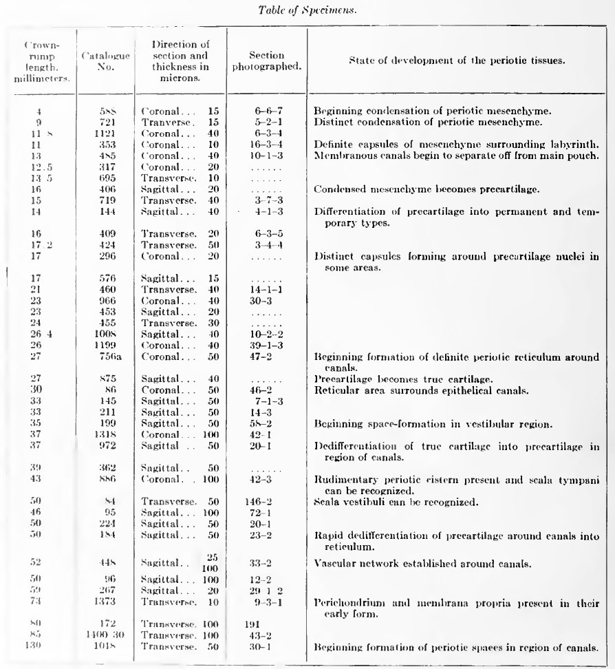
The observations recorded in this paper are made on human embryos and cover the period included between 4 mm. and 130 mm., crown-rump length, which is approximately equivalent to the period between the fourth and the sixteenth week of fetal life. The embryos were taken from the collection made by Professor Mall and that now belongs to the Department of Embryology of the Carnegie Institution of Washington. With two exceptions they had already been prepared in serial sections. In most of the stages the whole embryo is included in the sections, in some of the older ones the head alone is included, and in the two specimens that were especially prepared for this investigation the sections include only the region of the temporal bone. In the following table are listed the embryos that were found particularly suitable for the purpose at hand. They are arranged in the apparent order of development. The measurements given are those under which they are listed in the catalogue of the collection, and they all signify crown-rump measurement. Where sections were photographed this is indicated and the slide number, followed by the row and number of section, is given.
Table of Specimen
In studying the development of the cartilaginous capsule and the histogenesis of the periotic reticulum it was found necessary to i)repare enlarged pliotographs of the special regions studied. liy liaving tliesc all made on the same scale of enlargement it was possible to follow from stage to stage the change in volume and in form of the constituent tissue masses. Home of the photographs are reproduced on Plates I and II. In drawing conclusions from such photographs account was taken of the fact that the technique of preparing the .serial sections introduces an element of uncertainty in that some embryos in the process of embedding shrink more than others. This is particularly so in human embryos, where there is necessarily some difference in the freshness of the material at the time it is obtained. Furthermore, even in the same embryo some tissues are affected by the technique more than others. Due allowance was made for these factors.
In order to determine the form and relations of the periotic-tissue spaces, wax-plate models of the membranous labyrinth and the surrounding spaces were reconstructed after the Born method. Advantage was taken of the improvements in the method devised by Lewis (1915). The serial sections were photographed at a suitable enlargement on bromide paper. By means of a preliminary model of the membranous labyrinth the necessary reconstruction lines were established and transferred to the bromide prints. From these prints the membranous labyrinth and the periotic spaces were then traced on wax plates. After cutting out from the plates the areas corresponding to these structures the plates were piled and the resultant cavity was filled with plaster of paris. When the wax was finally melted off there remained a permanent plaster cast of the objects desired at a definite enlargement. Views of these models are shown on plate 4.
In outlining the periotic spaces it was found necessary to adopt an arbitrary rule as to how much should be included in the model. The smaller spaces of the reticulum surrounding the main cavities can be seen coalescing to form larger spaces, and these in turn coalesce with the main cavity as it advances into new territory. There is thus a considerable range in the size and completeness of the spaces in any one .section. The main spaces and the larger adjacent ones that communicate with them are outlined by a membrane-like border. This characteristic was adopted as the guide in determining which spaces to admit into the model; only those possessing a more or less complete border of this kind were included.
Carnegie Institution No.20 Otic Capsule: Introduction | Terminology | Historical | Material and Methods | Development of cartilaginous capsule of ear | Condensation of periotic mesenchyme | Differentiation of precartilage | Differentiation of cartilage | Growth and alteration of form of cartilaginous canals | Development of the periotic reticular connective tissue | Development of the perichondrium | Development of the periotic tissue-spaces | Development of the periotic cistern of the vestibule | Development of the periotic spaces of the semicircular ducts | Development of the scala tympani and scala vestibuli | Communication with subarachnoid spaces | Summary | Bibliography | Explanation of plates | List of Carnegie Monographs
| Historic Disclaimer - information about historic embryology pages |
|---|
| Pages where the terms "Historic" (textbooks, papers, people, recommendations) appear on this site, and sections within pages where this disclaimer appears, indicate that the content and scientific understanding are specific to the time of publication. This means that while some scientific descriptions are still accurate, the terminology and interpretation of the developmental mechanisms reflect the understanding at the time of original publication and those of the preceding periods, these terms, interpretations and recommendations may not reflect our current scientific understanding. (More? Embryology History | Historic Embryology Papers) |
Glossary Links
- Glossary: A | B | C | D | E | F | G | H | I | J | K | L | M | N | O | P | Q | R | S | T | U | V | W | X | Y | Z | Numbers | Symbols | Term Link
Cite this page: Hill, M.A. (2024, April 30) Embryology Book - Contributions to Embryology Carnegie Institution No.20 part 2. Retrieved from https://embryology.med.unsw.edu.au/embryology/index.php/Book_-_Contributions_to_Embryology_Carnegie_Institution_No.20_part_2
- © Dr Mark Hill 2024, UNSW Embryology ISBN: 978 0 7334 2609 4 - UNSW CRICOS Provider Code No. 00098G


