Book - Contributions to Embryology Carnegie Institution No.20
| Embryology - 27 Apr 2024 |
|---|
| Google Translate - select your language from the list shown below (this will open a new external page) |
|
العربية | català | 中文 | 中國傳統的 | français | Deutsche | עִברִית | हिंदी | bahasa Indonesia | italiano | 日本語 | 한국어 | မြန်မာ | Pilipino | Polskie | português | ਪੰਜਾਬੀ ਦੇ | Română | русский | Español | Swahili | Svensk | ไทย | Türkçe | اردو | ייִדיש | Tiếng Việt These external translations are automated and may not be accurate. (More? About Translations) |
Streeter GL. The histogenesis and growth of the otic capsule and its contained periotic tissue-spaces in the human embryo. (1918) Contrib. Embryol., Carnegie Inst. Wash. 8: 5-54.

| Historic Disclaimer - information about historic embryology pages |
|---|
 |
The Histogenesis and Growth of the Otic Capsule and its Contained Periotic Tissue-Spaces in the Human Embryo
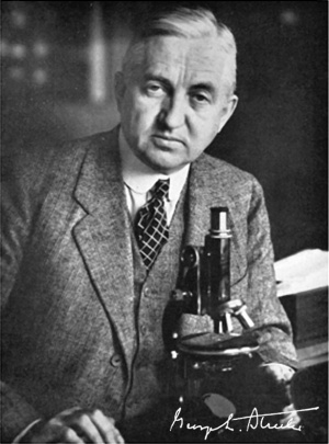
By George L. Streeter 4 text-figures and 4 plates (1918)
Monograph Contents
- Introduction
- Terminology
- Historical
- Material and Methods
- Development of cartilaginous capsule of ear
- Condensation of periotic mesenchyme
- Differentiation of precartilage
- Differentiation of cartilage
- Growth and alteration of form of cartilaginous canals
- Development of the periotic reticular connective tissue
- Development of the perichondrium
- Development of the periotic tissue-spaces
- Development of the periotic cistern of the vestibule
- Development of the periotic spaces of the semicircular ducts
- Development of the scala tympani and scala vestibuli
- Communication with subarachnoid spaces
- Summary
- Bibliography
- Explanation of plates
Introduction
During the past year the writer has published two shorter communications regarding the development of the connective tissue and cartilaginous coverings that inclose the membranous labyrinth, one of which dealt with the histogenesis of the cartilaginous capsule and the other with the periotic tissue-spaces that are formed in the interval between the cartilaginous capsule and the membranous labyrinth. In the present pajjer the same matter will be treated in more complete form and a general description will be given of the development of the otic capsule as a whole and of the problems involved in its growth.
In making this study the effort has not been so much toward the determination
of the exact form of the capsule as it has been toward the detection of some of the
factors that are involved in the production of this form. These two problems,
however, are not to be altogether separated. It is the distinctive form of the otic
capsule that makes it a particularly favorable place for determining the histological
features of the growth of such a structure. Owing to the fact that it is so well
provided with known landmarks, the changes in its size and form can be accurately
followed and it is therefore possible to determine deductively at what points, for
instance, new cartilage is being laid down and at what points it is being removed.
It was soon recognized that the growth of the otic capsule resolves itself into an increase in its external dimensions and a simultaneous hollowing-out and reshaping of its contained cavities, the latter being so managed that their general form and proportions are continuously maintained and a suitable space always provided for the enlarging membranous labyrinth. It is particularly the feature of cartilage excavation accompanying the increase in the total mass to which attention will be invited. It is quite evident that such growth can not be explained on the basis of a simple interstitial increase in the amount of cartilage, together with its passive rearrangement to allow for the enlarging cavities, due, one might say, to a mechanical expansive pressure from the growing membranous labyrinth with its surrounding tissue and fluid. Such a passive rearrangement could only occur in a tissue that is very plastic, whereas cartilage is one of the least plastic of the embryonic tissues. Moreover, the histological picture is not that of mechanical pressure; the cartilaginous chambers are always excavated slightly in advance of the space actually
required by the membranous labyrinth, and there is no evidence of the labyrinth being cramped or of the creation of pressure grooves in the margin of the cartilage.
Furthermore, it can not be the perichondrium that is the essential factor, either in
the deposit of new cartilage or in the excavation of the old, because the perichondrium, as we shall see, is not formed until after a considerable amount of the growth
and hollowing-out of the labyrinth is already completed. Therefore, in the development of the cartilaginous capsule there is something more than interstitial and
perichondrial growth.
As forming at least one element, and an important one, in this process it has
been found that there occurs a regression of certain areas of cartilaginous tissue to a
more embryonic form followed by its alteration into a different type of tissue. It is
this process of dedifferentiation that constitutes the essential factor in the hollowing-out and reshaping of the otic capsule which take place continuously during its
development. Though the significance and wide occurrence of dedifferentiation
and redifferentiation have been well known to botanists and to those investigators
who have worked with the simpler forms of animal life, this, as far as the writer
knows, is the first time that they have been shown to occur in the human embryo.
It is not unlikely that these principles will eventually enter into our conception of
the growth of other tissues and organs in human as well as in other mammalian
embryos. The establishment of this point, of the occurrence of retrogressive as
well as progressive differentiation in human embryos, is considered by the writer to
be the chief contribution of the following paper.
The fate of the periotic connective tissue that intervenes between the cartilage
and the membranous labyrinth and the formation of the characteristic periotic
spaces form problems that are naturally of a morphological character. These spaces
have been studied by modeling methods and a description will be given of the steps
by which the larger spaces acquire their adult form. It will be pointed out that
these spaces show a marked individuality. They have constant and definite characteristics, including their time and point of origin, the manner in which they spread,
and their eventual form and structure. They have a structural individuality which,
though less complicated, is just as definite as that of the other parts of this sense mechanism. All of this we will come to later.
Terminology

The writer is not unmindful of a certain feeling of distress that is aroused when it is found on reading a new paper that the author of it is adding to the already difficult matter of following another's description by making a new application of terms or by introducing a whole battery of freshly created ones. Nomenclature constitutes one field in which rock-bound conservatism bias many points of merit and where oiiginality may expect a cold and critical reception. It is therefore with some embarrassment that the writer approaches the subject of terminology, and it is al.so with some apprehension as to whether the " originality " in this instance will prove to be justified. It has in fact seemed best to avoid the incorporation of the term "lymphatic" in describing of the tissue-spaces surrounding the membranous labyrinth. It has been the custom to designate these as "perilymphatic" spaces since 1833, when the term was introduced by Brescliet, who thus distinguished them from the "endolymphatic" cavities of the membranous labyrinth. These terms, together with the terms "perilymph" and "endolymph" for their contained fluids, seemed particularly appropriate and in practical use have proved to be very convenient. Since Breschet's time, however, the lymphatic vascular system has taken on an increased and individual importance, due to researches in which American investigators have taken a particularly active part, and it now seems impottant to restrict the term "lymphatic" to it and its associated structures.
Inasmuch as the tissue-spaces surrounding the labyrinth have no known connection with the true lymphatic sj'stem, either in their origin or in their ultimate relations, it follows that the use of the term "Iymphatic" in connection with them is misleading. It therefore seems advisable to ehminate it, even at the expense of losing such a convenient terminology. As a substitute for " perilymphatic " the term "periotic" was finally decided upon and will be so used throughout this paper. In the formation of this adjective the Greek word ovs, from which it is derived, is used in the restricted sense of representing the essential sense-organ, that is, the otocyst itself and eventually the membranous labyrinth. Inasmuch as numerous words derived from the same source are in commtm use, it is felt that this term will be readily understood.
We shall speak of a periotic connective tissue that everywhere surrounds the membranous labyrinth. This periotic connective tissue includes in part the fine-meshed periotic reticulum, and in part the large walled-off periotic spaces with their contained periotic fluid, the most prominent of which are the scala vestibuli, scala tympani, and the vestibular cistern. For the term " endolymphatic " fluid one could substitute "otic" fluid; we would then have "Uquor perioticus" and "Hquor oticus." In all other instances, except when elsewhere specified, the Basillensis Nomina Anatomica (BNA) terms have been adhered to. The term "semicircular duct" is used to specify the epithelial or membranous canal as distinguished from the cartilaginous semicircular canal. This usage was recommended by Breschet and was adopted in the BNA. It was not taken advantage of, however, by Retzius (1884) in his monograph on the vertebrate ear, who used the term "semicircular canal" for the epithelial channel as well as for the cavity in which it lies. The influence of this
great monograph has delayed somewhat the adoption of the BXA recommendation, and one finds subsequent writers still following Retzius in this respect, among whom may be mentioned v. Ebner. R. Krau.se, Rothig, and myself in previous papers on the development of the membranous labyrinth. In a similar manner the usage by Retzius of the term "anterior" canal instead of "superior" canal, as recommended by the BNA, has occurred in relatively recent papers, including, it must be confessed, those of my own. In the present paper, however, this usage has been corrected. In the historical review which follows the various structures mentioned will be largely referred to in the older terms used by the respective authors.
Carnegie Institution No.20 Otic Capsule: Introduction | Terminology | Historical | Material and Methods | Development of cartilaginous capsule of ear | Condensation of periotic mesenchyme | Differentiation of precartilage | Differentiation of cartilage | Growth and alteration of form of cartilaginous canals | Development of the periotic reticular connective tissue | Development of the perichondrium | Development of the periotic tissue-spaces | Development of the periotic cistern of the vestibule | Development of the periotic spaces of the semicircular ducts | Development of the scala tympani and scala vestibuli | Communication with subarachnoid spaces | Summary | Bibliography | Explanation of plates | List of Carnegie Monographs
| Historic Disclaimer - information about historic embryology pages |
|---|
|
Glossary Links
- Glossary: A | B | C | D | E | F | G | H | I | J | K | L | M | N | O | P | Q | R | S | T | U | V | W | X | Y | Z | Numbers | Symbols | Term Link
Cite this page: Hill, M.A. (2024, April 27) Embryology Book - Contributions to Embryology Carnegie Institution No.20. Retrieved from https://embryology.med.unsw.edu.au/embryology/index.php/Book_-_Contributions_to_Embryology_Carnegie_Institution_No.20
- © Dr Mark Hill 2024, UNSW Embryology ISBN: 978 0 7334 2609 4 - UNSW CRICOS Provider Code No. 00098G