Book - Contributions to Embryology Carnegie Institution No.10
| Embryology - 27 Apr 2024 |
|---|
| Google Translate - select your language from the list shown below (this will open a new external page) |
|
العربية | català | 中文 | 中國傳統的 | français | Deutsche | עִברִית | हिंदी | bahasa Indonesia | italiano | 日本語 | 한국어 | မြန်မာ | Pilipino | Polskie | português | ਪੰਜਾਬੀ ਦੇ | Română | русский | Español | Swahili | Svensk | ไทย | Türkçe | اردو | ייִדיש | Tiếng Việt These external translations are automated and may not be accurate. (More? About Translations) |
Mall FP. The human magma reticule in normal and in pathological development. (1916) Contrib. Embryol., Carnegie Inst. Wash. Publ. 224, 4:5-26.
| Historic Disclaimer - information about historic embryology pages |
|---|
 |
The Human Magma Reticule in Normal and in Pathological Development
By Franklin P. Mall (1916)
Introduction
Students of embryology are familiar with the jelly-like substance found in the human exocoelom, which varies much in appearance in different specimens. Sometimes this substance is gelatinous, with, delicate fibers; at other times it is mixed with granules; and, in extreme cases, it forms quite a solid body. I think it was Giacomini who pointed out definitely that the morphological appearance of the magma determines, with considerable certainty, whether or not the contained embryo is normal or pathological. We are indebted to him for about a dozen papers on pathological embryology, a summary of which he published in Merkel-Bonnet's Ergebnisse. In this summary the following statement is made:
- " In the early stages of development we can determine by the extent of the exocoelom and its contained magma whether or not the embryo under consideration is normal. A large coelom, containing a rich magma, with its meshes sufficiently filled with a flaky precipitate to mask the embryo, is a certain sign of pathological development."
It is well known that the magma is least conspicuous in fresh specimens and becomes more pronounced after being hardened in alcohol or other preservative fluids. In recent years it is found that magma shows to the greatest advantage in specimens hardened in formalin; the fibrils are somewhat tougher, but the magma has usually the same appearance as in the fresh state. However, the experience of embryologists has been that the magma is more pronounced in pathological specimens, and for this reason it has been suspected that it does not exist in normal development. In fact, the illustration of the magma given by Velpeau in his monograph — in which he first uses the term "magma reticule" — is undoubtedly of a pathological specimen. A glance at the other plate which accompanies this handsome monograph shows clearly that most of the specimens he describes are decidedly pathological. During the 80 years which have elapsed since his time, embryologists, through comparative study, have been able to separate normal from pathological embryos with considerable precision; and in the abortion material, as collected in various laboratories, far over one-half of the specimens of the first 2 months of pregnancy are pathological, and in them we usually find a highly differentiated magma. However, if normal specimens are studied with care, we find that they too, contain some magma; therefore, magma must be viewed as a normal constituent of the human ovum.
It has been shown by Keibel that there is marked magma within the exocoelom of monkey embryos. In specimens containing embryos 1.3 mm. and 5 mm. in length, he describes it as a flaky, reticular mass outside the amnion, and speaks of it as coaguluni composed of reticular magma which had to be removed before the embryo could be seen. Undoubtedly he was dealing with normal specimens, thus showing quite conclusively that a delicate magma must be viewed as a normal constituent of the exocoelom. According to Keibel's figure 7, the magma appears to be denser in monkeys than is usually the case in normal human specimens. However, a very dense magma in human specimens invariably indicates, as was first demonstrated by Giacomini, that the ovum is pathological.
The best account of magma reticule is given by Retzius, who brought the subject up to 1890, and left it with the conclusion that magma reticule is a normal constituent of the human ovum. The statements of the earlier embryologists, from the time of Haller, are mainly of historical interest; but these investigators were at times inclined to view magma as a "middle embryonic layer" of the ovum, and, again, they believed it to represent the allantois of lower animals. Retzius reinvestigated the subject, taking into consideration normal as well as pathological embryos, and his conclusion is that magma is present in both kinds. His own words are as follows:
- " Bei der Oeffnung des Chorionsackes der Eier des ersten und zweiten Monats sah ich, wie in der Einlcitung crwalint wuitlo, und dies oft, am bcsten nach kurzer Erhartung in Ueberosmivimsiiure (von 3.1 Proc.) odor in Miillcr.sclRT Lusunfi ip'wcihiilich nach doiipeltor ^'ol•dunIunlg) , in dcni scliloiniigcn Irihalt, wclclicr zwisi-hcii dcni Chorion unil dcm Amnion, also im sulK'horionischfn Kaume vorhandcn war, diiiuioro odor dickere Fiidcn und J^triingc, (He mehr oder wcnigor dicht von der aus.*<orcn Fliiche des Amnion zur inncren Fliichc des Chorion hinul)orIicfen, um sicli dort mit ihrcn Enden an den beiden Hautcn zu licfcstigcn, indcm sie sich oft an ihnen vcrbreitcrtcn und in ihre U-kleidende Schicht iibcrgingen. Diese Faden und Striingo, wolehc im frischen Priiparate kaum sichtbar waren, traten nach der Behandlung mit den erwahnten Fliissigkciten deutUch hcrvor. In der Fig. 15 der Taf. X^'III habe ich ein seiches Ei abgebildet. Das stark zottige Chorion (ch) ist geoffnet, und man sieht im subchorionischen Raume den Amnion.sack (a) an weisslichcn Strangen (m) aufgehiingt liegen.
- " Die Strange, welche im unerhiirtotcnZustandecinc scliIcimigfasorigoConsistcnzhaben,sichaber ohne Schwierigkeit Stiickweise au.ssclineiden iiussen und dann in Mikroskope eine ileutlich f;iserige truc(ur darbieteii, zeigeu nach der JCrhiirtuug einen ausgcjjriigt fibrillaren Habitus. In einer homogen, structurlosen (iruud.substanz tretcn Zlige echtcr bindcgcwebiger Fibrillen hcrvor, welche oft eine Hauptrichtung ein schlagen, also ziemlich parallel verlaufen. Jedoch konimen auch viele sich kreuzende Fasern vor. Hier und da bemerkt man dickere Biindel verschiedenen Calibers welche aus dicht gedriingten Fibrillen besteiieii. Es sind also fibrillar-bindegewel)igo Balken, welche durch eine homogene, zahlreiche einzelne Fibrillen enthaltende Intercellularsubstanz ziehen. Zwischen den Balken und Fibrillenzugeii sieht man recht zahlreiche Zellen, welche theils und am moisten rutuUich oder oval, theils auch spin(lelf()rmig sind und in ihrem oft reichlichten Proto]ilasma gr().s.serc Klanzendc Korner enthalteii. Diese Zellen liegen in der (Irundsubstanz ohne Ix-soiidere anordnung zerstreut, bilden al.so keine Scheiden o. d. um die Filjiillenbiindel."
- "Man hat es hier ofTenbar mit einem iiurcifcn B i itdcgcwcbe zu ihun, inncm cmbryoiuilcn mucosen liindcgnvebf, irclchcg imlesmn in drr Kitlwickluiuj zum jlbrilUircn liindcycH'cbc schon tccit vorgcschritcn ist."
We have now in the literature a detailed description of a number of young litunan ova, smd, according to their clinical histories, some of them, at least, must be normal. The classic specimen is that described by Peters, which came from a woman who had committed suicide. The specimen was hardened in situ in an approv('<l manner, and was worked up and described under the best possible conditions. In it the co'loni is filled with a gelatinous substance, through which radiate delicate bands of fibrils, among which appear scattered nuclei. Near the embryo there is a small space, the interpretation of which was very difficult at the time the specimen was described.
Since Peters studied this specimen, the sections have been carefully reworked and discussed in a critical way by Grosser, who gives a new interpretation in two figures and states that the cavity of the ovum contains reticular magma which is partly made up of heavier strands of tissue accompanied by nuclei. In the neighborhood of the embryo there are two large spaces, lined with cells, which appear to be the primitive body-cavities. In his work on the comparative development of the embryonic membranes, Grosser describes this space in great detail and also gives us two new illustrations of the embryo in his plates 3 and 4. According to this authority these two body-cavities communicate b.y means of a slit-like canal just behind the umbilical vesicle (Grosser's figure 31, plate 4). This interpretation of the Peters specimen shows that the cavity of the ovum is first filled with a free mass of reticular magma, after which the coelom begins to form near the body of the embryo. As this cavity expands subsequently, it probably first destroys the more delicate strands of magma, leaving the heavier ones; thus in a short time the cavity of the ovum is lined b}- the endothelium of the coelom, which also must cover the stronger bands of magma radiating as trabeculae throughout this cavity (Grosser,
pp. 78 and 79).
Keibel explains the formation of the human coelom as follows:
- "It is, however, not quite clear how the cavity traversed by scattered strands of mesoblast and lying between the yolk-sac and the chorion in the Petersovum is to be interpreted. It may be supposed to represent the extraembryonic coelom; but it may also be imagined that it has arisen from
an extensive loosening up of ths tissus, and not by a splitting of the mesoderm, and that the triangular space between the caudal extremity of the embryo, which is lined with flat cells having an epithelial arrangement, is the first primordium of the coelom."
A condition similar to that found in the Peters specimen has been observed by Lewis in the Herzog specimen, which is of about the same stage of development. Lewis says (see his paper, p. 300) that there are occasional clefts in the mesoderm of the chorion of the Herzog embryo, but that they are of doubtful significance. His reconstruction shows a strand of mesoderm, more pronounced than m the Peters ovum, extending from the yolk-sac to the chorion and circumscribing a space on the ventral side of the embryo.
Eternod has written several papers in which he describes the formation of the exocoelom and the fate of the magma reticule. He says that it first fills the entire space between the primordium of the embryo and the chorionic wall. Later, larger spaces appear within the substance of the magma, leaving denser strands of magma fibrils to support the embrj-o wdthin the gradually expanding chorion. In general this coincides with the opinions just cited.
The relation of the exocoelom to the magma Ls strikingly shown by Waterston in a section of a small embryo in situ. The space between the embryo and the chorion is filled with a dense mass of fibrils, into which the exocoelom is burrowing. Waterston's figure 1 shows the relation of this cavity to the magma, and only near the embryo is the exocoelom lined with a layer of cells. "When this figure is compared with C.rosscr's figure of the Peters ovum, it becomes clear that the two spaces in the latter are in reality the beginning of the exocoelom.
The studies referred to above indicate that the space near the embryo i.s the primitive exocoelom and that the remainder of the so-called cavity of the chorion is simply the young normal ovum filled with delicate fibrils which communicate freely with the fibrils of the chorionic membrane. We have in our collection a young normal specimen, No. 763, containing an embryo anlage 0.2 mm. in length, which in general confirms the observations in the Peters ovum. A hst of the normal specimens in our collection discussed in this paper is given in table 1.
Table 1. — List of normal embryos

Specimen No. 763 was removed from a woman who was the mother of 6 children, the oldest being 10 years old. She had had one miscarriage. During the year before the operation she suffered much from headache and backache, but otherwise her health appeared to be normal. When she was admitted to the hospital she complained of abdominal enlargement and there was some urinary disturbance. At the operation for rupture of the perineum the uterus w'as scraped out ; subsequently the ovum was found in one of these scrapings. The fragments both of the mucous membrane and ovum appear to be normal.
Unfortunately we have only a few of the sections of this valuable specimen, but these show that we are undoubtedly dealing with a normal ovum of the same stage
of development as that described bj' Peters. The chorionic cavity is partly filled with mother's blood, put there are some strands of reticular magma, with nuclei and
protoplasm radiating through the blood. The specimen has been staimnl in hematoxyin and eosin, which is not esjiecially favorable for defining magma fiprils.
The specimen described by Ilerzog is also undoubtedly normal, as it was obtained from a woman who was killed by a stab-wound through the heart. The large colored plate publi.shed by Herzog shows the specimen to be quite identical with that of Peters. It shows free cells in the coclom, which contains no other foreign substance, but a photograph (figure 24, published by Herzog) shows that the coelom is filled by a very pronounced substance, reminding one very much of reticular magma. The same is true of a specimen recently described by Johnstone. A colored photograph which he published shows quite distinctly a pronounced magma throughout the coelom. (See, for instance, his figure 3.) This establishes definitely the presence of reticular magma in ova the size of the specimen of Peters. AVe have, however, the valuable specimen of Bryce and Teacher, which also shows the condition of the magma in an earlier stage. In this specimen the chorionic cavity is filled with a dense mass of fibrils, throughout which are scattered numerous nuclei, as shown in their plates 3 and 4. The specimen was not perfectly hardened and there is a small cleft between the chorionic wall and the mass of magma. As yet there is no exocoelom, showing that it is younger than the Peters specimen.
More advanced stages of the condition of the magma are represented in the specimens described by Jung and by Strahl and Beneke. In the Jung specimen the cavity of the ovum is filled with a very pronounced magma, running together in stronger bands, as in our own normal specimen. No. 836, to be described later. The larger cavity Jung marks "exocoelom," but it is not clear that this is lined with endothelium. From his large illustration one gains the impression that the specimen is somewhat pathological, for it is of the same type as numerous specimens in our collection with embryos that are usually found to be pathological. Taking the illustrations given in Jung's plates 1 and 2, the specimen again appears to be pathological, and I should be inclined to pronounce it such did not his plate 6, figure 17, show this same section on an enlarged scale, which gives a very sharp outUne of different embryo structures and scattered through them are numerous cells undergoing division. It would be impossible, with our present knowledge, to accept such sections as coming from a pathological embryo. The specimen described by Strahl and Beneke is of about the same stage as the Jung specimen, although the magma does not seem to be so well pronounced. It is unequal m nia.ss and has scattered through it deUcate strands, as shown in their figure 63. In fact, the above-described specimen underlies also the diagram on the form of the ccelom given by Strahl and Beneke on page 18 of their monograph.
Magma of uniform consistency, as seen in the Bryce and Teacher specimen, soon arranges itself in bands, which gradually become more and more pronounced in older specimens. Between these bands are spaces filled with fluid, and those spaces near the embryo become lined with endothelium to form the exocoelom. There are other spaces between the exocoelom and the chorionic wall. The sharper bands of magma fibrils — well shown in our embryo No. 836 (plate 1, figs. 3 and 4) — apparently support the embryo and the wall of the exocoelom within the chorion.
We have in our collection an excellent embryo. No. 391. which is a little larger than that described by Strahl and Beneke. This specimen came to us in formalin
and was opened with great care. It was found that the embryo and appendages
were suspended bj^ means of numerous delicate fibrils which radiated from them to
the chorionic wall. As the sections were stained with cochineal, the fibrils do not
show in them, so that this description is based entirely upon the apearance of the
uncut specimen. In general the specimen appears to be normal.
Our specimen No. 779, somewhat older than the one just mentioned, apparently contams no magma. It also was hardened in formalin. The ovum is entirety covered with vili, which branch twice, are of uniform size, and appear to be normal. In the main chorionic wall there is a pronounced fold. The specimen was bent
along the line of the fold, but the chorion was gradually dissected away with the
aid of direct sunlight. The chorion is entirely Imed by a smooth membrane, and
contains a cavity wliich is filled with a clear fluid and which apparently contains no magma, within there is a clear, worm-like body, which is bent upon itself,
with another body ari-sing from the middle of the bend. Apparently this is a flexed
empryo with the umbilical vesicle attached to it. The body is of uniform diameter,
mea.^^uring less than a millimeter. We are probably dealing here with a normal
ombryo. In opening this siiecimen great care was taken not to touch the embryo, so as to avoid injuring it. The embryo was taken out and cut into serial sections. It contains 14 somites and is without limb-buds. The sections give the impression that the embryo is pathological. There are no data in the history of the case which bear upon this point; therefore, for the present we may view it as a normal specimen without magma — or, if the embryo is taken into consideration, as a pathological specimen with dis.solution of the magma. Usually in pathological specimens the magma is greatly increased in quantity.
No. 104 is a somewhat older specimen. It came to us from an autolysis, with the entire uterus, and the sections of it indicate that the embryo is undoubtedly normal. The only record of the magma which we now have is given by several photographs which were taken at the time we received the specimen. These show a few strands of reticular magma, without any granular magma, radiating from the embryo. The photographs were taken while the sjiecimen was in formalin.
The next specimen, No. 463, is somewhat more advanced in development and contains a flexed embryo, 3.9 mm. in length. The ovum is covered completely on one side, and partly on the other, with villi 1.75 to 2.75 mm. long. On the partly covered side the villi leave relativelj^ bare one area, centrally situated, measuring 8 by 4.5 mm. Over it the villi occur only here and there, about 2 mm. apart, and are branched and apparently normal. On opening the ovum the reticular magma is found to fill the exocoelom. By carefully exploring with fine tweezers, an apparently normal embryo is seen with a yolk-sac measuring 3.5 bj- 4 mm. The embryo has anterior limb-buds and at least three gill-slits which are visible externally. No note was taken at the time regarding the condition of the magma, but sections of the entire chorion show that there is a very decided reticular magma between the embryo and the chorionic wall. There is no granular magma. The magma is composed mostly of fibrils, of much the same appearance as those of mesenchyme. Between the network of magma fibrils are denser strands accompanied by cells. In the fresh state imdoubtedly the denser strands would appear as filn-ils, while the rest would be transparent and jelly-like. The specimen came from a woman who was perfectly healthy and had given birth to 2 children durmg the last 4 years. This was her first miscarriage, and there was no indication of uterine disease.
Specimen No. 486, of the same degree of development as the one described above, is in a perfect state of preservation, but there is no history which would indicate whether or not the specimen is normal. However, the chorion is covered with villi about 3 mm. long, with a bare spot on one side about 4 mm. in diameter. The sections of the embryo do not show any attached fibrils of magma, but the chorionic wall, after hardening in alcohol, shows a decided layer of magma attached to it.
No. 470 is an interesting specimen, as it was found floating in a mass of bloodclotfi, which were sent to the laboratory in formalin. The ovum is covered with normal villi and contains a well-formed embryo within the amnion. It is apparently normal in every respect. No magma could be seen at the time, but drawings of the embryo subsequently made show delicate strands of fibrils forming a fuzzy layer around the umbilical cord and extending over the umbilical vesicle: undoubtedly these are magma fibrils. This seems to be the normal condition for this stage and is verified in specimen No. 836, to be described later. Sections through the mass and the chorion, stained with carmine, show the magma as a granular mass; only at points is there any indication of fibrils. However, this mass resolves itself into the most definite fibrils when colored with Van Gieson stain, in Mallory's stain, in hematoxylin, aurantia and orange G., or in iron hematoxylin. With Van Gieson stain the fibrils take on fuchsin color about as intensely as do the fibrils of the chorionic wall, with which thej' are continuous. The contrast obtained with Mallory 's stain is quite marked, as the endoplasm of the mesenchyme of the chorionic wall stains shghtly blue, while the exoplasm and the fibrils of the magma reticule remain unstained. This difference is not shown in sections stained in iron hematoxylin, as all fibrils are colored intensely black. However, it does not come out with the Oppels-Biondi method or with hematoxylin and eosin or aurantia. As the fibrils of the magma are continuous with those of the exoplasm of the chorionic wall, which do not stain in jNIallory's connective-tissue uiLxture, they can not be considered as white fibers, and from their failure to stain in Weigert's elastic-tissue mixture they are not elastic. As will be shown subsequently, they give the reactions of embryonic connective-tissue syncitium; and this is Retzius's opinion regarding their character. In specimen No. 486 the fibrils of the magma are not accompanied by any nuclei; so they must be viewed as belonging to the cells of the chorionic wall, from which they extend to bind the chorion with the primordium of the embryo.
Specimen No. 588 came from a woman who had 2 children living, aged 14 and 20 years respectively. Since the last birth she had aborted 11 times, and in the opinion of her physician all the abortions were due to mechanical means. This indicates that the specimen is normal. A figure of this embryo with strands of magma radiating from the umbilical cord and vesicle is shown in plate 3, figure 2.
Specimen No. 136 is of about the same stage of development as No. 588, although the chorion is covered with poorly defined villi. For an embryo of this stage it is unusually small, and I have therefore Usted it with the pathological specimens in my paper on monsters. A photograph of the ovum after it had been cut open shows that the chorion is completely filled with reticular magma, so that the embryo is practically obscured. A block of the whole ovum encircling the embryo was cut in serial sections. These show that there are strands of tissue accompanied by cells which form partitions in the exocoelom. The quantity of the magma appears to be somewhat excessive for a normal ovum of this stage of development.
Specimen No. 836, a perfect specimen containing an embryo 4 mm. ia length, settles definitely the condition of the magma at this stage of development (plate 1, figures 3 and 4). In this ovum the exocoelom, measuring 9 by 4 mm., contains a delicate spiderweb-like reticular magma, several of -the strands being considerably larger than the others. ^lost of this magma occurs between the yolk-sac and the amnion
Miul the adjacent embryonic viili which the fibrils are unusually abuiulant. This specimen was ohtaiiieil from a hysterectomy upon a woman, 20 years old, for a fibrous tumor of the uterus. She had been married 4 years, this being her first pregnancy. There were no special symptoms bearing upon the case, excepting the discomfort which accompanied the tumor of the uterus. Her last menstrual period had been delayed, and as it had been more jjrofuse than usual she believed that she had had a miscarriage; otherwise, everything ai^peared normal. This w-as confirmed by a careful examination of the specimen, which showed it to be normal in every respect. The uterus was opened by the surgeon at the time of the operation, but fortunately the site of the ovum was not injured. The specimen was sent to the laboratory immediately. where it was fixed in- Dr. Evans, who made the following record:
- "The specimen consists of a myomatous uterus which has l)ccu opened (apparently in a midline anterior incision) so as to disclose an abundant deciduous endometrium thrown into large folds. At the upper posterior surface of the uterus an oval mass, about 251)y 20 by 20 mm., projects. It is a sjic and is covered with a rather smooth membrane (decidua reflexa), beneath which tortuous vessels are apparent. On one side the sac (the implanted chorion) is adherent to the uterine mucosa (decidua vera). With a sharp .«calpel the entire mass was dissected away fiom the uterus and brought under a iiinocular microscope in warm salt solution. The middle portion of the free surface was opened carefully, beautiful villi being found, and then the delicate wall of the chorion was divided. Within, a transparent young embryo and its umbilical vesicle were seen, the embryo appearing to be about 5 mm. in length. Through this opening in the chorion, warm (40° C.) saturated aciueous solution of HgCl;, containing o per cent glacial acetic acid, was gently introduced and the entire mass placed in .500 c.c. of this fixation fluid. The main body of the uterus was dissected from the mj-omatous nodule and fixed in 10 per cent formalin, the site of the imjilanted ovum being marked by a short wooden rod."
The fixed and hardened specimen had undergone a readily appreciable shrinkage from the condition seen in warm salt solution. All of the tis.sues were beautifully preserved. The implanted ovum, covered with the decidua capsularis, measures approximately 22 by 18 by 11 mm. The adjacent decidua parietalis is thrown into large folds, which are themselves marked by numerous tiny elongated crack-like depressions, as well as by more circular pit-like apertures. The relatively smooth but irregular surface of the decidua capsularis is marked here and there by very conspicuous, small, oval pits, which may attain 0.5 mm. in diameter. The four flaps of this coat at its highest point, where it was opened directly over the middle of the ovum, and rather smooth on their inner surface and stand apart from the subjacent chorionic villi (intervillous space) to which they were originally adherent. The villi are about 2.5 mm. in length and possess one or two large branches and many "stub-like" tiny bulbous ones on the main stem. The villi are uniformly distributed in the small area exposed. With a slender scalpel the ovum was carefully divided under the dissecting micro.scope, the embryo and yolk-sac being visible. The yolk-sac appears to be almost 2 cm. in diameter and the embryo is surrounded by its amnion, its head (visible from above) being about 3 cm. in length and showing the fourth ventricle covered by a transparent ependyma. Two gill-arches are visible. The yolk-sac surface presents an exquisite picture of irregidar. clear vascular channels and a uniform pattern of small, opaque white blood-islands. The preservation seen is perfect.
After the embryo had been carefully removed, the ovum was cut into blocks which included its implantation. A block 1 mm. thick, which included the largest circumference of the embryo, was embedded in celloidin, the sections being stained in various ways. A photograph of this block is represented in plate 1, figure 4, which shows strikingly the extent of the magma. Sections which have been stained in hematoxylin and aurantia show the magma much as it appears in the other embryos that have just been considered. There is a denser magma just under the chorionic wall, and heavy strands radiate in every direction, with a fine network resembling spider-web, among the main strands. A number of loose nuclei accompany these strands, but they do not have the appearance of the nuclei of the main wall of the chorion. They are mostly round and are of unequal thickness, simulating very much the blood-cells. Occasionally there is a large nucleus. Sections which have been treated by the Weigert fibrin method do not show these fibrils. This confirms a previous experience which I have published elsewhere in my paper on monsters, namely, that magma fibrils do not give the reaction of fibrin, nor do these fibrils stain well in Van Gieson's mixture; however, they take on color similar to the mesenchyme of the chorion.
At points it appears as though these fibrils arise directly from the chorionic wall. They stain intensely blue by the Mallory method, and in sections treated in this way the nuclei of the mesenchyme of the vilU look much like the accompanying nuclei of the magma fibrils. On one side of the ovum a denser mass of the magma is directly continuous with the mesenchyme of the chorionic wall. However, just in this region the magma contams no nuclei. It, therefore, appears that the magma fibrils must be associated, at least partly, with the nuclei of the chorionic wall. Exceedingly good histological pictures were obtained from sections stained by Heidenhain's method, which show all the transition stages between magma containing no nuclei and magma very rich in nuclei. It would seem that there is quite a free wandering of the nuclei along the magma fibrils, and whenever they come in contact with the chorionic wall the fibrils enter it, showing direct continuity. The most instructive specimens are obtained by the Weigert elastic-tissue stain, which gives a sUght blue-black tinge to the mesenchyme fibrils of the chorionic wall, as well as to those of the centers of some of the villi. The magma itself takes on a very light stain, but where it is in contact with the chorionic wall it grades over into its blue network. It appears, then, that the centers of the villi, which represent their older portion, stain somewhat with elastic-tissue stain; and, if we view the chorionic wall as the more differentiated portion of the chorion, we must conclude that the older mesenchyme fibrils behave more like elastic-tissue fibrils than do the younger. At any rate, the magma fibrils do not take on elastic-tissue stain.
From all that has been said it is clear that the mesenchyme of the chorionic wall and the magma fibrils are continuous and, as I have pointed out elsewhere, they together form a common syncytium. I have already demonstrated that very young connective tissue arises directly from the mesenchyme, the earlier stages of which I have designated r^s the connective-tissue syncytium. Towards digestive reagents the connective-tissue syncytium gives somewhat the reaction of yellow elastic tissue, just as do the mesenchyme and the magma of No. 836 when treated with Weigert's elastic tissue stain. 1 have also shown that tho younger the connective-tissue syncytium is. tlie more difficult it is to digest it in jx'jjsin. Frozen sections shrink Injt little when treated with acetic acid, while white fibers become transparent. The syncytium itself is somewhat elastic, as shown by pressure upon the coverglass over a frozen section. If treated for 2-i hours with pepsin, the fibrils disintegrate. They are tlierefore much more resistant to the action of pepsin than are white fibrils.
The action of pancreatin is, in a measure, the opposite of that of pepsin. When the main mass of syncytium is formed by exoplasm, it digests readily in pancreatin. The more the syncytium is developed, the more resistant it is towards pancreatin very young syncytium fibrils, therefore, react towards pancreatin and jiejisin much like elastic fibers and this is confirmed in a measure, by tinctorial methods, when applied to sections of the chorion and magma, in specimen No. 836.
I have discussed the denser strands of tissue within the main mass of the magma. In the fresh state it appears that these are distinct fibrils, as shown in plate 3, figure 2. They are, also, observed in plate 1, figure 3. It is not quite so clear that there are fibrils in the magma as shown on plate 1, figure 4. In fact, it appears as though we have compartments separated by membranes, and that at the junction of several of these membranes the fibrils become denser, and therefore often appear as distinct fibers. It would be more appropriate than to state that the exoccelum is broken up into compartments the walls of which arc composed of membranes, and that where several of the membranes come together the increased amount of tissue gives the point of juncture the appearance of fibers to the naked eye and under the enlarging lens.
I liavc taken great pains to follow the cells which mark the stronger bands of magma, and it is difficult to arrive at any conclusion, for, in a measure, they seem to be related to the endothelial lining of the exoccelom. In the Peters ovum the spaces near the embryo are lined by a distinct layer of cells, but otherwise there is no indication of endothelial lining in any other portion of the chorionic cavity, nor is there any indication of such a lining in the figures given by Herzog, Johnstone, Jung, or Strahl and Beneke. It would seem that what corresponds to the exocoelom of the chorion in the later stages is represented by a diffuse mass in the specimen of Bryce and Teacher where the nuclei are scattered through it. The mode of the destruction of the mesenchyme is well indicated in figures on page 18 of a monograph by Strahl and Beneke. These irregular cells are first of all attached to the heavier strands of magma, and they must, therefore, correspond to the endothelial lining of the exoca'lom. For the present, however, it appears as if the exoccrlom of the human chorion is lined only in part by a layer of endothelium; these cells also accompany the magma fibers and line the inner side of the chorion near the embryo.
As tlie amnion expands, it naturally pushes these strands of magma up against the chorion, and in a short time we can recognize only a few fibrils in the exocoelom which encircle the umbilical cord. The.se are well seen in specimen No. 145, and their remnants are shown in No.'jTO, of which I give an illustration on plate 2, figure 2. Xo. 14S is vmdoubtedly normal, for it was obtained by mechanical means. and No. .')7fi is also a normal specimen obtained from a tubal pregnancy.
The conclusion regarding the condition of the magma of normal development is that the cavity of the ovum is filled with delicate fibrils which are intorpersed with numerous nuclei and which form one continuous network, extending from the embryo to the chorionic wall, and blending with its connective-tissue network. It forms one continuous syncytium, and as the ovum grows the magma reticule differentiates somewhat. Stronger bands of membranes soon form, breaking the cavity of the chorion into compartments. This process continues until the amnion begins to expand, and then these fibrils are pushed up against the chorionic wall. The exococlom begins as two larger spaces near the embryo, and in this portion of the ovum its cavity is lined with a layer of endothelium. It is quite certain that this sharply defined cavity does not extend to include the whole cavity of the ovum, but the cells lining it arise in common with those which accompany the magma fibrils. The exact extent and the fate of the two small spaces near the embryo in the Peters specimen is still undetermined, but Waterston's specimen indicates that they do not extend to fill the entire chorionic cavity. The examination of numerous specimens, however, indicates very definitely that the exocoelom of the ovum at 2 months does not contain a complete endothelial lining.
The Magma In Pathological Ova
Since the publications by Giacomini it has become well known that an increased quantity of magma within the coelom indicates with certainty that the embryo is pathological. When the magma is pictured or described, it is quite easy to determine whether or not the embryos and ova published in the literature are normal or pathological. This is demonstrated in the plates accompanying Yelpeau's work. His was able to separate most of the normal from the pathological embryos, but he relied mainly upon the external form of the specimens, which he compared with other mammalian embryos. Unless an embryo appeared much like those of other mammals and was not transparent and sharply defined, he decided that it was not normal but pathological. The work of Hochstetter, who limited his study to embryos obtained through hysterectomy, has been used to advantage by Keibel and Else in the preparation of their Normentafel, so that now we have adequate tables and plates which enable us to recognize with considerable certainty whether or not an embryo is normal, without paying much attention to the magma or the chorion. However, embrj^ologists are well aware that they can predict whether a specimen is normal or pathological by the quantity of the magma which masks the embryo when the ovum is opened.
By the contents of the exocoelom it is quite easj' to classify pathological ova into three chief groups. In the first group, which includes most pathological specimens, the magma is changed into an organized mass of reticular fibrils, intermingled more or less with granular substance.
To the second group belong specimens in which the exoccelom is large and contains only a fluid mass — that is, a liquid substance which does not coagulate in either formalin or alcohol. I have pictured a number of specimens of this sort in my paper on monsters. Specimen No. 512, of which I give an illustration on plate 2. figure 1, belongs to this group. The embryo is atrophic, and it is questionable whether or not if is oiicircled l)y tlic amnion. In these specimens the ocrlom is usually enlarged and sometimes it is greatly distended. Often there is a small granular precipitate in older specimens, but this is not of sufficient quantity or density to form a continuous mass. The histories of these specimens show that they are considerably older than their sizes indicate, and I am inclined to view them as having once had a dense mass of magma within the coelom, which subsequently underwent dissolution, leaving a more or less flaky deposit that finally disappeared altogether.
In the third group, the coelom is greatly distended, the ainiiioii is usually absent, all the ovum is filled with a gelatinous substance. This is well illustrated by specimen No. 604, plate 1, figures 1 and 2.
Table 2.— List of specimens containing pathological magma
I will now review several specimens illustrating these three varieties of pathological magma. The specimens considered are arranged in table 2. The list could
easily be increased to several hundred, but as the specimens with catalogue numbers less than 403 have been iiublished in detail with illustrations in my monograph on monsters, I will allude only to some of them. The pathological specimens with numbers over 402 are being prepared for publication, so that a few selected specimens with higher numbers are illustrated. Pathological sjiecimens from tubal pregnancy with numbers up to 1,000 will be found described in detail in my monograph on tubal pregnancy.
The first specimen which 1 shall consider (No. 278) consists of an entire ovum, measuring 6 by 4 mm. It was sent me by Dr. Stanton, of Albany, New York. The specimen might be viewed as normal, but it contains no embryo, and as it was obtained from a diseased uterus, it is probably pathological, the magnia having undergone minor changes.
This ovum was found accidentally in curct lings from a woman supposed to have chronic endometritis following pregnancy. There is nothing in the history from
ul,;,l, ti„. a^e of the specimen could be estimated. Part of the specimen had been cut into sections before it was received at the laboratory, with the statement that
no embryo had been found, it having faUen out. I found that the half sent contained a coelom, 3 by 2.5 mm., filled with magma, in which there was a cavity about
1.5 by 1 mm. 1^'ections showed that the cavity was natural and not sharply defined,
with nothing to indicate that it had contained an embryo. On the contrary, it
was found that the magma reticule was composed of a loose network of mesoderm
cells, which bound one side of the chorion with the other. These cells are directly
continuous with those of the mesoderm and resemble them in every particular.
At one point there is a small grouj) of epithelial cells, which may represent what was
originally the embryo. Otherwise, the chorion and its villi are normal in appearance, being encapsulated in decidua which has in it some uterine glands. All in
all, this specimen reminds one very much of the Peters ovum. There are some leucocytes in the decidua, but no accumulation of them indicating inflammation of the
uterus. Several figures, illustrating this specimen, may be found in my monograph
on monsters.
Specimen No. 531 is in many respects similar to the one just described (No. 278 ) .
It came from a woman who had been pregnant 6 times, her periods having been 17
days overdue before this abortion. The ovum is spherical, 19 mm. in diameter, and
is covered only by a mass of villi which appear normal. The ccclom within contains many magma fibrils, the meshes of which arc more or less filled with dense
granules, as is i^hown in plate 1, figure 8. Within this mass there is a detached
vesicle, 1.5 mm. in diameter, which no doubt represents the umbilical vesicle.
A specimen intermediate between the two just described is No. 250, of which several illustrations are published in my paper on monsters. The specimen came embedded in a mass of decidua and was obtained by scraping the uterus. When opened it was found filled with magma reticule just beneath the chorion, in which could be seen a small embryo, and farther away towards the center of the coelom was the umbilical vesicle. The whole ovum was cut hito sections. The chorion and the villi are apparently normal in shape and structure, being also rich in bloodvessels, which are filled with embryo blood. The villi are bathed in mother's blood and covered with an active trophoblast. The decidua is somewhat infiltrated with leucocytes, but there are no abscesses. The front end of the amnion is absent, and its free edge and the embryo are embedded in reticular magma, indicating that the amnion was destroyed before the abortion took place. The general shape of the embryo and its degree of development are practically normal. The heart is well formed and, including the blood-vessels, is filled with blood. The alimentary canal, brain, spinal cord, otic and eye vesicles, myotomes, and branchial arches are much like those of embryo No. 12, to be described presently. The septum transversum is well marked and the thyroid gland is just beginning. The tissues of the embryo, however, and the cavity of the front end of the brain are filled with numerous small round cells with fragmented nuclei. All stages of fragmentation are seen, just as may be observed in the leucocytes in small abscesses. IMost of the red blood-cells are within the blood-vessels, but those within the tissues appear perfectly normal. On account of the dimmished number of mesoderm cells, which, in fact, diminish in proportion as the fragmented cells increase, the conclusion must bo drawn tliat the fragmented cells arise from the mesoderm cells. Tlie epidormis covers the whole embryo. The primary change in this specimen is no doubt in the mesoderm, for all the rest of the embryo appears normal. That the c(|uililM-ium was overthrown is indicated by the necrotic amnion and the great amount of reticular magma in the exoccrlom. What is especially interesting in this specimen is the partial destruction of the amnion, which brings the embryo directly in contact with the pathological magma of the coelom.
Embryo No. 12, which has been just referred to, may also be discussed in this connection. It was questionable for a long time whether or not the eml)ryo was normal, as the villi and contents of the ccelom and embryo are beautifully preserved and show no pathological change. However, more careful consideration of the specimen shows that there are a few fibrinous masses between the villi, with every indication of uterine inflammation and infection. The extent of the reticular magma is more pronounced than usual, and it w'as necessary to dis.sect it away before the embryo could Ije isolated sufficiently so that it could be well seen. The head is no doubt atrophic, and I am fully convinced that this part of the embryo must have undergone pathological changes a short time before the abortion.
Specimen No. 318 is much like No. 250. The ovum, measuring 20 by 18 b.\- 1 1 mm., is covered with villi which appear to be perfectly normal. Upon opening, it was found to be filled with stringy magma, on one side of which was embedded an
embryo 2.5 mm. in length. The head is sharply outlined, but the embryo seems to
continue directly with the uml)ilical vesicle, leaving an atrophic tail. Sections .show'
that the amnion over the head has dissolved, leaving a picture very much like that
shown in No. 250. ^^'e have here a small embryo with a very large coelom, the ovum
being moderately filled with reticular magma and a small embryo only partly
covered with the amnion. No. 543 is another embryo of the same type. The
magma is a little den.ser than in No. 318. The chorionic villi are developed, but
markedly pathological, as the photograph show^s. The embryo within is 3 nmi.
long, lying (juite free within the mass of magma. It is covered by a ragged anmion;
that is, the amnion is jjartly destroyed.
An interesting specimen in this connection is No. 402, which is partly described in my paper on monsters, since the issue of which the embryo and chorion have been cut into serial sections. The villi of the ovum are not well developed, and they are distributed irregularly over the surface. The coelom is filled with reticular magma. The embryo is club-shaped, the head being much too far advanced for the body. The umbilical vesicle is normal in .size; the heart is well outlined, and the extremities are just appearing. Sections .show that the amnion is greatly distended. Sections of the chorion were stained with cochineal and Van Gieson, and show beautifully the fibrillated structure of the chorionic membrane. These fibers take im red stain, as do thoseof the reticular magma. The two are continuous, as .shown by the illustration on plate 3, figure 3. In fact, this continuity is much more pronounced in pathological than in normal specimetis.
Specimen No. 533 (plate 2, figure 3) .shows a more atlvanced stage ol an extensive
development of reticular magma. The villi of the ovum appear to be norinal and
tl,,. iLiioular magma is very dense. Hehveeii tlie meshes there are a number of opaque nodules about 0.5 mm. in diameter. \\ illi much difficult^' the embryo was
teased out, but it was practically impossible to clear it entirely of the magma fibrils.
The embryo is long and slender, looking more like that of a dog than a human specimen, the head being unusually small and thin for a human embryo of 0.5 mm. long.
The fibers are irregularly^ stuck together by small granules, and there is a gap in the
center which represents the place in which the embryo was located. The illustration
shows this condition beautifully. The specimen was sent me bj' Dr. Fewsmith, of
Trenton, New Jersey, who obtained it from a woman whose menstrual period had
been a month overdue.
An extremely interesting specimen is No. 545, well illustrated in figure 1, plate 3.
The magma is not extensive, but it is pronounced. The embryo is atrophic, and the
chorion is only partly covered with vilU. The specimen was sent me by Dr. Rand,
of New Haven, Connecticut. It was obtained from a woman who is the mother of
one healthy child. The last menstrual period began on September 2. Bleeding
began on October 22 and ended with the abortion on October 25. The ovum was
found embedded in the clots of blood attached to the cervix of the uterus.
An extreme case of degeneration of the magma is shown in No. 660, also well
illustrated in figures 4 and 5, plate 3. There is a tendency towards membrane
formation, tough strands of fibrils, spaces, and clumps of granules. The chorionic
wall is hemorrhagic and degenerated; within there is a collapsed amnion containing
a cheesy granular mass.
I shall use two more specimens to illustrate the nature of granular mass in more
advanced stages. The first is No. 605 and the second is No. 584a. No. 605 is a white
transparent specimen, covered with a uniform laj'er of \'\\\i which branch two or
three times. The entire specimen measures 45 by 40 by 25 mm. ; a small patch of
decidua adheres to the outside. The interior is partly filled with coarse strands of
reticular magma, having numerous granules attached. On one side of the specimen
the umbilical cord is seen, surrounded b}- a ragged amnion. The tip of the cord has
a piece of intestine and stomach hanging from it. The larger masses of tissue which
are intermingled with the reticular magma must be the renmants of the embryo,
parts of which appear to be normal, and judging from the form and size of the arms
and legs the embryo is about 10.5 mm. long. The second specimen is unusually
interesting because it contains a normal embryo with hernia of the Uver. The
exoccelom is unusually large and is filled with a more extensive layer of reticular
magma than should be found in an ovum containing a normal embrj^o of this size.
The remaining three specimens are given because they well illustrate various
degrees of reticular magma within the ovum.
No. 560 (plate 1, figure 6) shows very pronounced reticular magma intermingled with much granidar. Two stages of somewhat later development are given
in Nos. 636 and 991. In the former (plate 1, figure 10) the magma is more pronounced than in normal development, and in the latter (plate 1, figure 7) it is in an
extreme amount.
Finally, a unique specimen (No. 1189) throws some light upon the formation of the reticular magma. The ovum came to us within the uterus, having been removed by an operation. At first it seemed to be normal, but on opening it the embryo was found encircled by a large mass of transparent, tough, stringy reticular
magma, which was removed only with great difficulty. It behaved much like the
vitreous humor of the eye. On account of its great (juantity we at once suspected
that the sju'cimen was pathological, and after the embryo was removed it proved to
pe so. Ahhough (juite advanced in development, its head was found to be smaller
than normal, the tissues of the face were dissociated, and the borders of the eye were
not sharp but ragged. No doubt the specimen had continued to develop normally
until shortly before the operation, and the magma increased in quantity and became
tough and fibrous. It is an interesting specimen, showing changes in the magma
late in di'velopment. Sections of the implanted ovum have not .yet been made.
The specimen is from a negress, 45 years of age, who had had 9 previous jjregnaiicies. Her last menstrual jjeriod was 67 days before the operation. Pregnancy was
suspected before the removal of the uterus, but a hysterectomy was performed
because her periods had become verj' severe, lasting 8 days and causing faint ness
and weakness.
The two types of degeneration which the reticular magma undergoes have been
considered above. The magma becomes granular and denser as it lessens and becomes li(|uid. The liquid again either coagulates or remains fluid when the specimen is fixed in formalin. The two fluid types maj' be related to the destruction of
the amnion, but as yet I have been unable to reach a conclusion regarding this point.
The beginning of the formation of granular magma is shown in specimens No. otJO and 991 (plate 1, figures 6 and 7) as well as in Nos. 533 (plate 2, figure 3) and 6G0 ( plate 3, figure 5) . It appears to extend into the cavity of the amnion, and often forms great crusts, which surrovmd the embryo, as shown in several specimens
pictured in my monograjjh on monsters (e. g., Nos. 79, 94, 104, 230, and 261). An extreme specimen of granular magma within the exococlom is shown in specimen
No. 651</ (plate 1, figure 9).
It is extremely difficult to determine with certainty the structure of the granular
magma, but in studying pathological ova (especially those obtained from tul)al pregnancy) I have fre(|uentiy ol)served that there are large masses of granular magma
which take on hematoxylin stain. The granules are mixed with a slimy mass
which also takes on hematoxylin stain. My attention was called to these granules
becau.se they have a characteristic circular stratification and contain within their
centers small granules which also stain intensely. 1 am by no means certain
whether all granular magma stains in this way with hematoxylin, and what T have
just stated may apply only to a portion of the granular magma.
Specimen No. 531 (shown in figure 8, plate 1) has its ccrlom filled with a liquid mass in which there is a granular dejwsit that surrounds the embryo anlage. Such specimens are numerous and, without opening them, they may frequently be recognized by the transparency of the chorionic wall, which is covered with but few atrophic villi. A more advanced embryo, showing the same condition, is shown in specimen No. 512. In it the embryo is atrophic and macerated, without the presence of an amnion. The chorion is thin and is fully covered with delicate degenerated villi. Other specimens which come within this group are Nos. 21, 7S. 122, and 244f/. These are all illustrated in my monograph on monsters.
Sometimes the entire specimen is filled with a gelatinous mass, which becomes firmer when fixed in formalin and separates into a more sohd mass, and into a liquid when preserved in formalin. This mass appears to lie within the amnion in most specimens, as in cases where it fills the whole ovum the amnion is missing. Specimen Xo. G04 (plate 1 . figures 1 and 2) is quite typical, as is also No. 130. In both the embryos are atrophic and necrotic, and the jelly-like fluid fell out with ease when the ovum was cut open. The chorion is atrophic in both of them and is covered only with a few atrophic villi. Specimen No. 604 came to me without a history, and measures 70 by 50 by 50 mm. It is fully covered with fibrinous clots, between which there are few large villi, as the picture shows. The chorionic wall is 3 to 4 mm. in thickness, and its interior is entirely filled with jelly-like magma of uniform consistency. On one side of the specimen, lying free witliin the hyaline magma, is a straight embryo, 17 mm. in length, with atrophic head, arms, and legs. The same description applies equally well to Xo. 135. Specimens like these are quite numerous in our collection of human ova, but usually the jelly is lost when the specimen is ojjened. Figures illustrating embryos of this sort maj' be .seen in my paper on monsters, under the descriiition of embryos X'^os. 79, 94, 230, 261, and 270.
No. 1117 (plate 1, figure 5) contains an embryo well packed in the jelly-like magma. The cavity of the ovum is small and its wall is very hemorrhagic. The specimen came from a woman, age 26 years, who was married at 15. She had two births at term and one previous abortion. She believed she became pregnant about 3 months before the operation, although she had not missed her regular periods.
Another specimen belonging to this category is No. 813. It consists of a fleshy mole, well filled with tough, jelly-like magma. All the vilh are destroyed and its surface shows ulceration. Further study of this magma is necessarj' before it can be related to the granular magma which forms with the reticular magma in the exocoelom. I am inclined to believe that the hyaline substance which is so often found within the amnion of pathological specimens arises from the amniotic liquid, which has become richer in albumen, and therefore congeals into a jelly-like mass when preserved in formalin.
Conclusion
The fibrils forming reticular magma are always in direct continuity with those of the mesenchyme of the chorionic wall. This can easily be demonstrated by means of Van Gieson stain, and reticular magma must therefore be viewed as embryonic connective tissue extending into the cavity of the ovum. The stronger strands are accompanied more or less by mesenchyme nuclei, show^ing that the magma itself must be viewed as independent connective tissue identical with the mesenchyme of the chorion. As the amnion extends these strands are pushed aside, their final remnants being seen in that portion of the exoccelom which encircles the umbihcal cord.
In pathological specimens the reticular magma increases in quantity in the earlier stages of development, continumg for a number of months of pregnancy. Frequently the meshes between the reticular fibrils are filled with pecuhar stratified granules which take on an extensive hematoxjdin stain. Often the amnion is destroyed e:irl>- in (k'veK)pnient, in wliicli case the magniii may dissolve, but sometimes it inrreases {greatly in (luantity, forming a gelatinous mass. Freciuently pathological ova are encountered in which the development of the embryo is retarded, and the amnit)n is often found filled with a flaky deposit that, as time goes on. increases greatly in quantity and finally forms large crusts which invest the embryo. In other cases there is marked hydramnios, and in certain instances, where the amnion is destroyed, the magma dissolves, leaving only the embryo floating in the fluid encircled by the chorionic wall. Specimens are also found in which the ca\ity of the amnion is greatly enlarged and is filled with a jelly-like substance, which in later stages may form crusts encircling the embryo. The true relation between the pathol()gical changes of the contents of the exocoelom and of the cavitv of the amnion remains to be determined.
Bibliography
Bryce and Teachre: Contributions to the study of the early development and imbedding of the human ovum. Glasgow, 1908.
Eternod, a. C F. : La gastrulc dans la .seric animalc et plus speeialemeut ehcz I'hommc et les mammif6rcs. Tirage a part du Bull. Soc. Vaud. So. Nat., 1906, xi.ii, 150, Lausanne, 1906.
. Dea premiers stades de I'ocuf liumain et de son implanation dans I'uterus. M6moirc present^ au premier Congrfcs ffdfratif international d'anatomic (Geneve, 6-10 aout 190.5), Nancy, 1906.
. L'ceuf humain. Implantation et Ke.?tation trophoderme et placenta. Mf-moirc publi6 ii I'occasion du Jubilfi de rUniversitt, l,i.59 1909, Geneve, 1909.
. In6galit6s de croissance du chorion ovulaire humain et localisations consecutivcs en chorion laive et chorion frondosum. C R. de la Reunion de Association des Anatomistes (Nancy, 5-7 avril 1909). Lille, 1909.
Fkassi, L.; Ueber ein jungcs menschlicheaEi in situ. Archiv fiir mikroskopische Anatomic und Entwicklungsgcschichte, Bd. 70, 1907.
GiACoMiNi, C: Probleme aus Entwickelungsanomalien d. Menschl. Embryo. Mcrkel and Bonnet "Ergeljnisse." iv, 1894.
CJrosser, O. : Eihiiute und der Placenta. Wien und Leipzig, 1909.
. The development of the egg membranes and the placenta; menstruation. Keibel and Mall, Human Embryology, i, 1911.
Herzog, M.: a contribution to our knowledge of *he earliest known stages of placcntation and embryonic development in man. American Journal of Anatomy, ix, 1909.
His, W. Anatomie menschlicher Embryonen. Leipzig, 1880-1885.
HocHSTETTER, : Bildcr der ausseren Korpcrform einigcr menschlicher Embryonen avis den beidcn crsten Monaten der Entwicklung. Mimchen, 1907.
Ingalls, N. W.: Beschrcibung cines menschlichen Embryos von 4:9 mm. Archiv. fQr mikroskopische Anatomie und EntwicklungsKcschichte, Bd. 70, 1907.
Johnstone, R. W.: Contribution to the study of the early human ovum. Journal of Obstetrics and Gynccologj' of the British Empire, 1914. Juno, P.: Ei-Einbettung Iieim menschlichen Weibo. Berlin,
1908.
Keibel, F. : Die aussere Korpcrform von Affenembryonen.
.Selenka, Entwickelungsgeschichte, xiv, Wiesbaden,
1900.
. The formation of the germ layers and the gastnilation problem. Keibel and Mall, Human Embrology, I, chapter v, 1911. Keibel und Elsa: Normentafel zur Entwicklungsgeschichtc des Mcnschen. Normentafel zur Entwicklungsgcschichte der Wirbcltiere. Achtcs Heft. Jena, 1908.
Lewis, F. T.: The development of the intestinal tract and respiratory organs. Keibel and Mall, Human embryology, ii, 1912.
Mall., F. P.; Origin of human monster. Journal of Morphology, XIX. Published also as a monograph by the Wistar institute of Anatomy, Philadelphia, 1908.
. Development from the connective tissue of the syncytium .Vmerican Journal of Anatomy, i, 1901.
. On the fate of the human embryo in tubal pregnancy. Publication No. 221, Carnegie Institution of Washington, 1915.
Peters, H.: Einbcttung des menschlichen Eies. Leipzig und Wien, 1S99.
Retzius, G.: Ueber das Magma rfcticulfe des menschlichen Eies. Biologische Untersuchungen, I, Stockholm,
1890.
Stbahl and Beneke: Ein junger menschlicher Embryo. Wiesbaden, 1910.
Velpeau, a. L. M.: Embryologie ou Ovologic Humaine. Paris, 1833.
Waterston. a young embryo with 27 somites. Journal of Anatomy and Physiologj', xlix, 1914.
Explanation of Plates
| Historic Disclaimer - information about historic embryology pages |
|---|
|
- Mall 1916 Links: Table 1 Normal Embryos | Table 2 Abnormal Embryos |Plate 1 | Fig. 1+2 | Fig. 1 | Fig. 2 | Fig. 3 | Fig. 4 | Fig. 5 | Fig. 6 | Fig. 7 | Fig. 8 | Fig. 9 | Fig. 10 | Plate 2 | Fig. 1 | Fig. 2 | Fig. 3 | Plate 3 | Fig. 1 | Fig. 2 | Fig. 3 | Fig. 4+5 | Fig. 4 | Fig. 5 | Contributions to Embryology No.10
Plate 1

|
Fig. 1, 2. Photographs of the two halves of ovum No. 604. Natural size. The cavity of the ovum is filled with a jelly-like substance in which a pathological embryo is embedded. Fig. 3. Section through a normal ovum No. 836, encapsulated in the decidua. X3}. Drawn by Mr. Didusch. The embryo lies within the coelom, and bands of -magma fibrils radiate from the amnion to the chorionic wall. The head of the embryo shines through the more transparent portion of the amnion. Fig. 4. Photograph of a block of the ovum, Xo. S36, in situ after the embryo had been removed. X25. The suporting strands of magma are strikingly shown. Fig. 5. Pathological embryo No. 1117, embedded in hyaline magma. X4. From a tubal pregnancy following gonorrhea (?). Fig. 6. Pathological ovum No. 560, containing a great quantity of reticular magma. X2i. The embryo is normal in form. From a case of retroversion of the uterus. Fig. 7. Pathological ovum No. 991, with the cavity completely filled with reticular magma. Natural size. The embryo is normal inform. From a negro woman. Sect ions of the embryo indicate that it is macerated. Fig. 8. Pathological ovum No. 531, containing a granular deposit around a nodular embryo. X 1 . Fig. 9. Pathological ovum with a nodular embryo (651?). X2. The exocteloni is gorged with granular magma. Fig. 10. Specimen No. 636. X. The embryo and chorion are normal in form, but the reticular magma is markedly increased in quantity. |
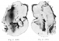
Fig. 1+2
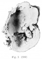
Fig. 1
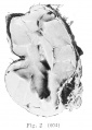
Fig. 2
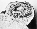
Fig. 3
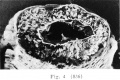
Fig. 4
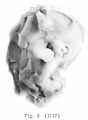
Fig. 5
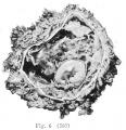
Fig. 6
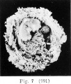
Fig. 7
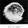
Fig. 8
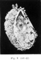
Fig. 9
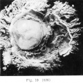
Fig. 10


Plate 2

|
Fig. 1. Pathological embryo No. 512, lying free within the ovum. X6. The villi are tliin and scattered and the embryo is atrophic. There is no formed magma.
Fig. 2. Ovum No. 576, obtained from tubal pregnancy, showing a delicate layer of magma fibrils around the attachment of the umbilical cord to the chorion. X3. Fig. 3. Ovum No. 533, showing very extensive magma. x6. |
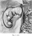
Fig. 1
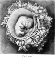
Fig. 2
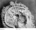
Fig. 3


Plate 3

|
Fig. 1. Ovum No. 545. X7. There is a delicate network of fibrils below the amnion and the chorion.
Fig. 2. Embryo No.588. X5. Delicate strands are shown radiating from the umbilical cord and yolk-sac. This figure is given to show the appearance of magma in vesicle development. From a woman who has had numerous mechanical abortions performed upon herself. Uterus badly inflamed. Fig. 3. Section through the chorion and magma of No. 402. X280. The specimen was stained with Van Gieson stain and shows that the fibrils of the magma are continuous witli those of the mesenchyme of the chorionic wall. It came from a case with subinvolution and symptoms of endometritis. Fig. 4. Outline of the ovum of No. 660. natural size. The diagram indicates the part of the specimen shown enlarged in figure 5. Fig. 5. No. 060, showing very extensive changes in the magma. X6. The upper tip of the amnion is shown. The magma is fibrillar and granular, and at places the fibrils seem to form membranes. The chorionic wall is very hemorrhagic. |
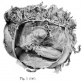
Fig. 1
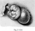
Fig. 2
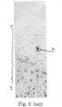
Fig. 3
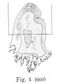
Fig. 4

Fig. 5
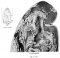
Fig. 4 and Fig 5


| Historic Disclaimer - information about historic embryology pages |
|---|
|
Glossary Links
- Glossary: A | B | C | D | E | F | G | H | I | J | K | L | M | N | O | P | Q | R | S | T | U | V | W | X | Y | Z | Numbers | Symbols | Term Link
Cite this page: Hill, M.A. (2024, April 27) Embryology Book - Contributions to Embryology Carnegie Institution No.10. Retrieved from https://embryology.med.unsw.edu.au/embryology/index.php/Book_-_Contributions_to_Embryology_Carnegie_Institution_No.10
- © Dr Mark Hill 2024, UNSW Embryology ISBN: 978 0 7334 2609 4 - UNSW CRICOS Provider Code No. 00098G