Paper - A comparison of the Herzog and Strahl-Beneke embryos
| Embryology - 27 Apr 2024 |
|---|
| Google Translate - select your language from the list shown below (this will open a new external page) |
|
العربية | català | 中文 | 中國傳統的 | français | Deutsche | עִברִית | हिंदी | bahasa Indonesia | italiano | 日本語 | 한국어 | မြန်မာ | Pilipino | Polskie | português | ਪੰਜਾਬੀ ਦੇ | Română | русский | Español | Swahili | Svensk | ไทย | Türkçe | اردو | ייִדיש | Tiếng Việt These external translations are automated and may not be accurate. (More? About Translations) |
Lewis FT. A comparison of the Herzog and Strahl-Beneke embryos. (1917) Anat. Rec, 11: 386.
| Online Editor |
|---|
 This 1917 paper presented by Lewis at the 33rd Meeting of the American Association of Anatomists in New York (Dec 27-29, 1916). Describes two early human embryos (Carnegie stage 6) that had been previously serially sectioned. This 1917 paper presented by Lewis at the 33rd Meeting of the American Association of Anatomists in New York (Dec 27-29, 1916). Describes two early human embryos (Carnegie stage 6) that had been previously serially sectioned.
The Herzog embryo was also described in Herzog MA. A contribution to our knowledge of the earliest known stages of placentation and embryonic development in man. (1909) Amer. J Anat., 9(3): 361-400.
|
| Historic Disclaimer - information about historic embryology pages |
|---|
| Pages where the terms "Historic" (textbooks, papers, people, recommendations) appear on this site, and sections within pages where this disclaimer appears, indicate that the content and scientific understanding are specific to the time of publication. This means that while some scientific descriptions are still accurate, the terminology and interpretation of the developmental mechanisms reflect the understanding at the time of original publication and those of the preceding periods, these terms, interpretations and recommendations may not reflect our current scientific understanding. (More? Embryology History | Historic Embryology Papers) |
A Comparison of the Herzog and Strahl-Beneke Embryos
Harvard Medical School.
(Lantern) Presented at the 33rd Meeting of the American Association of Anatomists in New York (Dec 27-29, 1916).
Abstract
The Herzog and Strahl-Beneke embryos are of particular value to embryologists since they are the youngest human embryos of which satisfactory drawings of serial sections have heretofore been published (22 sections of the Herzog embryo and 60 of the Strahl-Beneke specimen). Of the Peters embryo, apparently only a single section is thus available, which has been reproduced everywhere; and although 30 sections of the Bryce-Teacher specimen have bcen published, they are fragments defying interpretation, at least apart from the specimens themselves. Bryce and Teacher estimate that their embryo is three days younger, and Peters two days younger, than the Bencke specimen; but that these figures are merely approximations is indicated by the fact that Bryce and Teacher consider Peters embryo to be thirteen and one-half to fourteen and one-half days old on p. 53 of their work, but fourteen to fifteen days on p. 59. Although the difference in age of the four specimens under discussion is perhaps not greater than three days, thcre is the widest difference in the amount of information available about their structure. The Strahl-Beneke and Herzog embryos are the ones to which at present we must resort for an approximately adequate description.
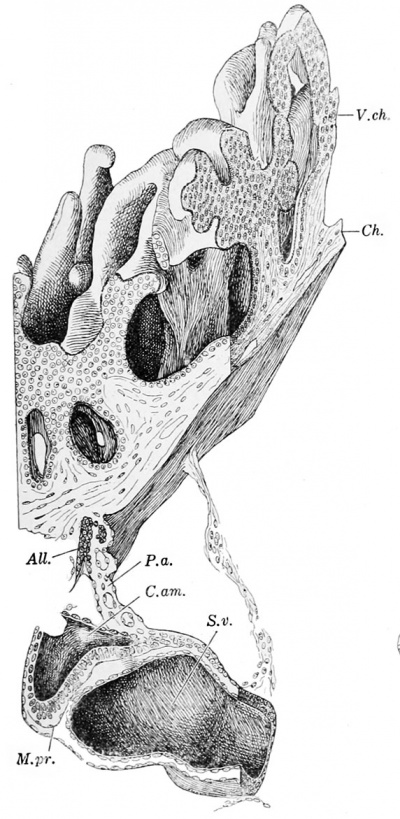
Herzog’s embryo is not now in a good state of preservation and its reconstruction is in part a matter of restoration. Thus the detached tube in the body-stalk, which was originally intcrpretcd as an allantois, is clearly an amniotic duct. The place where it was torn off from the amnion can be definitely located. (This fundamental correction, reported at the 1913 meeting of this association, involves a change in labelling rather than any other modification of the model figured by the writer in Keibel and Mall’s Embryology, vol. 2.) The Strahl-Beneke embryo is in far better condition, and helps to explain the Herzog specimen, whereas the latter is the most interesting commentary on Strahl and Beneke’s important publication. This was appreciated by Strahl and Beneke who expressed regret that Herzog’s paper was received too latc for them to make a detailed comparison between his sections and their own, and they merely note that his sections differ in many respects very strikingly from theirs. We can now supply such a comparison and find that the embryos are so nearly alike that they may be said to establish the essential features of a certain early stage in hunian development. The conditions in younger human embryos are still subjects for diagram and conjecture.
Online Editor - Images of the embryos below are from other publications and do not appear in the above abstract.
Wax reconstruction of Herzog's embryo
Cite this page: Hill, M.A. (2024, April 27) Embryology Paper - A comparison of the Herzog and Strahl-Beneke embryos. Retrieved from https://embryology.med.unsw.edu.au/embryology/index.php/Paper_-_A_comparison_of_the_Herzog_and_Strahl-Beneke_embryos
- © Dr Mark Hill 2024, UNSW Embryology ISBN: 978 0 7334 2609 4 - UNSW CRICOS Provider Code No. 00098G

