Musculoskeletal System - Skull Development: Difference between revisions
| Line 41: | Line 41: | ||
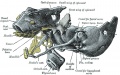
Fetal head section | Fetal head section | ||
| This mid-line section through the fetal head shows features of the brain, face and mouth. | | This mid-line section through the fetal head shows features of the developing skull and the brain, face and mouth. | ||
* Neural | * Neural | ||
** developing brain and brainstem. | ** developing brain and brainstem. | ||
| Line 47: | Line 47: | ||
** fourth ventricle. | ** fourth ventricle. | ||
** developing pituitary sitting in the sella turcica. | ** developing pituitary sitting in the sella turcica. | ||
Musculoskeletal | * Musculoskeletal | ||
* cartilage - septum of the nose. | ** cartilage - septum of the nose. | ||
* bone - ossifying nasal concha. | ** bone - ossifying nasal concha. | ||
* bone - palate roof of mouth. | ** bone - palate roof of mouth. | ||
* cartilage - soft palate back of mouth. | ** cartilage - soft palate back of mouth. | ||
* cartilage - base of skull and vertebra. | ** cartilage - base of skull and vertebra. | ||
* muscle - tongue, attached note foramen cecum. | ** muscle - tongue, attached note foramen cecum. | ||
* bone - mandible. | ** bone - mandible. | ||
* cartilage - developing hyoid and thyroid bones. | ** cartilage - developing hyoid and thyroid bones. | ||
Revision as of 10:37, 18 March 2012
Introduction
The Skull is a unique skeletal structure in several ways: embryonic cellular origin (neural crest), form of ossification (intramembranous and endochondrial) and flexibility (fibrous sutures). The cranial vault (which encloses the brain) bones are formed by intramembranous ossification. While the bones that form the base of the skull are formed by endochondrial ossification. The bones enclosing the brain have large flexible fibrous joints (sutures) which allow firstly the head to pass through the birth canal and secondly postnatal brain growth.
In humans, ossification continues postnatally, through puberty until mid 20s and in old age the sutures separating the vault plates are often completely ossified.
In the entire skeleton, early ossification occurs in the jaw and at the ends of long bones (More? see movie developing mouse). Osteoblasts manufacture bone and are derived from ectomesenchymal in origin. (More? see lineage below). Flexible fibrous sutures allow growth of the brain to be accomodated by calvarial plate growth. Recent studies have show that noggin (a BMP antagonist) is involved in closure of these sutures.
| Historic - Human Fetus (CRL 43mm) Skull | Category:Skull
Some Recent Findings

|
Fetal Skull
The Images below show the combined endochondral and intramembranous ossification that is occurring in early fetal development (week 12).
In the first 2 images the bone cartilage is shown in blue and the new bone in red.
Note the difference in appearance between the upper and lower jaw (maxilla and mandible), the currently cartilage base of the skull (chondrocranium) and the cranial vault (neurocranium).

Fetal head lateral view |

Fetal head medial view |

Fetal head section |
This mid-line section through the fetal head shows features of the developing skull and the brain, face and mouth.
|
Skull Views

|

|

|

|
| anterior view | superior view | lateral view | lateral view |
| showing anterior fontenelle, sutures, mandible | showing anterior fontenelle, sutures | showing suture, mandible | newborn skull |
Skull Sutures

The bones enclosing the brain have large flexible fibrous joints (sutures) which allow firstly the head to compress and pass through the birth canal and secondly to postnatally expand for brain growth. (More? Molecular Skull Sutures) These sutures gradually fuse at different times postnatally, firstly the metopic suture in infancy and the others much later. Abnormal fusion (synostosis) of any of the sutures will lead to a number of different skull defects, leading to disruption of brain development. (More? Abnormal Synostosis) In old age all these sutures are generally completely fused and ossified.
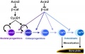
At the molecular level, accelerated suture intramembranous ossification can be mediated through a dual role of β-catenin in both the expansion of osteoprogenitors and the maturation of osteoblasts.[4] These researchers also show that disruption of Axin2/β-catenin signaling alters the regulation of the downstream transcription target, cyclin D1, in the canonical Wnt pathway.[5]
Computed Tomography Views

Skull CT Vertex, later and basal views.[6] |
Sutures and Fontanels
|
coronal suture

lambdoid suture

metopic suture begins at nose and runs superiorly to meet sagittal suture and fuses during infancy (fusion beginning at 3 months and completes by 6 to 8 months of age) before all other cranial sutures.
sagittal suture
Fetal Head Growth


Abnormalities
There are several skull deformities caused by premature fusion (synostosis) of different developing skull sutures. Suture abnormalities are classified as either "simple" (only one suture involved) or "compound" (two or more sutures involved).
 |
* craniosynostosis premature cranial suture fusion, results in an abnormal skull shape, blindness and mental retardation.
|
Craniosynostosis
Attenuation of signaling pathways stimulated by pathologically activated FGF-receptor 2 mutants prevents craniosynostosis.[7] "Craniosynostosis, the fusion of one or more of the sutures of the skull vault before the brain completes its growth, is a common (1 in 2,500 births) craniofacial abnormality, approximately 20% of which occurrences are caused by gain-of-function mutations in FGF receptors (FGFRs). ...These experiments show that attenuation of FGFR signaling by pharmacological intervention could be applied for the treatment of craniosynostosis or other severe bone disorders caused by mutations in FGFRs that currently have no treatment."
Dolichocephaly and scaphocephaly

|
| Dolichocephaly and scaphocephaly
(premature fusion of the sagittal suture) |
Brachycephaly and anterior plagiocephaly
(Greek, brakhu = short) (Greek plagios = oblique)
- brachycephaly - premature bicoronal fusion
- anterior plagiocephaly - unicoronal fusion
Leads to a restriction of anterior-posterior calvarial growth and relatively unaffected biparietal growth.


Skull Turricephaly

Skull Trigonocephaly

(Greek, trigonos = three angles) This abnormality results from the premature fusion of the metopic suture occurring before 6 months (3-9 months) of age.
Skull Oxycephaly

Images show oxycephaly from severe sagittal and coronal synostoses (arrowheads).
Craniofrontonasal Syndrome
Craniofrontonasal syndrome (CFNS) is a human X-linked developmental disorder caused by a mutation in ephrin-B1 affecting mainly females. Characterised by abnormal development of cranial and nasal bones, craniosynostosis (premature coronal suture fusion), and other extracranial anomalies (limb polydactyly and syndactyly).

|
(a) Facial view showing marked hypertelorism, divergent squint, and central nasal groove (subject age, 1 year).
|
| Craniofrontonasal syndrome[8] | Links: OMIM - Craniofrontonasal Syndrome |
Skull Bone Histology
A histological image of a skull bone formation by Intramembranous ossification.

References
Reviews
<pubmed>1522265</pubmed> <pubmed>9482048</pubmed> <pubmed>7813156</pubmed> <pubmed>7813157</pubmed> <pubmed>8266985</pubmed>
Articles
<pubmed>14504503</pubmed>
Search PubMed
Search July 2010 "Skull Development" All (15473) Review (1231) Free Full Text (1634)
Search Pubmed: Skull Development
Additional Images

Adult axial skeletonon

Endochondral bone
Skull - osteoblast lineage model


Historic Images
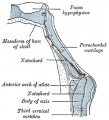
Sagittal section of cephalic end of notochord
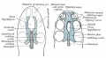
Cartilaginous cranium (chondrocranium)
Human Embryo (CRL 8 cm) embryo model
Human Embryo (CRL 8 cm) embryo model from the left side


Terms
- anterior fontanel - closes by about 20 months.
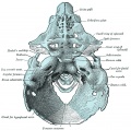
- basion - anatomical region on the basiocciput located at the midpoint between the anterior margin and posterior margin (opisthion) of the foramen magnum.
- compound craniosynostosis premature suture fusion involving two or more sutures.
- craniosynostosis - (craniostenosis) the premature fusion of cranial sutures.
- harlequin eye - a term used to describe the prominent bilateral elliptical orbits of the skull seen in brachycephaly.
- endochondral ossification - bone formation from a pre-existing cartilage template, such as the chondrocranium.
- intramembranous ossification - bone formation from a membrane where no pre-existing cartilage is found, such as the calvarial vault component.
- neurocranium - the portion of the skull that surrounds the brain, endochondral and intramembranous ossification in origin.
- opisthion - anatomical region located on the occipital bone, located at the midpoint of the posterior margin of the foramen magnum.
- posterior fontanel - closes by about 3 months.
- primary craniosynostosis - an intrinsic defect in a suture.
- secondary craniosynostosis - premature closure of normal sutures due to systemic and metabolic (hyperthyroidism, hypercalcemia, hypophosphatasia, vitamin D deficiency, renal osteodystrophy, Hurler's Syndrome, sickle cell disease and thalassemia) and those that can affect brain growth.
- simple craniosynostosis - premature fusion involving only one suture.
- synostosis - premature fusion.
External Links
External Links Notice - The dynamic nature of the internet may mean that some of these listed links may no longer function. If the link no longer works search the web with the link text or name. Links to any external commercial sites are provided for information purposes only and should never be considered an endorsement. UNSW Embryology is provided as an educational resource with no clinical information or commercial affiliation.
- PubMed Health Craniosynostosis
Glossary Links
- Glossary: A | B | C | D | E | F | G | H | I | J | K | L | M | N | O | P | Q | R | S | T | U | V | W | X | Y | Z | Numbers | Symbols | Term Link
Cite this page: Hill, M.A. (2024, May 2) Embryology Musculoskeletal System - Skull Development. Retrieved from https://embryology.med.unsw.edu.au/embryology/index.php/Musculoskeletal_System_-_Skull_Development
- © Dr Mark Hill 2024, UNSW Embryology ISBN: 978 0 7334 2609 4 - UNSW CRICOS Provider Code No. 00098G