Movie - Embryo stage 14: Difference between revisions
From Embryology
No edit summary |
(Redirected page to Carnegie Stage 14 Surface Movie) |
||
| (4 intermediate revisions by the same user not shown) | |||
| Line 1: | Line 1: | ||
#REDIRECT [[Carnegie_Stage_14_Surface_Movie]] | |||
{| border='0px' | {| border='0px' | ||
|- | |- | ||
| < | |<wikiflv height="475" width="600" autostart="true">Stage14_model3.flv|Stage14modelicon.jpg</wikiflv> | ||
| valign="top" |'''Stage 14 Embryo''' | | valign="top" |'''Stage 14 Embryo''' | ||
| Line 22: | Line 23: | ||
{{Model stage 14 links}} | |||
|- | |- | ||
| | |- | ||
| [[File:Bailey087.jpg|600px]] | |||
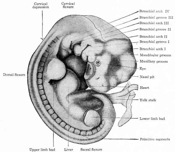
| valign=top|The stage 14 model shows the embryo at about the midpoint of embryonic development when the external surface has not yet formed the adult anatomy and has a number of transient structures that are remodelled through the later embryonic stages. It therefore provides an ideal teaching model for demonstrating features such as sensory placodes, pharyngeal arches, limb buds, axial skeleton development, heart and liver surfaces bulges and the umbilicus. | |||
The lefthand labelled drawing is by Mall showing a 7 mm embryo. | |||
|} | |||
Latest revision as of 09:44, 7 March 2013
Redirect to:
| Stage14modelicon.jpg</wikiflv> | Stage 14 Embryo
This is a rotating model of the Stage 14 embryo. The animation begins with the right hand side view and rotates ventrally. Firstly, note the curvature of the embryo and the relative proportions of the head and body. Next, identify the following visible external features some of which represent the growth and development of internal structures.
|

|
The stage 14 model shows the embryo at about the midpoint of embryonic development when the external surface has not yet formed the adult anatomy and has a number of transient structures that are remodelled through the later embryonic stages. It therefore provides an ideal teaching model for demonstrating features such as sensory placodes, pharyngeal arches, limb buds, axial skeleton development, heart and liver surfaces bulges and the umbilicus.
|