Integumentary System - Nail Development: Difference between revisions
mNo edit summary |
mNo edit summary |
||
| Line 1: | Line 1: | ||
{{Header}} | |||
== Introduction == | == Introduction == | ||
This page introduces the development of nails, fingernails and toenails, these integumentary specializations in other species have been specialised as claws and hooves. | This page introduces the development of nails, fingernails and toenails, these integumentary specializations in other species have been specialised as claws and hooves. | ||
| Line 6: | Line 7: | ||
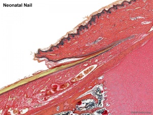
[[File:Neonatal_nail.jpg|thumb|Neonatal nail]] | [[File:Neonatal_nail.jpg|thumb|Neonatal nail]] | ||
{{Integumentary Links}} | |||
== Some Recent Findings == | == Some Recent Findings == | ||
{| | {| | ||
| Line 106: | Line 108: | ||
* Key steps in the development of three major ectodermal appendages [http://www.ncbi.nlm.nih.gov/pmc/articles/PMC2561923/figure/F1/ Tooth, Hair and Mammary Gland] | * Key steps in the development of three major ectodermal appendages [http://www.ncbi.nlm.nih.gov/pmc/articles/PMC2561923/figure/F1/ Tooth, Hair and Mammary Gland] | ||
{{ | |||
{{Glossary}} | |||
{{Footer}} | |||
[[Category:Nail]] [[Category:Integumentary]] | [[Category:Nail]] [[Category:Integumentary]] | ||
Revision as of 10:20, 30 May 2014
| Embryology - 4 May 2024 |
|---|
| Google Translate - select your language from the list shown below (this will open a new external page) |
|
العربية | català | 中文 | 中國傳統的 | français | Deutsche | עִברִית | हिंदी | bahasa Indonesia | italiano | 日本語 | 한국어 | မြန်မာ | Pilipino | Polskie | português | ਪੰਜਾਬੀ ਦੇ | Română | русский | Español | Swahili | Svensk | ไทย | Türkçe | اردو | ייִדיש | Tiếng Việt These external translations are automated and may not be accurate. (More? About Translations) |
Introduction
This page introduces the development of nails, fingernails and toenails, these integumentary specializations in other species have been specialised as claws and hooves.
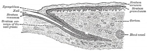
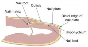
Each nail is convex on its outer surface, concave within, and is implanted by a portion, called the root, into a groove in the skin; the exposed portion is called the body, and the distal extremity the free edge.
Some Recent Findings
|
| More recent papers |
|---|
|
This table allows an automated computer search of the external PubMed database using the listed "Search term" text link.
More? References | Discussion Page | Journal Searches | 2019 References | 2020 References Search term: Nail Embryology <pubmed limit=5>Nail Embryology</pubmed> |
Textbooks
- Human Embryology (2nd ed.) Larson Chapter 14 p443-455
- The Developing Human: Clinically Oriented Embryology (6th ed.) Moore and Persaud Chapter 20: P513-529
- Before We Are Born (5th ed.) Moore and Persaud Chapter 21: P481-496
- Essentials of Human Embryology Larson Chapter 14: P303-315
- Human Embryology, Fitzgerald and Fitzgerald
- Color Atlas of Clinical Embryology Moore Persaud and Shiota Chapter 15: p231-236
Development Overview
- Forelimb before hindlimb - week 10 fingernails, week 14 toe nails
- nail field - appears at tip and migrates to dorsal surface
- thickened epidermis - surrounding cells form nail fold
- keratinization of proximal nail fold forms nail plate
Nails reach Digit Tip
- week 32 fingernails
- week 36 toenails
Note that nail growth can be used as an indicator of prematurity.
Abnormalities
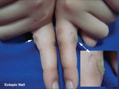
Ectopic Nail
Congenital ectopic nails are an extremely rare deformity.[3]
Anonychia
A very rare abnormality resulting in an absence of nails. Can result from both genetic and environmental defects and be associated with a range of other abnormalities. Simple anonychia is the congenital absence of the nails without any other coexisting major congenital anomaly.
Hyponychia
Nail-patella syndrome
Ectodermal dysplasias
Witkop syndrome
An autosomal dominant genetic disorder characterized by the absence of several teeth and abnormalities of the nails.
References
- ↑ <pubmed>20579456</pubmed>
- ↑ <pubmed>18429815</pubmed>
- ↑ <pubmed>19736447</pubmed>| Indian J Dermatol Venereol Leprol.
Journals
Reviews
<pubmed>19932326</pubmed> <pubmed>18459147</pubmed> <pubmed>15468149</pubmed> <pubmed>12949775</pubmed> <pubmed>11710767</pubmed>
Articles
<pubmed>21387539</pubmed>
Search PubMed
Search Pubmed: Nail Development | Nail embryology
Additional Images
Terms
- cuticle - tissue that overlaps the plate and rims the base of the nail
- hyponychium - the epithelium located beneath the nail plate
- lunula - half-moon at the base of the nail
- matrix - hidden part of the nail unit under the cuticle
- nail bed - skin beneath the nail plate
- nail folds - skin folds that frame and support the nail on three sides
- nail plate - visible part of the nail
- trachyonychia - nail plate roughness, pitting, and ridging affecting one or all twenty nails.
External Links
External Links Notice - The dynamic nature of the internet may mean that some of these listed links may no longer function. If the link no longer works search the web with the link text or name. Links to any external commercial sites are provided for information purposes only and should never be considered an endorsement. UNSW Embryology is provided as an educational resource with no clinical information or commercial affiliation.
- Embryo Images - Human (day 64) primary nail fields
- Key steps in the development of three major ectodermal appendages Tooth, Hair and Mammary Gland
Glossary Links
- Glossary: A | B | C | D | E | F | G | H | I | J | K | L | M | N | O | P | Q | R | S | T | U | V | W | X | Y | Z | Numbers | Symbols | Term Link
Cite this page: Hill, M.A. (2024, May 4) Embryology Integumentary System - Nail Development. Retrieved from https://embryology.med.unsw.edu.au/embryology/index.php/Integumentary_System_-_Nail_Development
- © Dr Mark Hill 2024, UNSW Embryology ISBN: 978 0 7334 2609 4 - UNSW CRICOS Provider Code No. 00098G


