Integumentary System - Nail Development
| Embryology - 22 Jul 2026 |
|---|
| Google Translate - select your language from the list shown below (this will open a new external page) |
|
العربية | català | 中文 | 中國傳統的 | français | Deutsche | עִברִית | हिंदी | bahasa Indonesia | italiano | 日本語 | 한국어 | မြန်မာ | Pilipino | Polskie | português | ਪੰਜਾਬੀ ਦੇ | Română | русский | Español | Swahili | Svensk | ไทย | Türkçe | اردو | ייִדיש | Tiếng Việt These external translations are automated and may not be accurate. (More? About Translations) |
Introduction

This page introduces the development of nails, fingernails and toenails, these integumentary specializations in other species have been specialised as claws and hooves.
Each nail is convex on its outer surface, concave within, and is implanted by a portion, called the root, into a groove in the skin; the exposed portion is called the body, and the distal extremity the free edge.
Some Recent Findings
|
| More recent papers |
|---|
 This table allows an automated computer search of the external PubMed database using the listed "Search term" text link.
More? References | Discussion Page | Journal Searches | 2019 References | 2020 References Search term: Nail Embryology | Nail Development | |
| Older papers |
|---|
| These papers originally appeared in the Some Recent Findings table, but as that list grew in length have now been shuffled down to this collapsible table.
See also the Discussion Page for other references listed by year and References on this current page.
|
Textbooks

- Human Embryology (2nd ed.) Larson Chapter 14 p443-455
- The Developing Human: Clinically Oriented Embryology (6th ed.) Moore and Persaud Chapter 20: P513-529
- Before We Are Born (5th ed.) Moore and Persaud Chapter 21: P481-496
- Essentials of Human Embryology Larson Chapter 14: P303-315
- Human Embryology, Fitzgerald and Fitzgerald
- Color Atlas of Clinical Embryology Moore Persaud and Shiota Chapter 15: p231-236
Development Overview

- Forelimb before hindlimb - week 10 fingernails, week 14 toe nails
- nail field - appears at tip and migrates to dorsal surface
- thickened epidermis - surrounding cells form nail fold
- keratinization of proximal nail fold forms nail plate
Nails reach Digit Tip
- week 32 fingernails
- week 36 toenails
Note that nail growth can be used as an indicator of prematurity.
Abnormalities
In a study of 500+ neonates, the most frequent nail alteration identified was the incomplete development of the hallux (big toe) nail, appearing as either triangular or trapezoidal - shaped.[2]
Ectopic Nail

Congenital ectopic nails are an extremely rare deformity.[4]
Anonychia
A very rare abnormality resulting in an absence of nails. Can result from both genetic and environmental defects and be associated with a range of other abnormalities. Simple anonychia is the congenital absence of the nails without any other coexisting major congenital anomaly.
Hyponychia
Nail Patella Syndrome

(NPS) rare (1/50,000) Autosomal dominant disorder characterized by hypoplastic or absent patellae, dystrophic nails, dysplasia of the elbows and iliac horns. Potentially due to mutations in LMX1B, a LIM-homeodomain transcription protein.
Ectodermal dysplasias
Witkop syndrome
An autosomal dominant genetic disorder characterized by the absence of several teeth and abnormalities of the nails.
References
- ↑ Cui CY, Klar J, Georgii-Heming P, Fröjmark AS, Baig SM, Schlessinger D & Dahl N. (2013). Frizzled6 deficiency disrupts the differentiation process of nail development. J. Invest. Dermatol. , 133, 1990-7. PMID: 23439395 DOI.
- ↑ 2.0 2.1 Milano A, Cutrone M, Laforgia N & Bonifazi E. (2010). Incomplete development of the nail of the hallux in the newborn. Dermatol. Online J. , 16, 1. PMID: 20579456
- ↑ Okada M, Nishimukai H, Okiura T & Sugino Y. (2008). Lyonization pattern of normal human nails. Genes Cells , 13, 421-8. PMID: 18429815 DOI.
- ↑ Feily A, Ayoobi A, Yaghoobi R & Kheradmand P. (2009). A case of congenital ectopic nails on the bilateral second fingers without bone deformity. Indian J Dermatol Venereol Leprol , 75, 525-7. PMID: 19736447 DOI.
- ↑ Senguttuvan NB, Sivaraman A, Kandasamy D & Marimuthu K. (2011). Nail patella syndrome: a rare cause of renal failure in a young adult. Pan Afr Med J , 9, 31. PMID: 22145064
Journals
Reviews
Smith RJ & Rubin AI. (2020). Pediatric nail disorders: a review. Curr. Opin. Pediatr. , 32, 506-515. PMID: 32692049 DOI.
Bergqvist C, Ramia P, Abbas O & Kurban M. (2017). Genetics of syndromic and non-syndromic hereditary nail disorders. Clin. Genet. , 91, 813-823. PMID: 27613389 DOI.
Piraccini BM & Starace M. (2014). Nail disorders in infants and children. Curr. Opin. Pediatr. , 26, 440-5. PMID: 24886951 DOI.
Harrison S & Bergfeld WF. (2009). Diseases of the hair and nails. Med. Clin. North Am. , 93, 1195-209. PMID: 19932326 DOI.
Duverger O & Morasso MI. (2008). Role of homeobox genes in the patterning, specification, and differentiation of ectodermal appendages in mammals. J. Cell. Physiol. , 216, 337-46. PMID: 18459147 DOI.
van Steensel MA, van Geel M & Steijlen PM. (2004). Molecular genetics of hereditary hair and nail disease. Am J Med Genet C Semin Med Genet , 131C, 52-60. PMID: 15468149 DOI.
Hamrick MW. (2003). Evolution and development of mammalian limb integumentary structures. J. Exp. Zool. B Mol. Dev. Evol. , 298, 152-63. PMID: 12949775 DOI.
Hamrick MW. (2001). Development and evolution of the mammalian limb: adaptive diversification of nails, hooves, and claws. Evol. Dev. , 3, 355-63. PMID: 11710767
Articles
Cai J & Ma L. (2011). Msx2 and Foxn1 regulate nail homeostasis. Genesis , 49, 449-59. PMID: 21387539 DOI.
Search PubMed
Search Pubmed: Nail Development | Nail embryology
Additional Images


Historic Images
| Historic Disclaimer - information about historic embryology pages |
|---|
|
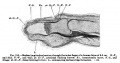
Fig. 215 Fetal Finger 8.5 cm 12 weeks (14 weeks GA)

Terms
- Beau lines - nail grooved transverse lines appearing like indentations or ridges in the nail plate. Named in 1846 by Joseph Honoré Simon Beau (1806–1865), a French physician.
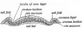
- cuticle - tissue that overlaps the plate and rims the base of the nail
- hyponychium - the epithelium located beneath the nail plate
- lunula - half-moon at the base of the nail
- matrix - hidden part of the nail unit under the cuticle
- Muehrcke lines - paired transverse white lines appearing in the nails secondary to edematous nail bed, resulting abnormal nail bed vasculature, forming transverse lines that disappear when the nail bed is compressed. Seen in patients with chronic hypoalbuminemia and malnutrition.
- nail bed - skin beneath the nail plate
- nail folds - skin folds that frame and support the nail on three sides
- nail plate - visible part of the nail
- trachyonychia - nail plate roughness, pitting, and ridging affecting one or all twenty nails.
External Links
External Links Notice - The dynamic nature of the internet may mean that some of these listed links may no longer function. If the link no longer works search the web with the link text or name. Links to any external commercial sites are provided for information purposes only and should never be considered an endorsement. UNSW Embryology is provided as an educational resource with no clinical information or commercial affiliation.
- Embryo Images - Human (day 64) primary nail fields
- Key steps in the development of three major ectodermal appendages Tooth, Hair and Mammary Gland
Glossary Links
- Glossary: A | B | C | D | E | F | G | H | I | J | K | L | M | N | O | P | Q | R | S | T | U | V | W | X | Y | Z | Numbers | Symbols | Term Link
Cite this page: Hill, M.A. (2026, July 22) Embryology Integumentary System - Nail Development. Retrieved from https://embryology.med.unsw.edu.au/embryology/index.php/Integumentary_System_-_Nail_Development
- © Dr Mark Hill 2026, UNSW Embryology ISBN: 978 0 7334 2609 4 - UNSW CRICOS Provider Code No. 00098G