Integumentary System - Mammary Gland Development
| Embryology - 13 May 2024 |
|---|
| Google Translate - select your language from the list shown below (this will open a new external page) |
|
العربية | català | 中文 | 中國傳統的 | français | Deutsche | עִברִית | हिंदी | bahasa Indonesia | italiano | 日本語 | 한국어 | မြန်မာ | Pilipino | Polskie | português | ਪੰਜਾਬੀ ਦੇ | Română | русский | Español | Swahili | Svensk | ไทย | Türkçe | اردو | ייִדיש | Tiếng Việt These external translations are automated and may not be accurate. (More? About Translations) |
Introduction
The mammary gland is the functional structure of the female breast and develops initially as a skin specialization. Breast growth and appearance in male and female children are virtually identical prior to puberty.
At puberty females, under the influence of mainly sex hormone signaling, undergo a series of growth changes that can be defined by a series of "Tanner Stages".
In pregnancy, an additional series of signals leads to further changes in breast structure. The key function of this process is to prepare the maternal breast for lactation and providing nutrition through milk to the newborn. (More? Normal Development - Milk)
At menopause, changes in sex hormone secretion can once again alter breast structure.
The breast also associated with oncogenesis (breast cancer). Research in this area has been aided by the discovery in 1994 of the two breast cancer susceptibility genes (BRCA1, BRCA2). There is some developing evidence that modification of stem cells (progenitor cells) that exist in the mammary gland may also contribute to neoplasms (cancer).
Some Recent Findings
|
| More recent papers |
|---|
 This table allows an automated computer search of the external PubMed database using the listed "Search term" text link.
More? References | Discussion Page | Journal Searches | 2019 References | 2020 References Search term: Mammary Embryology <pubmed limit=5>Mammary Embryology</pubmed> |
Textbooks
- Human Embryology (2nd ed.) Larson Chapter 14 p443-455
- The Developing Human: Clinically Oriented Embryology (6th ed.) Moore and Persaud Chapter 20: P513-529
- Before We Are Born (5th ed.) Moore and Persaud Chapter 21: P481-496
- Essentials of Human Embryology Larson Chapter 14: P303-315
- Human Embryology, Fitzgerald and Fitzgerald
- Color Atlas of Clinical Embryology Moore Persaud and Shiota Chapter 15: p231-236
Development Overview
- week 6 epidermis downgrowth into dermis, modified sweat glands
- epithelia/mesenchyme inductive interaction, mesenchyme forms connective tissue and fat
- mammary ridges - mammary bud formation, pair of ventral regions axilla to inguinal
- pectoral regions generate breasts
- buds branch to form lactiferous ducts, only main duct formed at birth
- mammary pit - forms fetal period
- areola - depressed region at gland, proliferation of connective tissue postnatally
- prior to puberty male and female glands the same
Anatomy
| The mamma consists of gland tissue; of fibrous tissue, connecting its lobes; and of fatty tissue in the intervals between the lobes. The gland tissue, when freed from fibrous tissue and fat, is of a pale reddish color, firm in texture, flattened from before backward and thicker in the center than at the circumference.
(from Gray's Anatomy) |

|
Puberty
- sex hormone estrogen stimulate growth, full development approx 20 years
- growth also influenced by other hormones - progereterone, prolactin, corticoids, growth hormone
- mainly fat and connective tissue deposition
Tanner Mammary Development Stages
In 1976 Tanner and Whitehouse established a series of descriptive stages for primary and secondary sexual characteristic development at puberty. The female secondary sex characteristics of breast development were divided into five numbered (1 - 5) "Tanner Stages".[4]
Mammary Glands Pregnancy
During pregnancy raised estrogens and progesterone stimulate gland development, secretory alveolar structures form and differentiate, leading to milk production in late pregnancy and milk secretion during lactation. Breasts are hemispherical in shape due to fat deposition. After birth, neonatal lactation supports further growth/development.
Mammary Glands Weaning
After the infant ceases breast feeding, weaning, the mammary gland milk-producing epithelial cells undergo a process called "involution", that requires cell apoptosis.

Mammary involution[5]
Mouse Mammary

E10 - milk line first formed by a slight thickening and stratification of the surface ectoderm.
E11.5 - the milk line breaks up into individual placodes and the underlying mammary mesenchyme begins to condense.
E15.5 - mammary epithelium begins to proliferate at the tip and the primary sprout pushes through the mammary mesenchyme towards the underlying fat pad.
E18.5 - elongating duct has now grown into the fat pad and has branched into a small ductal system. Cells of the mammary mesenchyme have formed the nipple, which is made of specialized epidermal cells.
Timeline data from review[7]
Wnt Signaling
Canonical Wnt signals are transduced through a Frizzled receptor and the LRP5 or LRP6 co-receptor. Loss of Lrp6 compromises Wnt/beta-catenin signaling and interferes with mammary placode, fat pad, and branching development during embryogenesis.[8]
- Links: Mouse Timeline Detailed
Abnormalities
Abnormalities occur in approximately 1% of female population and include in both sexes:
- polymastia - extra breast
- polytheli - extra nipple
- supernumerary nipple (relatively common in males)
- gynecomastia (Greek, gyne = woman, mastos = breast) is the excessive development of the male breast, which can occur transiently in puberty or due to other (hormonal) abnormalities.
International Classification of Diseases
Q83 Congenital malformations of breast
Excl.: absence of pectoral muscle (Q79.8)
- Q83.0 Congenital absence of breast with absent nipple
- Q83.1 Accessory breast Supernumerary breast
- Q83.2 Absent nipple
- Q83.3 Accessory nipple Supernumerary nipple
- Q83.8 Other congenital malformations of breast Hypoplasia of breast
- Q83.9 Congenital malformation of breast, unspecified
Breast Cancer
In 1994, two breast cancer susceptibility genes were identified BRCA1 on chromosome 17 and BRCA2 on chromosome 13.
When an individual carries a mutation in either BRCA1 or BRCA2, they are at an increased risk of being diagnosed with breast or ovarian cancer at some point in their lives. Normal function of these genes was to participate in repairing radiation-induced breaks in double-stranded DNA. It is though that mutations in BRCA1 or BRCA2 might disable this mechanism, leading to more errors in DNA replication and ultimately to cancerous growth. (text modified from: NCBI genes and disease)
- Links: OMIM - BRCA1 | OMIM - BRCA2
References
- ↑ <pubmed>20382343</pubmed>
- ↑ <pubmed>20926579</pubmed>
- ↑ <pubmed>20824491</pubmed>
- ↑ <pubmed>952550</pubmed>
- ↑ <pubmed>16677411</pubmed>| Breast Cancer Res.
- ↑ <pubmed>16933995</pubmed>| PLoS Genet.
- ↑ <pubmed>18007652</pubmed>
- ↑ <pubmed>19503830</pubmed>
Journals
Reviews
<pubmed>20565255</pubmed> <pubmed>20484386</pubmed> <pubmed>19889198</pubmed> <pubmed>19020961</pubmed> <pubmed>18556345</pubmed>
Articles
<pubmed>20689821</pubmed> <pubmed>20584313</pubmed> <pubmed>20552032</pubmed>| PLoS One. <pubmed>18826651</pubmed> <pubmed>15960763</pubmed>
Search PubMed
Search Pubmed: Mammary Gland Development
Additional Images
Historic
The Skin and its Appendages (1902)
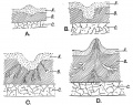
Fig. 57. Showing the various stages in the development of the Mamma
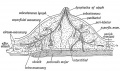
Fig. 58. Diagrammatic Section of the Breast to show the arrangement of its Capsule and Lymphatics.


External Links
External Links Notice - The dynamic nature of the internet may mean that some of these listed links may no longer function. If the link no longer works search the web with the link text or name. Links to any external commercial sites are provided for information purposes only and should never be considered an endorsement. UNSW Embryology is provided as an educational resource with no clinical information or commercial affiliation.
Glossary Links
- Glossary: A | B | C | D | E | F | G | H | I | J | K | L | M | N | O | P | Q | R | S | T | U | V | W | X | Y | Z | Numbers | Symbols | Term Link
Cite this page: Hill, M.A. (2024, May 13) Embryology Integumentary System - Mammary Gland Development. Retrieved from https://embryology.med.unsw.edu.au/embryology/index.php/Integumentary_System_-_Mammary_Gland_Development
- © Dr Mark Hill 2024, UNSW Embryology ISBN: 978 0 7334 2609 4 - UNSW CRICOS Provider Code No. 00098G