File:Vestibular labyrinth cartoon.jpg
Original file (600 × 657 pixels, file size: 44 KB, MIME type: image/jpeg)
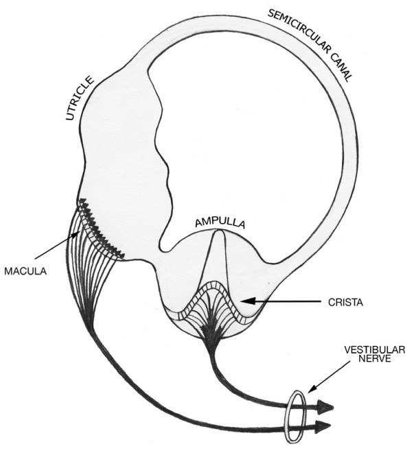
Scheme of the Vestibular Labyrinth
After Melville-Jones [1] This schematic highlights the serial organization of the three component chambers in the basic vestibular labyrinth.
- Semicircular canal
- Utricle
- Ampulla
Reference
- ↑ <pubmed>19723316</pubmed>
Copyright
© 2009 Pender; licensee BioMed Central Ltd. This is an Open Access article distributed under the terms of the Creative Commons Attribution License (http://creativecommons.org/licenses/by/2.0), which permits unrestricted use, distribution, and reproduction in any medium, provided the original work is properly cited.
Pender Theoretical Biology and Medical Modelling 2009 6:19 doi:10.1186/1742-4682-6-19
Original file name: 1742-4682-6-19-2.jpg http://www.tbiomed.com/content/6/1/19/figure/F2
Cite this page: Hill, M.A. (2024, April 27) Embryology Vestibular labyrinth cartoon.jpg. Retrieved from https://embryology.med.unsw.edu.au/embryology/index.php/File:Vestibular_labyrinth_cartoon.jpg
- © Dr Mark Hill 2024, UNSW Embryology ISBN: 978 0 7334 2609 4 - UNSW CRICOS Provider Code No. 00098G
File history
Click on a date/time to view the file as it appeared at that time.
| Date/Time | Thumbnail | Dimensions | User | Comment | |
|---|---|---|---|---|---|
| current | 01:12, 11 June 2010 | | 600 × 657 (44 KB) | S8600021 (talk | contribs) | Scheme of the Vestibular Labyrinth (after Melville-Jones). This schematic highlights the serial organization of the three component chambers in the basic vestibular labyrinth. Pender Theoretical Biology and Medical Modelling 2009 6:19 doi:10.1186/174 |
You cannot overwrite this file.
File usage
The following page uses this file: