File:Supernumerary renal vein 01.jpg: Difference between revisions
No edit summary |
mNo edit summary |
||
| Line 13: | Line 13: | ||
<pubmed>20461189</pubmed>| [http://www.ncbi.nlm.nih.gov/pmc/articles/PMC2864862 PMC2864862] | [http://www.kjronline.org/DOIx.php?id=10.3348/kjr.2010.11.3.346 Korean J Radiol] | <pubmed>20461189</pubmed>| [http://www.ncbi.nlm.nih.gov/pmc/articles/PMC2864862 PMC2864862] | [http://www.kjronline.org/DOIx.php?id=10.3348/kjr.2010.11.3.346 Korean J Radiol] | ||
====Copyright==== | |||
This is an Open Access article distributed under the terms of the Creative Commons Attribution Non-Commercial License (http://creativecommons.org/licenses/by-nc/3.0) which permits unrestricted non-commercial use, distribution, and reproduction in any medium, provided the original work is properly cited. | This is an Open Access article distributed under the terms of the Creative Commons Attribution Non-Commercial License (http://creativecommons.org/licenses/by-nc/3.0) which permits unrestricted non-commercial use, distribution, and reproduction in any medium, provided the original work is properly cited. | ||
{{Footer}} | |||
[[Category:Human]] [[Category:Adult]] [[Category:Renal]] [[Category:Cardiovascular]] [[Category:Venous]] [[Category:Computed Tomography]] | [[Category:Human]] [[Category:Adult]] [[Category:Renal]] [[Category:Cardiovascular]] [[Category:Venous]] [[Category:Computed Tomography]] | ||
Revision as of 10:13, 11 May 2016
Supernumerary Renal Vein
- most common venous anomalies are multiple renal veins seen in approximately 15-30% of patients
- more common on the right side and these occur in up to 30% of individuals
- Supernumerary right renal vein in 30-year-old male voluntary kidney donor.
- Anterior oblique volume rendered image show two right renal veins crossing each other and draining into inferior vena cava (arrows).
Renal Vascular Anomalies: Multiple renal arteries | Accessory renal artery | Supernumerary right renal vein 1 | Supernumerary right renal vein 1 | Multiple right renal veins 2 | Multiple right renal veins 2 | Cardiovascular System Development
Original file name: Fig. 8 kjr-11-346-g008.jpg (panel B cropped from full image)
Reference
<pubmed>20461189</pubmed>| PMC2864862 | Korean J Radiol
Copyright
This is an Open Access article distributed under the terms of the Creative Commons Attribution Non-Commercial License (http://creativecommons.org/licenses/by-nc/3.0) which permits unrestricted non-commercial use, distribution, and reproduction in any medium, provided the original work is properly cited.
Cite this page: Hill, M.A. (2024, May 19) Embryology Supernumerary renal vein 01.jpg. Retrieved from https://embryology.med.unsw.edu.au/embryology/index.php/File:Supernumerary_renal_vein_01.jpg
- © Dr Mark Hill 2024, UNSW Embryology ISBN: 978 0 7334 2609 4 - UNSW CRICOS Provider Code No. 00098G
File history
Click on a date/time to view the file as it appeared at that time.
| Date/Time | Thumbnail | Dimensions | User | Comment | |
|---|---|---|---|---|---|
| current | 12:23, 3 September 2011 | 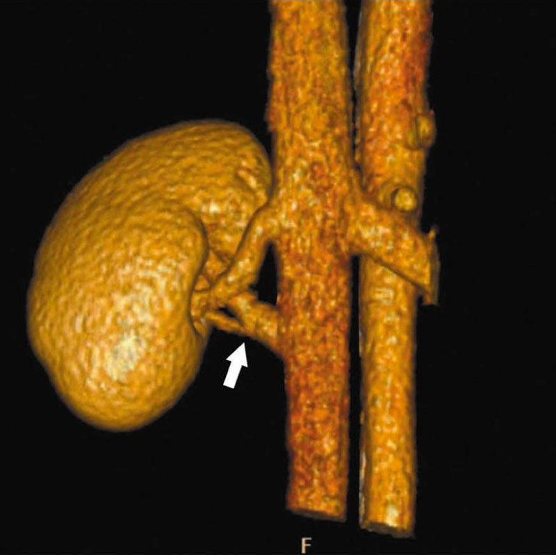 | 800 × 798 (72 KB) | S8600021 (talk | contribs) | ==Supernumerary Renal Vein== * most common venous anomalies are multiple renal veins seen in approximately 15-30% of patients * more common on the right side and these occur in up to 30% of individuals * Supernumerary right renal vein in 30-year-old male |
You cannot overwrite this file.
File usage
The following 3 pages use this file: