File:Stage14 sem2l.jpg
Original file (630 × 1,000 pixels, file size: 96 KB, MIME type: image/jpeg)
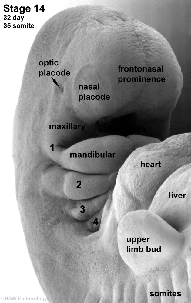
Human Embryo Carnegie stage 14
Carnegie stage 14, day 32, 35 somites, week 5, right ventrolateral view upper embryo.
This image most clearly shows the upper limb bud and the pharyngeal arches.
- Links: Carnegie stage 14
Image Source: Scanning electron micrographs of the Carnegie stages of the early human embryos are reproduced with the permission of Prof Kathy Sulik, from embryos collected by Dr. Vekemans and Tania Attié-Bitach. Images are for educational purposes only and cannot be reproduced electronically or in writing without permission.
Image Code: Stage14= carnegie stage sem= scanning em 2 = image 2 1000px size l=labeled
Original file name: Stage14day32somite35-ventral-sem2-1000px.jpg
Cite this page: Hill, M.A. (2024, May 4) Embryology Stage14 sem2l.jpg. Retrieved from https://embryology.med.unsw.edu.au/embryology/index.php/File:Stage14_sem2l.jpg
- © Dr Mark Hill 2024, UNSW Embryology ISBN: 978 0 7334 2609 4 - UNSW CRICOS Provider Code No. 00098G
File history
Click on a date/time to view the file as it appeared at that time.
| Date/Time | Thumbnail | Dimensions | User | Comment | |
|---|---|---|---|---|---|
| current | 10:16, 3 September 2009 | | 630 × 1,000 (96 KB) | S8600021 (talk | contribs) | Human Embryo Carnegie stage 14, day 32, 35 somites Original file name: Stage14day32somite35-ventral-sem2-1000px.jpg Image Code: Stage14= carnegie stage sem= scanning em 2 = image 2 1000px size l=labeled {{Template:SEM}} {{Template:Footer}} |
You cannot overwrite this file.
File usage
The following 18 pages use this file:
- 2011 Lab 10 - Early Embryo
- 2011 Lab 6 - Early Embryo
- AACP Meeting 2013 - Face Embryology
- ANAT2341 Lab 10 - Early Embryo
- ANAT2341 Lab 3 - Week 4
- Abnormal Development - Thalidomide
- B
- BGDA Lecture - Development of the Embryo/Fetus 2
- Human Embryo SEM
- K12 Comparative Embryology
- K12 Thalidomide
- Lecture - Head Development
- Lecture - Sensory Development
- Musculoskeletal System - Appendicular Skeleton Development
- Musculoskeletal System - Limb Development
- Pharyngeal arches
- Sensory System Development
- Template:Carnegie stage 11-14 image table