File:Somite cartoon5.png
Somite_cartoon5.png (400 × 300 pixels, file size: 27 KB, MIME type: image/png)
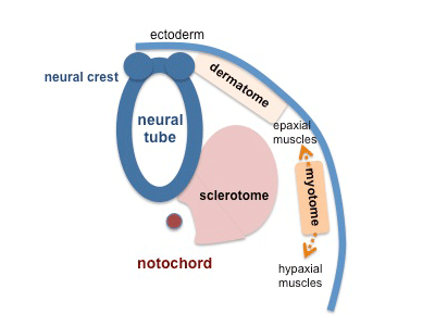
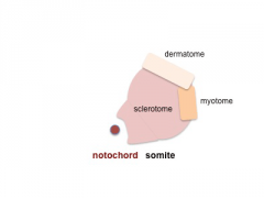
Somite Development cartoon
The myotome differentiates to form 2 components dorsally the epimere and ventrally the hypomere, which in turn form epaxial and hypaxial muscles respectively.
The bulk of the trunk and limb muscle coming from the hypaxial mesoderm.
Different structures will be contributed depending upon the somite level. Note that neural crest cells migrate beside and through somite.
Note - the cartoons show just the embryo righthand side mesoderm development (the same events occur on the lefthand side).
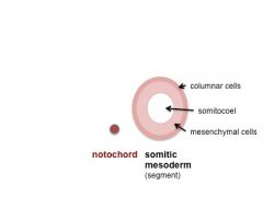
- Somite Links: 1 paraxial | 2 early somite | 3 sclerotome and dermomyotome | 4 dermatome and myotome | 5 somite spreading | SEM image - Human Embryo (week 4) showing somites | Movie - somitogenesis Hes expression
- Somite Cartoons
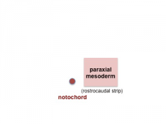
paraxial
early somite
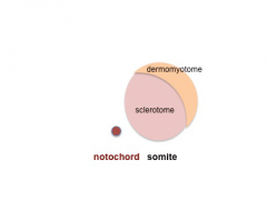
sclerotome and dermomyotome
dermatome and myotome

somite spreading


Cite this page: Hill, M.A. (2024, May 19) Embryology Somite cartoon5.png. Retrieved from https://embryology.med.unsw.edu.au/embryology/index.php/File:Somite_cartoon5.png
- © Dr Mark Hill 2024, UNSW Embryology ISBN: 978 0 7334 2609 4 - UNSW CRICOS Provider Code No. 00098G
File history
Click on a date/time to view the file as it appeared at that time.
| Date/Time | Thumbnail | Dimensions | User | Comment | |
|---|---|---|---|---|---|
| current | 18:01, 16 May 2014 | | 400 × 300 (27 KB) | Z8600021 (talk | contribs) | |
| 10:43, 10 August 2009 | 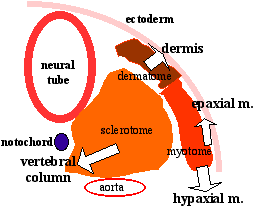 | 270 × 209 (6 KB) | MarkHill (talk | contribs) | Somite Development cartoon 5 Neural crest cells migrate beside and through somite. The myotome differentiates to form 2 components dorsally the epimere and ventrally the hypomere, which in turn form epaxial and hypaxial muscles respectively. The bulk of |
You cannot overwrite this file.
File usage
The following 43 pages use this file:
- 2009 Lecture 13
- 2009 Lecture 5
- 2010 BGD Lecture - Development of the Embryo/Fetus 1
- 2010 BGD Lecture - Development of the Embryo/Fetus 2
- 2010 BGD Practical 6 - Week 3
- 2010 Lab 3
- 2010 Lecture 13
- 2010 Lecture 5
- 2011 Lab 3 - Week 3
- 2014 Group Project 8
- ANAT2341 Lab 3 - Week 3
- BGDA Lecture - Development of the Embryo/Fetus 1
- BGDA Lecture - Development of the Embryo/Fetus 2
- BGDA Practical 7 - Week 3
- Developmental Mechanism - Epithelial Mesenchymal Transition
- Integumentary System Development
- Lecture - Limb Development
- Lecture - Mesoderm Development
- Lecture - Musculoskeletal Development
- Mesoderm
- Musculoskeletal System - Bone Development
- Musculoskeletal System - Limb Development
- Musculoskeletal System - Muscle Development
- Musculoskeletal System Development
- Notochord
- S
- Somite Musculoskeletal Movie
- Somitogenesis
- Talk:2010 BGD Practical 6 - Week 3
- Talk:2011 Lab 3
- Talk:2014 Group Project 8
- File:Mesoderm cartoon 05.jpg
- File:Mesoderm cartoon 06.jpg
- File:Mesoderm cartoon 07.jpg
- File:Mesoderm cartoon 08.jpg
- File:Mesoderm cartoon 09.jpg
- File:Somite cartoon1.png
- File:Somite cartoon2.png
- File:Somite cartoon3.png
- File:Somite cartoon4.png
- File:Somite cartoon5.png
- Template:Somite cartoon
- Category:Skeletal Muscle