File:Somite cartoon5.png
From Embryology
No higher resolution available.
Somite_cartoon5.png (400 × 300 pixels, file size: 27 KB, MIME type: image/png)
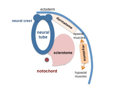
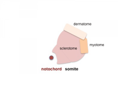
5. Somite Development - Loss of the Somite
Neural crest cells will migrate beside and through somite and finally the somite structure is lost and spreads as the 3 component parts.
- Sclerotome - from the left and right somite at each level will engulf the notochord. This transient structure is then resegmented to form the axial skeleton, vertebra and intervertebral discs.
- Dermatome - forms a thick band in the dorsal region of the embryo. This will then spread ventrally under the surface ectoderm (epidermis) to form the dermis of the skin.
- Myotome - from the ventrolateral lip of the dermomyotome, spreads both dorsally and ventrally to eventually form skeletal muscle cells.
- Dorsally - the epimere which in turn forms epaxial muscles, located behind the vertebral column.
- Ventrally - the hypomere, which in turn forms hypaxial muscles, located on the ventral body wall and somites at the level of the limbs will also form limb muscles.
Note - the cartoons show just the embryo righthand side mesoderm development (the same events occur on the lefthand side).
- Somite Links: 1 paraxial | 2 early somite | 3 sclerotome and dermomyotome | 4 dermatome and myotome | 5 somite spreading | SEM image - Human Embryo (week 4) showing somites | Movie - somitogenesis Hes expression
- Somite Cartoons
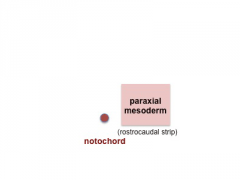
paraxial
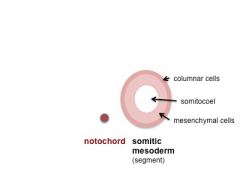
early somite
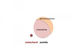
sclerotome and dermomyotome
dermatome and myotome

somite spreading


Cite this page: Hill, M.A. (2024, April 27) Embryology Somite cartoon5.png. Retrieved from https://embryology.med.unsw.edu.au/embryology/index.php/File:Somite_cartoon5.png
- © Dr Mark Hill 2024, UNSW Embryology ISBN: 978 0 7334 2609 4 - UNSW CRICOS Provider Code No. 00098G
File history
Click on a date/time to view the file as it appeared at that time.
| Date/Time | Thumbnail | Dimensions | User | Comment | |
|---|---|---|---|---|---|
| current | 18:01, 16 May 2014 | | 400 × 300 (27 KB) | Z8600021 (talk | contribs) | |
| 10:43, 10 August 2009 | 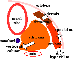 | 270 × 209 (6 KB) | MarkHill (talk | contribs) | Somite Development cartoon 5 Neural crest cells migrate beside and through somite. The myotome differentiates to form 2 components dorsally the epimere and ventrally the hypomere, which in turn form epaxial and hypaxial muscles respectively. The bulk of |
You cannot overwrite this file.
File usage
The following 43 pages use this file:
- 2009 Lecture 13
- 2009 Lecture 5
- 2010 BGD Lecture - Development of the Embryo/Fetus 1
- 2010 BGD Lecture - Development of the Embryo/Fetus 2
- 2010 BGD Practical 6 - Week 3
- 2010 Lab 3
- 2010 Lecture 13
- 2010 Lecture 5
- 2011 Lab 3 - Week 3
- 2014 Group Project 8
- ANAT2341 Lab 3 - Week 3
- BGDA Lecture - Development of the Embryo/Fetus 1
- BGDA Lecture - Development of the Embryo/Fetus 2
- BGDA Practical 7 - Week 3
- Developmental Mechanism - Epithelial Mesenchymal Transition
- Integumentary System Development
- Lecture - Limb Development
- Lecture - Mesoderm Development
- Lecture - Musculoskeletal Development
- Mesoderm
- Musculoskeletal System - Bone Development
- Musculoskeletal System - Limb Development
- Musculoskeletal System - Muscle Development
- Musculoskeletal System Development
- Notochord
- S
- Somite Musculoskeletal Movie
- Somitogenesis
- Talk:2010 BGD Practical 6 - Week 3
- Talk:2011 Lab 3
- Talk:2014 Group Project 8
- File:Mesoderm cartoon 05.jpg
- File:Mesoderm cartoon 06.jpg
- File:Mesoderm cartoon 07.jpg
- File:Mesoderm cartoon 08.jpg
- File:Mesoderm cartoon 09.jpg
- File:Somite cartoon1.png
- File:Somite cartoon2.png
- File:Somite cartoon3.png
- File:Somite cartoon4.png
- File:Somite cartoon5.png
- Template:Somite cartoon
- Category:Skeletal Muscle