File:Respiratory histology 05.jpg: Difference between revisions
| Line 14: | Line 14: | ||
* tracheal cartilage - hyaline cartilage, 16 to 20 C-shaped cartilages. | * tracheal cartilage - hyaline cartilage, 16 to 20 C-shaped cartilages. | ||
* trachealis muscle - (smooth muscle) Not visible in this section, together with connective tissue fibres, join ends of the cartilages together. | * trachealis muscle - (smooth muscle) Not visible in this section, together with connective tissue fibres, join ends of the cartilages together. | ||
'''Hyaline Cartilage Development''' | |||
* forms from mesenchymal cells. | |||
* precursor cells become rounded and form densely packed cellular masses, chondrification centres. | |||
* chondroblasts - (cartilage-forming cells) begin secreting the extracellular matrix components of cartilage. | |||
** extracellular matrix - ground substance (hyaluronan, chondroitin sulfates and keratan sulfate) and tropocollagen (polymerises into fine collagen fibres, not visible). | |||
Latest revision as of 15:01, 10 March 2013
Respiratory Trachea - Layers
Mucosa - formed by epithelium and underlying lamina propria.
- respiratory epithelium - (pseudostratified columnar and ciliated) ciliated cells, goblet cells, brush cells, endocrine cells, surfactant-producing cells (Clara cells), serous cells, basal cells, basement membrane.
- lamina propria - loose connective tissue, many elastic fibres
- elastic lamina - forming the border between the mucosa and submucosa is not visible in H&E stained slides.
Submucosa - connective tissue and submucosal glands
- submucosal gland - serous (dark) and mucous (light) parts have different staining appearance.
Cartilage
- perichondrium - surface of cartilage.
- tracheal cartilage - hyaline cartilage, 16 to 20 C-shaped cartilages.
- trachealis muscle - (smooth muscle) Not visible in this section, together with connective tissue fibres, join ends of the cartilages together.
Hyaline Cartilage Development
- forms from mesenchymal cells.
- precursor cells become rounded and form densely packed cellular masses, chondrification centres.
- chondroblasts - (cartilage-forming cells) begin secreting the extracellular matrix components of cartilage.
- extracellular matrix - ground substance (hyaluronan, chondroitin sulfates and keratan sulfate) and tropocollagen (polymerises into fine collagen fibres, not visible).
- Trachea Histology Links: Overview HE | Overview VG | Detail 1 HE Detail 2 HE | Respiratory Histology | Histology Stains | Histology
- Respiratory Histology: Bronchiole | Alveolar Duct | Alveoli | EM Alveoli septum | Alveoli Elastin | Trachea 1 | Trachea 2 | labeled lung | unlabeled lung | Respiratory Bronchiole | Lung Reticular Fibres | Nasal Inferior Concha | Nasal Respiratory Epithelium | Olfactory Region overview | Olfactory Region Epithelium | Histology Stains
Links: Histology | Histology Stains | Blue Histology images copyright Lutz Slomianka 1998-2009. The literary and artistic works on the original Blue Histology website may be reproduced, adapted, published and distributed for non-commercial purposes. See also the page Histology Stains.
Cite this page: Hill, M.A. (2024, May 18) Embryology Respiratory histology 05.jpg. Retrieved from https://embryology.med.unsw.edu.au/embryology/index.php/File:Respiratory_histology_05.jpg
- © Dr Mark Hill 2024, UNSW Embryology ISBN: 978 0 7334 2609 4 - UNSW CRICOS Provider Code No. 00098G
File history
Click on a date/time to view the file as it appeared at that time.
| Date/Time | Thumbnail | Dimensions | User | Comment | |
|---|---|---|---|---|---|
| current | 19:51, 27 February 2012 | 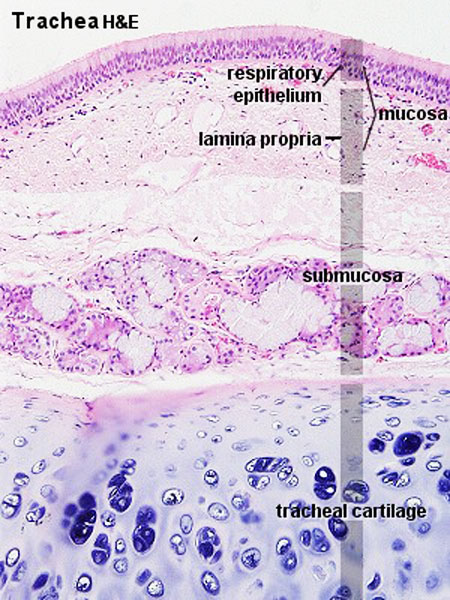 | 450 × 600 (96 KB) | Z8600021 (talk | contribs) | |
| 15:59, 2 March 2011 | 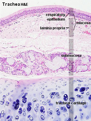 | 300 × 400 (54 KB) | S8600021 (talk | contribs) | ||
| 15:57, 2 March 2011 |  | 300 × 400 (61 KB) | S8600021 (talk | contribs) | ==Respiratory Trachea== Respiratory histology 05.jpg Original file name: Tra11he.jpg {{Template:Blue Histology}} Category:Respiratory |
You cannot overwrite this file.
File usage
The following 10 pages use this file: