File:Respiratory histology 03.jpg: Difference between revisions
From Embryology
(uploaded a new version of "File:Respiratory histology 03.jpg") |
|||
| (11 intermediate revisions by the same user not shown) | |||
| Line 1: | Line 1: | ||
==Respiratory Alveoli== | ==Respiratory Alveoli== | ||
[[File:Alveolar-sac-01.jpg|thumb|300px|Alveous cartoon]] | |||
{{HE}} | |||
Structure of the functional unit of the lung, note the capillary embedded in wall. Compare the structure with [[:File:Alveolar-sac-01.jpg|alveoli cartoon]]. | |||
===Alveolar type I cells=== | |||
* small alveolar cells or type I pneumocytes | |||
* are extremely flattened (the cell may be as thin as 0.05 µm) and branched | |||
* form the bulk (95%) of the surface of the alveolar walls. | |||
* The shape of the cells is very complex, and they may actually form part of the epithelium on both faces of the alveolar wall. | |||
* multiple '''cytoplasmic plates''' and relatively devoid of organelles | |||
** plates represent the gas exchange surface in the alveolus. | |||
===Alveolar type II cells=== | |||
* large alveolar cells or type II pneumocytes | |||
* about as many type II cells as type I cells (cell shape accounts for small contribution to alveolar area). | |||
* irregularly (sometimes cuboidal) shaped. | |||
* form small bulges on the alveolar walls. | |||
* contain are large number of granules called cytosomes (or multilamellar bodies) | |||
** consist of precursors to pulmonary surfactant (mixture of phospholipids that keep surface tension in the alveoli low). | |||
* can act as a progenitor cell for both type I and type II cells. | |||
===Alveolar Macrophage=== | |||
* dust cell | |||
* monocyte-derived immune system cell | |||
* removes debris and microorganisms from the alveoli. | |||
** subset of pulmonary macrophages. | |||
{{ | |||
{{Respiratory Histology}} | |||
{{Blue Histology}} | |||
[[Category:Respiratory]] | [[Category:Respiratory]] | ||
Latest revision as of 11:55, 8 April 2013
Respiratory Alveoli

(Stain - Haematoxylin Eosin)
Structure of the functional unit of the lung, note the capillary embedded in wall. Compare the structure with alveoli cartoon.
Alveolar type I cells
- small alveolar cells or type I pneumocytes
- are extremely flattened (the cell may be as thin as 0.05 µm) and branched
- form the bulk (95%) of the surface of the alveolar walls.
- The shape of the cells is very complex, and they may actually form part of the epithelium on both faces of the alveolar wall.
- multiple cytoplasmic plates and relatively devoid of organelles
- plates represent the gas exchange surface in the alveolus.
Alveolar type II cells
- large alveolar cells or type II pneumocytes
- about as many type II cells as type I cells (cell shape accounts for small contribution to alveolar area).
- irregularly (sometimes cuboidal) shaped.
- form small bulges on the alveolar walls.
- contain are large number of granules called cytosomes (or multilamellar bodies)
- consist of precursors to pulmonary surfactant (mixture of phospholipids that keep surface tension in the alveoli low).
- can act as a progenitor cell for both type I and type II cells.
Alveolar Macrophage
- dust cell
- monocyte-derived immune system cell
- removes debris and microorganisms from the alveoli.
- subset of pulmonary macrophages.
- Respiratory Histology: Bronchiole | Alveolar Duct | Alveoli | EM Alveoli septum | Alveoli Elastin | Trachea 1 | Trachea 2 | labeled lung | unlabeled lung | Respiratory Bronchiole | Lung Reticular Fibres | Nasal Inferior Concha | Nasal Respiratory Epithelium | Olfactory Region overview | Olfactory Region Epithelium | Histology Stains
Links: Histology | Histology Stains | Blue Histology images copyright Lutz Slomianka 1998-2009. The literary and artistic works on the original Blue Histology website may be reproduced, adapted, published and distributed for non-commercial purposes. See also the page Histology Stains.
Cite this page: Hill, M.A. (2024, May 18) Embryology Respiratory histology 03.jpg. Retrieved from https://embryology.med.unsw.edu.au/embryology/index.php/File:Respiratory_histology_03.jpg
- © Dr Mark Hill 2024, UNSW Embryology ISBN: 978 0 7334 2609 4 - UNSW CRICOS Provider Code No. 00098G
File history
Click on a date/time to view the file as it appeared at that time.
| Date/Time | Thumbnail | Dimensions | User | Comment | |
|---|---|---|---|---|---|
| current | 19:45, 27 February 2012 |  | 450 × 600 (29 KB) | Z8600021 (talk | contribs) | |
| 15:48, 2 March 2011 | 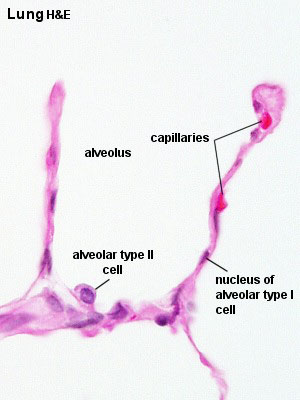 | 300 × 400 (21 KB) | S8600021 (talk | contribs) | ||
| 15:47, 2 March 2011 |  | 300 × 400 (27 KB) | S8600021 (talk | contribs) | ==Respiratory Alveoli== Respiratory histology 03.jpg Original file name: Lun40he.jpg {{Template:Blue Histology}} Category:Respiratory |
You cannot overwrite this file.