File:Periosteum.jpg: Difference between revisions
From Embryology
No edit summary |
No edit summary |
||
| Line 1: | Line 1: | ||
Bone | ==Bone Periosteum== | ||
A thin layer of cell-rich connective tissue, the endosteum, lines the surface of the bone facing the marrow cavity. Both the periosteum and the endosteum possess osteogenic potency. Following injury, cells in these layers may differentiate into osteoblasts (bone forming cells) which become involved in the repair of damage to the bone. | * Bone is surrounded by a layer of dense connective tissue, the periosteum. | ||
* A thin layer of cell-rich connective tissue, the endosteum, lines the surface of the bone facing the marrow cavity. | |||
* Both the periosteum and the endosteum possess osteogenic potency. | |||
* Following injury, cells in these layers may differentiate into osteoblasts (bone forming cells) which become involved in the repair of damage to the bone. | |||
{{Template:Blue Histology}} | |||
Original file name: Pos20he.jpg | |||
[[Category:Musculoskeletal]] [[Category:Histology]] | [[Category:Musculoskeletal]] [[Category:Histology]] | ||
Revision as of 14:57, 5 September 2011
Bone Periosteum
- Bone is surrounded by a layer of dense connective tissue, the periosteum.
- A thin layer of cell-rich connective tissue, the endosteum, lines the surface of the bone facing the marrow cavity.
- Both the periosteum and the endosteum possess osteogenic potency.
- Following injury, cells in these layers may differentiate into osteoblasts (bone forming cells) which become involved in the repair of damage to the bone.
Links: Histology | Histology Stains | Blue Histology images copyright Lutz Slomianka 1998-2009. The literary and artistic works on the original Blue Histology website may be reproduced, adapted, published and distributed for non-commercial purposes. See also the page Histology Stains.
Cite this page: Hill, M.A. (2024, May 5) Embryology Periosteum.jpg. Retrieved from https://embryology.med.unsw.edu.au/embryology/index.php/File:Periosteum.jpg
- © Dr Mark Hill 2024, UNSW Embryology ISBN: 978 0 7334 2609 4 - UNSW CRICOS Provider Code No. 00098G
Original file name: Pos20he.jpg
File history
Click on a date/time to view the file as it appeared at that time.
| Date/Time | Thumbnail | Dimensions | User | Comment | |
|---|---|---|---|---|---|
| current | 14:40, 18 February 2013 | 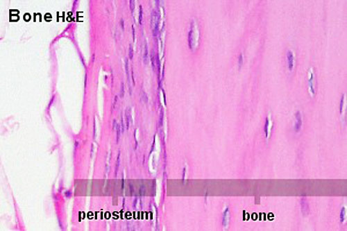 | 500 × 333 (34 KB) | Z8600021 (talk | contribs) | Increase image size and adjust contrast. |
| 11:26, 11 September 2009 | 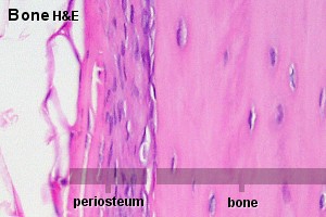 | 300 × 200 (18 KB) | S8600021 (talk | contribs) | Bone is surrounded by a layer of dense connective tissue, the periosteum. A thin layer of cell-rich connective tissue, the endosteum, lines the surface of the bone facing the marrow cavity. Both the periosteum and the endosteum possess osteogenic potenc |
You cannot overwrite this file.