File:Osteoclast.jpg
Osteoclast.jpg (500 × 333 pixels, file size: 41 KB, MIME type: image/jpeg)
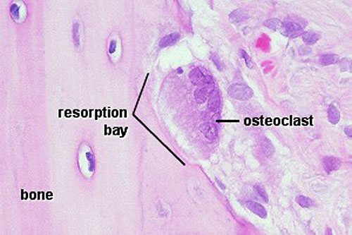
Osteoclasts
Very large (up to 100 µm), multi-nucleated (about 5-10 visible in a histological section, but up to 50 in the actual cell) bone-resorbing cells.
They arise by the fusion of monocytes (macrophage precursors in the blood) or macrophages. Osteoclasts attach themselves to the bone matrix and form a tight seal at the rim of the attachment site. The cell membrane opposite the matrix has deep invaginations forming a ruffled border. Osteoclasts empty the contents of lysosomes into the extracellular space between the ruffled border and the bone matrix. The released enzymes break down the collagen fibres of the matrix. Osteoclasts are stimulated by parathyroid hormone (produced by the parathyroid gland) and inhibited by calcitonin (produced by specialised cells of the thyroid gland). Osteoclasts are often seen within the indentations of the bone matrix that are formed by their activity (resorption bays or Howship's lacunae).
Original File Name: Ocl41he.jpg
File history
Click on a date/time to view the file as it appeared at that time.
| Date/Time | Thumbnail | Dimensions | User | Comment | |
|---|---|---|---|---|---|
| current | 14:37, 18 February 2013 | | 500 × 333 (41 KB) | Z8600021 (talk | contribs) | increased image size and adjusted contrast. |
| 11:28, 11 September 2009 |  | 300 × 200 (21 KB) | S8600021 (talk | contribs) | Osteoclasts Very large (up to 100 µm), multi-nucleated (about 5-10 visible in a histological section, but up to 50 in the actual cell) bone-resorbing cells. They arise by the fusion of monocytes (macrophage precursors in the blood) or macrophages. Oste |
You cannot overwrite this file.
File usage
The following 5 pages use this file: