File:Osteoclast.jpg: Difference between revisions
(Osteoclasts Very large (up to 100 µm), multi-nucleated (about 5-10 visible in a histological section, but up to 50 in the actual cell) bone-resorbing cells. They arise by the fusion of monocytes (macrophage precursors in the blood) or macrophages. Oste) |
No edit summary |
||
| (9 intermediate revisions by 2 users not shown) | |||
| Line 1: | Line 1: | ||
==Osteoclast== | |||
Very large (up to 100 µm), multi-nucleated (about 5-10 visible in a histological section, but up to 50 in the actual cell) bone-resorbing cells. | Very large (up to 100 µm), multi-nucleated (about 5-10 visible in a histological section, but up to 50 in the actual cell) bone-resorbing cells. | ||
* Cells arise by the fusion of [[M#monocyte|monocytes]] (macrophage precursors in the blood) or [[M#macrophage|macrophages]]. | |||
* Osteoclasts attach themselves to the bone matrix and form a tight seal at the rim of the attachment site. | |||
* The cell membrane opposite the matrix has deep invaginations forming a ruffled border. | |||
* Osteoclasts empty the contents of lysosomes into the extracellular space between the ruffled border and the bone matrix. | |||
* The released enzymes break down the collagen fibres of the matrix. | |||
* Osteoclasts are stimulated by parathyroid hormone (produced by the parathyroid gland) | |||
* Osteoclasts are inhibited by calcitonin (produced by specialised cells of the thyroid gland). | |||
* Osteoclasts are often seen lying over the indentations of the bone matrix that are formed by their activity (resorption bays or [[H#Howship.27s_lacuna|Howship's lacunae]]). | |||
{{Bone Histology}} | |||
{{Blue Histology}} | |||
Original File Name: Ocl41he.jpg | Original File Name: Ocl41he.jpg | ||
[[Category:Musculoskeletal]] [[Category:Histology]] [[Category:Bone]] | |||
Latest revision as of 14:43, 18 February 2013
Osteoclast
Very large (up to 100 µm), multi-nucleated (about 5-10 visible in a histological section, but up to 50 in the actual cell) bone-resorbing cells.
- Cells arise by the fusion of monocytes (macrophage precursors in the blood) or macrophages.
- Osteoclasts attach themselves to the bone matrix and form a tight seal at the rim of the attachment site.
- The cell membrane opposite the matrix has deep invaginations forming a ruffled border.
- Osteoclasts empty the contents of lysosomes into the extracellular space between the ruffled border and the bone matrix.
- The released enzymes break down the collagen fibres of the matrix.
- Osteoclasts are stimulated by parathyroid hormone (produced by the parathyroid gland)
- Osteoclasts are inhibited by calcitonin (produced by specialised cells of the thyroid gland).
- Osteoclasts are often seen lying over the indentations of the bone matrix that are formed by their activity (resorption bays or Howship's lacunae).
- Bone Histology: Cartilage Histology | Histology Stains | Histology | cartilage | bone | bone timeline
- Trabecular bone trabecular | lamellar | trabecular - overview HE | trabecular - low HE | trabecular - med HE
- Endochondral ossification primary ossification | endochondral ossification
- Intramembranous ossification intramembranous - VG low | intramembranous - VG high | intramembranous - HE low | intramembranous - HE high
Links: Histology | Histology Stains | Blue Histology images copyright Lutz Slomianka 1998-2009. The literary and artistic works on the original Blue Histology website may be reproduced, adapted, published and distributed for non-commercial purposes. See also the page Histology Stains.
Cite this page: Hill, M.A. (2024, May 28) Embryology Osteoclast.jpg. Retrieved from https://embryology.med.unsw.edu.au/embryology/index.php/File:Osteoclast.jpg
- © Dr Mark Hill 2024, UNSW Embryology ISBN: 978 0 7334 2609 4 - UNSW CRICOS Provider Code No. 00098G
Original File Name: Ocl41he.jpg
File history
Click on a date/time to view the file as it appeared at that time.
| Date/Time | Thumbnail | Dimensions | User | Comment | |
|---|---|---|---|---|---|
| current | 14:37, 18 February 2013 | 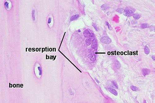 | 500 × 333 (41 KB) | Z8600021 (talk | contribs) | increased image size and adjusted contrast. |
| 11:28, 11 September 2009 |  | 300 × 200 (21 KB) | S8600021 (talk | contribs) | Osteoclasts Very large (up to 100 µm), multi-nucleated (about 5-10 visible in a histological section, but up to 50 in the actual cell) bone-resorbing cells. They arise by the fusion of monocytes (macrophage precursors in the blood) or macrophages. Oste |
You cannot overwrite this file.
File usage
The following 5 pages use this file: