File:Mouse organ of corti 03.jpg

Original file (1,280 × 1,024 pixels, file size: 207 KB, MIME type: image/jpeg)
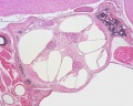
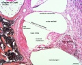
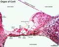
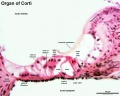
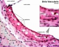
Organ of Corti (mouse)
This histology image is a section through one turn of the cochlea showing the organ of court.
- Deiters' cells - outer phalangeal cells
claudius cells - (cells of Claudius) columnar cells with microvilli overlying the basilar membrane and extend from Hensen's cells to the spiral prominence. Barrier cells that lie external to the organ of corti in endolymph.
Within the cochlea, the specialised structure required for converting mechanical vibration into an electrical signal occurs at the organ of Corti. Named after Alfonso Giacomo Gaspare Corti (1822–1876), an Italian anatomist who discovered this structure in 1851.
Inner Ear Histology: image - cochlea | image - cochlear duct | image - organ of corti | image - organ of corn detaili | image - stria vascularis | Inner Ear | Histology
cochlea
cochlear duct
organ of corti
organ of corti (detail)
stria vascularis


Links: Histology | Histology Stains | Blue Histology images copyright Lutz Slomianka 1998-2009. The literary and artistic works on the original Blue Histology website may be reproduced, adapted, published and distributed for non-commercial purposes. See also the page Histology Stains.
Cite this page: Hill, M.A. (2024, May 28) Embryology Mouse organ of corti 03.jpg. Retrieved from https://embryology.med.unsw.edu.au/embryology/index.php/File:Mouse_organ_of_corti_03.jpg
- © Dr Mark Hill 2024, UNSW Embryology ISBN: 978 0 7334 2609 4 - UNSW CRICOS Provider Code No. 00098G
Cite this page: Hill, M.A. (2024, May 28) Embryology Mouse organ of corti 03.jpg. Retrieved from https://embryology.med.unsw.edu.au/embryology/index.php/File:Mouse_organ_of_corti_03.jpg
- © Dr Mark Hill 2024, UNSW Embryology ISBN: 978 0 7334 2609 4 - UNSW CRICOS Provider Code No. 00098G
File history
Click on a date/time to view the file as it appeared at that time.
| Date/Time | Thumbnail | Dimensions | User | Comment | |
|---|---|---|---|---|---|
| current | 13:28, 18 May 2016 | | 1,280 × 1,024 (207 KB) | Z8600021 (talk | contribs) | ==Organ of Corti (mouse)== Within the cochlea, the specialised structure required for converting mechanical vibration into an electrical signal occurs at the organ of Corti. Named after Alfonso Giacomo Gaspare Corti (1822–1876), an Italian anatomist... |
You cannot overwrite this file.
File usage
The following 10 pages use this file:
- BGDB Face and Ear - Fetal
- Hearing - Inner Ear Development
- New 2016
- Paper - On the development of the membrana tectoria with reference to its structure and attachments
- File:Mouse organ of corti 01.jpg
- File:Mouse organ of corti 02.jpg
- File:Mouse organ of corti 03.jpg
- File:Mouse organ of corti 04.jpg
- File:Mouse organ of corti 05.jpg
- Template:Inner Ear Histology