File:Mouse organ of corti 01.jpg: Difference between revisions
From Embryology
mNo edit summary |
mNo edit summary |
||
| Line 1: | Line 1: | ||
==Organ of Corti (mouse)== | ==Organ of Corti (mouse)== | ||
Within the cochlea, the specialised structure required for converting mechanical vibration into an electrical signal occurs at the organ of Corti. Named after Alfonso Giacomo Gaspare Corti (1822–1876), an Italian anatomist who discovered this structure in 1851. | |||
This histology image is a section through one turn of the cochlea showing the organ of corti. | |||
{{Hearing Links}} | |||
| Line 6: | Line 12: | ||
{{Footer}} | {{Footer}} | ||
[[Category:Mouse]][[Category:Senses]] [[Category:Hearing]] [[Category:Histology]] [[Category:Inner Ear]] | |||
Revision as of 10:51, 16 May 2016
Organ of Corti (mouse)
Within the cochlea, the specialised structure required for converting mechanical vibration into an electrical signal occurs at the organ of Corti. Named after Alfonso Giacomo Gaspare Corti (1822–1876), an Italian anatomist who discovered this structure in 1851.
This histology image is a section through one turn of the cochlea showing the organ of corti.
Cite this page: Hill, M.A. (2024, May 2) Embryology Mouse organ of corti 01.jpg. Retrieved from https://embryology.med.unsw.edu.au/embryology/index.php/File:Mouse_organ_of_corti_01.jpg
- © Dr Mark Hill 2024, UNSW Embryology ISBN: 978 0 7334 2609 4 - UNSW CRICOS Provider Code No. 00098G
File history
Click on a date/time to view the file as it appeared at that time.
| Date/Time | Thumbnail | Dimensions | User | Comment | |
|---|---|---|---|---|---|
| current | 13:30, 18 May 2016 | 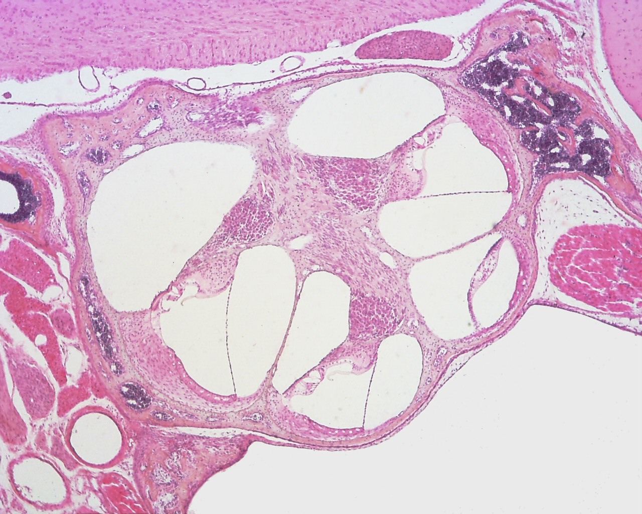 | 1,280 × 1,024 (339 KB) | Z8600021 (talk | contribs) | |
| 10:13, 18 May 2016 | 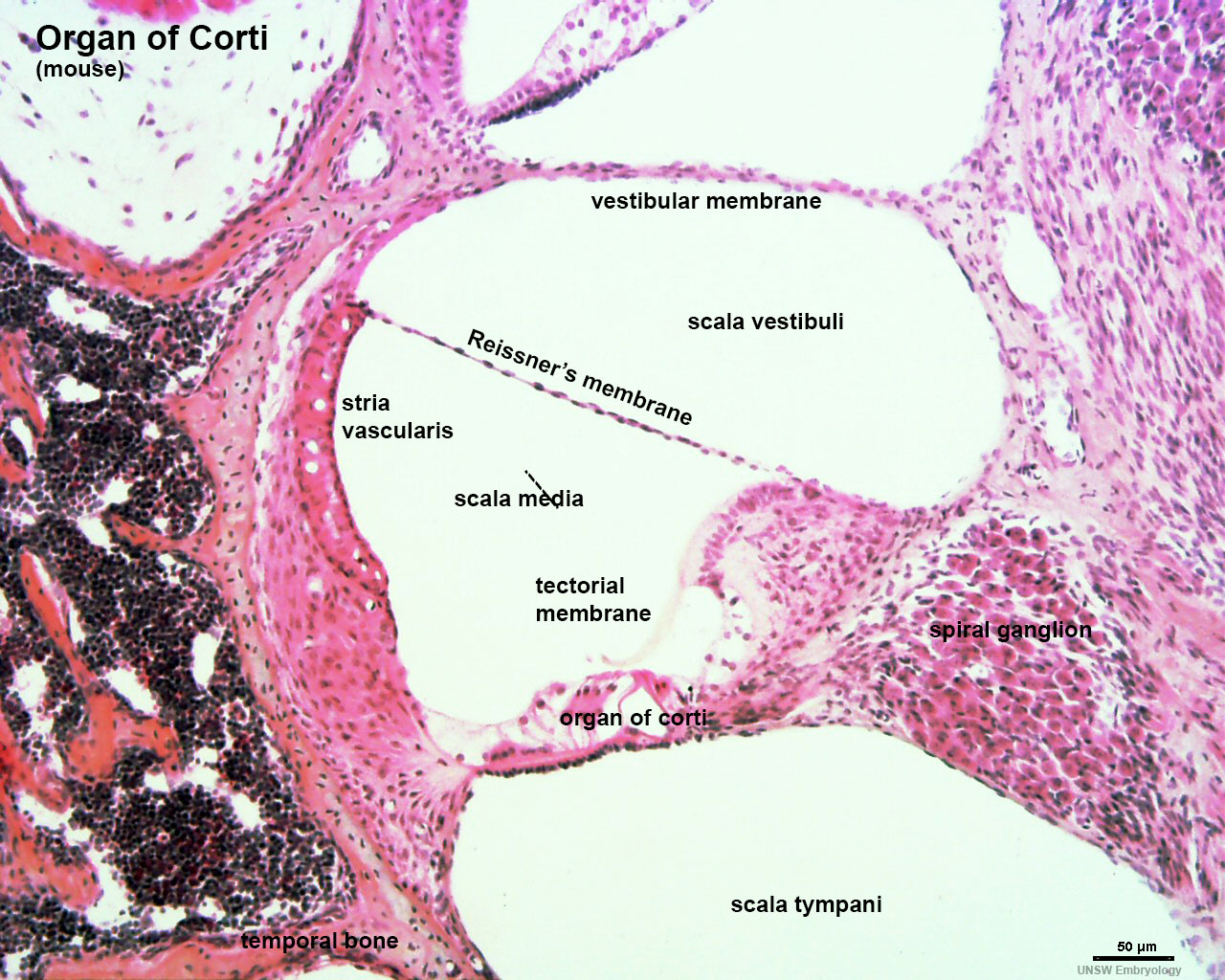 | 1,280 × 1,024 (320 KB) | Z8600021 (talk | contribs) | ||
| 10:46, 16 May 2016 | 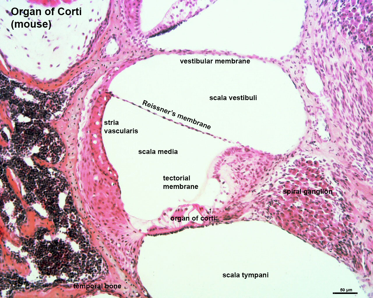 | 1,280 × 1,024 (320 KB) | Z8600021 (talk | contribs) | ==Organ of Corti (mouse)== |
You cannot overwrite this file.