File:Keibel Mall 261.jpg: Difference between revisions
From Embryology
({{KM Axial Skeleton}} {{Keibel_Mall Images}} Category:Human Category:Bone Category:Axial Skeleton) |
No edit summary |
||
| Line 1: | Line 1: | ||
==Fig. 261. The Development of the Cartilaginous Thorax== | |||
(After Charlotte Muller, Morpholog. Jahrb., 1906.) The development of the cartilaginous thorax. | |||
[[:File:Keibel Mall 260-263.jpg|Fig. 260-263.]] | |||
* [[:File:Keibel Mall 260.jpg|Fig. 260.]] Embryo 13 mm long. | |||
* [[:File:Keibel Mall 261.jpg|Fig. 261.]] Embryo 17 mm long. | |||
* [[:File:Keibel Mall 262.jpg|Fig. 262.]] Embryo 16 mm long. | |||
* [[:File:Keibel Mall 263.jpg|Fig. 263.]] Embryo 32 mm long. | |||
{{KM Axial Skeleton}} | {{KM Axial Skeleton}} | ||
{{Keibel_Mall Images}} | {{Keibel_Mall Images}} | ||
[[Category:Human]] [[Category:Bone]] [[Category:Axial Skeleton]] | [[Category:Human]] [[Category:Cartilage]] [[Category:Bone]] [[Category:Axial Skeleton]] | ||
Latest revision as of 21:03, 2 October 2012
Fig. 261. The Development of the Cartilaginous Thorax
(After Charlotte Muller, Morpholog. Jahrb., 1906.) The development of the cartilaginous thorax.
- Fig. 260. Embryo 13 mm long.
- Fig. 261. Embryo 17 mm long.
- Fig. 262. Embryo 16 mm long.
- Fig. 263. Embryo 32 mm long.
- Axial Skeleton: Fig. 231 to Fig. 273 | Vertebral Column and Thorax | Occipital Region | XI. Development of the Skeleton and of the Connective Tissues
- KM Figure Links: The Germ Cells | Segmentation | First Primitive Segment | Gastrulation | External Form | Placenta | Axial Skeleton | Limb Skeleton | Skull | Muscular System
| Historic Disclaimer - information about historic embryology pages |
|---|
 |
Glossary Links
- Glossary: A | B | C | D | E | F | G | H | I | J | K | L | M | N | O | P | Q | R | S | T | U | V | W | X | Y | Z | Numbers | Symbols | Term Link
Cite this page: Hill, M.A. (2024, May 19) Embryology Keibel Mall 261.jpg. Retrieved from https://embryology.med.unsw.edu.au/embryology/index.php/File:Keibel_Mall_261.jpg
- © Dr Mark Hill 2024, UNSW Embryology ISBN: 978 0 7334 2609 4 - UNSW CRICOS Provider Code No. 00098G
File history
Click on a date/time to view the file as it appeared at that time.
| Date/Time | Thumbnail | Dimensions | User | Comment | |
|---|---|---|---|---|---|
| current | 20:43, 2 October 2012 | 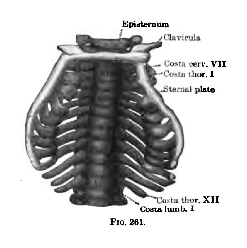 | 506 × 508 (32 KB) | Z8600021 (talk | contribs) | {{KM Axial Skeleton}} {{Keibel_Mall Images}} Category:Human Category:Bone Category:Axial Skeleton |
You cannot overwrite this file.
File usage
The following 3 pages use this file: